The relationship of the main foramen to the anatomic root apex has been the subject of several studies. Although they are anatomically close, they rarely coincide, and their distance can vary according to age or tooth type, ranging from 0.2 to 3.0 mm. The aim of this short communication was to evaluate the distance between the main foramen of independent middle mesial canals (MMCs) and the anatomical mesial root apex of mandibular first molars using the micro-computed tomography. Twenty-five mandibular first molars with MMCs were scanned (resolution of 9.9 µm), and the distance from its main foramen to the anatomical apex was evaluated. Overall, the distance ranged from 0.2 to 2.4 mm; however, in 3 specimens the distance was greater than 3 mm. This report demonstrates that the exit of the main foramen of the MMC varies considerably and could approach a substantial distance from the anatomical apex greater than previously reported.
Unusual Deviation of the Main
F o r a m e n f r o m t h e R o o t A p e x
Marco Aurélio Versiani1, Hany Mohamed Aly Ahmed2, Manoel Damião de Sousa-Neto1, Gustavo De-Deus3, Paul Michael Howell Dummer4
1Department of Restorative Dentistry, School of Dentistry of Ribeirão Preto, USP - Universidade de São Paulo, Ribeirão Preto, SP, Brazil 2Department of Conservative Dentistry, School of Dental Sciences, Universiti Sains Malaysia, Kelantan, MY, Malaysia 3Department of Endodontics, School of Dentistry, UNIGRANRIO - Universidade Grande Rio, Rio de Janeiro, Brazil
4School of Dentistry, College of Biomedical and Life Sciences, Cardiff University, Cardiff, UK
Correspondence: Prof. Dr. Marco A. Versiani, Av. do Café, s/nº, 14049-904 Ribeirão Preto, SP, Brasil. Tel: +55-16-3603-6783. e-mail: [email protected]
Key Words: anatomy, dental pulp cavity, microcomputed tomography, root canal preparation, tooth apex.
ISSN 0103-6440
Brazilian Dental Journal (2016) 27(5): 589-591 http://dx.doi.org/10.1590/0103-6440201601106
Introduction
An understanding of the common and unusual morphology of the root canal system, especially in the apical third, is a fundamental prerequisite for successful root canal treatment (1-3). Therefore, an overall comprehension of the anatomy of the apical root canal and its variations is necessary to understand the requirements for treatment, to avoid damage to periapical tissues and to ensure adequate disinfection of the canal. The literature describes the terminal portion of a tooth root by distinct landmarks namely the minor apical foramen, apical constriction, major apical foramen, root apex, and cemento-dentinal junction (CDJ) (2). The apical constriction is the apical part of the root canal with the narrowest diameter, and is generally located 0.5 – 1.5 mm from the apical foramen, the major apical foramen is the main exit of the root canal onto the external root surface, while the CDJ is the line of union between dentin and cementum at which pulpal tissue ends (4). The relation of the major apical foramen with the anatomic root apex has been the subject of several studies. Although they are anatomically close, they rarely coincide (4,5), and their distance can vary according to age or tooth type, ranging from 0.2 to 3.0 mm (2,6).
The mesial root of mandibular molars commonly has a mesiobuccal and a mesiolingual canal; however, other anatomical configurations such as the presence of an extra canal, termed middle mesial canal (MMC), have also been reported (1,7,8). Currently, improvements in digital imaging systems and the use of magnification in clinical practice
has led to an increased reporting on the incidence of MMCs in mandibular molars (1,8), when compared with previous publications using conventional methodologies. This anatomical variation has several endodontic implications. Radiographically, the MMC is usually superimposed over the root structure and the other mesial canals, making its orifice and terminus impossible to identify (1). Even though considerable amount of information regarding MMC has been published, there is no information available on the distance from its major foramen to the anatomic apex in mandibular molars. Therefore, the aim of this study was to evaluate the distance from the major foramen of the MMC to the anatomic apex of the mesial root of mandibular first molars, using a micro-computed tomographic imaging system (micro-CT).
Material and Methods
Braz Dent J 27(5) 2016
590
M
.A
.V
ersiani et al.
slices (NRecon v.1.6.9; Bruker-microCT), polygonal surface representations of the internal anatomy of mesial roots were obtained (CTAn v.1.14.4; Bruker-microCT). Then, the distance from the major foramen of the MMC to the anatomic apex of the mesial root was measured using DataViewer v.1.4.4 software (Bruker-microCT).
Results
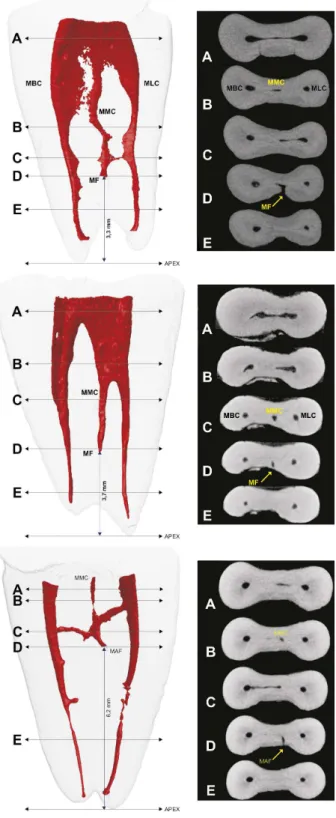
In 22 mandibular first molars, the distance from the main foramen of the MMCs to the anatomic apex of the mesial root ranged from 0.2 to 2.4 mm; however, in 3 specimens this distance was greater than previously reported in the literature (Fig. 1).
Discussion
The incidence of MMCs using conventional clearing and radiographic techniques has been reported to range from 0.82% to 37.5% (1). Unfortunately, the lack of accuracy of conventional tools in studying certain features of the root canal system is prone to a wide range of interpretation. Therefore, these inherent limitations encouraged the search for newer methodologies (1,9,10). In recent years, the development of non-invasive high-resolution micro-CT has gained increasing significance for three-dimensional assessment of the root canal system (9). Using this new imagery technology, several authors have confirmed the high level of complexity of the root canal system regarding apical anatomy and the presence of additional canals encased in the roots of maxillary and mandibular posterior teeth (1,9-12). Using this highly accurate contemporary non-destructive tool, Versiani et al. (1) reported that the incidence of MMCs in mandibular first molars was 18.6% (48 out of 258 molars), and that it merged with the other mesial canals in most of these teeth (73.3%).
The MMC orifice is often hidden by a dentinal projection on the mesial aspect of pulp chamber walls making its detection challenging. In order to find the MMC orifice, this dentin projection must be carefully removed and an extensive exploration of the grooves between mesial orifices should be done preferably under good illumination and magnification, using ultrasonic tips or long shank rounded burs (8). However, the length and depth of this groove must be taken into consideration during the troughing because of the limited dentin thickness toward the furcation side in relation to the MMC orifice, which increases the risk of root perforation (1).
It is well known that pulp and periodontium have embryonic and functional inter-relationships giving rise to anatomical connections that remain throughout the life of the tooth. In the lateral aspect of the roots, large accessory canals have been considered the main factor responsible for the progress of pulpal disease into the periodontal tissues,
with the development of lateral radiolucent lesions (7). This report revealed that the exit of the main foramen of the MMC could reach a greater distance from the anatomical apex than previously reported. In some instances, its exit
Braz Dent J 27(5) 2016
591
Unusual deviation of the main foramen.
may show considerable variations, reaching distances more than 3 mm from the anatomical apex (Fig. 1). Therefore, because of this significant displacement, the use of electronic foramen locators is of utmost importance to help the detection of the main foramen. This will confine the root canal treatment procedures within the root canal system thus preventing potential errors including incorrect measurement of canal length and subsequent over-instrumentation (5).
Awareness for the potential occurrence of this variation in the position of the main foramen in the middle mesial canal and its impact in the development of combined periodontal-endodontic lesions in the furcation area of mandibular molars is important. It would be advisable for clinicians to explore extensively the grooves between mesiobuccal and mesiolingual orifices in order to locate additional canals and to use electronic foramen locators to help the detection of the main foramen.
Acknowledgements
The authors deny any conflicts of interest. This study was supported by FAPESP (2013/03695-0, 2012/16072-2) and CNPq (168179/2014-8).
Resumo
A relação do forame principal com o ápice anatômico da raiz tem sido objeto de vários estudos. Embora estejam anatomicamente próximos, eles raramente coincidem, e a sua distância pode variar de acordo com a idade ou o tipo de dente, oscilando entre 0,2 e 3,0 mm. O objetivo deste artigo é relatar que a saída foraminal do canal mediano (MMC), presente na raiz mesial de molares inferiores, pode apresentar variações significativas, atingindo distâncias acima de 3 mm do ápice anatômico. Vinte e cinco primeiros molares inferiores com MMC foram escaneados (resolução de 9,9 mm) e a distância do forame principal ao ápice anatômico avaliada. Em geral, a distância mostrou variações consideráveis; no entanto, em 3 espécimes esta distância ficou acima de 3 mm. Este estudo teve como objetivo relatar que a saída do forame principal de canais medianos de primeiros molares inferiores pode apresentar variações consideráveis, podendo atingir distâncias maiores do que as relatadas previamente na literatura.
References
1. Versiani MA, Ordinola-Zapata R, Keles A, Alcin H, Bramante CM, Pécora JD et al. Middle mesial canals in mandibular first molars: A micro-CT study in different populations. Arch Oral Biol 2016;61:130-137. 2. Vertucci FJ. Root canal morphology and its relationship to endodontic
procedures. Endod Topics 2005;10:3-29.
3. Ahmed HMA, Hashem AA. Accessory roots and root canals in human anterior teeth: a review and clinical considerations. Int Endod J 2016;49:724-736.
4. Kuttler Y. Microscopic investigation of root apexes. J Am Dent Assoc 1955;50:544-52.
5. Nekoofar MH, Ghandi MM, Hayes SJ, Dummer PMH. The fundamental operating principles of electronic root canal length measurement devices. Int Endod J 2006;39:595-609.
6. Dummer PM, McGinn JH, Rees DG. The position and topography of the apical canal constriction and apical foramen. Int Endod J 1984;17:192-198.
7. Ahmed HMA, Luddin N. Accessory mesial roots and root canals in mandibular molar teeth: case reports, SEM analysis and literature
review. Endod Practice 2012;6:195-205.
8. Nosrat A, Deschenes RJ, Tordik PA, Hicks ML, Fouad AF. Middle mesial canals in mandibular molars: incidence and related factors. J Endod 2015;41:28-32.
9. Versiani MA, Pécora JD, Sousa-Neto MD. Root and root canal morphology of four-rooted maxillary second molars: a micro-computed tomography study. J Endod 2012;38:977-982.
10. Versiani MA, Pécora JD, Sousa-Neto MD. Microcomputed tomography analysis of the root canal morphology of single-rooted mandibular canines. Int Endod J 2013;46:800-807.
11. Ahmad IA, Al-Jadaa A. Three root canals in the mesiobuccal root of maxillary molars: case reports and literature review. J Endod 2014;40:2087-2094.
12. Ahmed HMA, Fadhli MF, Gutmann JL. Seven root canals in a deciduous maxillary molar tooth detected by the dental operating microscope and micro-computed tomography. Scanning 2016 [Epub Ahead of Print. doi:10.1002/sca.21299].