Identification of phenolic compounds and biologically related activities
from
Ocotea odorifera
aqueous extract leaves
Douglas Costa Gontijo
a,d, Geraldo Célio Brandão
b, Pablo Costa Gontijo
c, Alaíde Braga de Oliveira
a,
Marisa Alves Nogueira Diaz
d, Luciano Gomes Fietto
d, João Paulo Viana Leite
d,⇑aDepartamento de Produtos Farmacêuticos, Universidade Federal de Minas Gerais, MG, Brazil bEscola de Farmácia, Universidade Federal de Ouro Preto, MG, Brazil
cDepartamento de Entomologia, Universidade Federal de Lavras, MG, Brazil
dDepartamento de Bioquímica e Biologia Molecular, Universidade Federal de Viçosa, MG, Brazil
a r t i c l e
i n f o
Article history:
Received 15 November 2016
Received in revised form 11 March 2017 Accepted 14 March 2017
Available online 18 March 2017
Keywords:
Phenolic compounds Flavonoids Tannins Antioxidant Antibacterial Antimutagenic
a b s t r a c t
Ocotea odorifera(Vell.) Rohwer is popularly used as food and flavoring. The aim of this study was to deter-mine the chemical composition of the aqueous extract fromO. odoriferaleaves and evaluate the correla-tion of their phytochemical composicorrela-tion and biological activities. The antioxidant effect was determined by DPPH radical scavenging,b-carotene-linoleic acid and lipid peroxidation assays; the antibacterial activity was evaluated by the hole plate and MIC techniques and the antimutagenic activity was evalu-ated by the Ames test. Identification of phytochemicals was performed by LC–ESI/MS and the correlation between the phytochemical composition of the extract and the evaluated activities. The results allowed the identification of 13 phenolic compounds in the extract that exhibited high antioxidant activity and moderate antibacterial and antimutagenic action. Statistical analyses showed correlation of the total phe-nolic content with biologically related activities. The phytochemical analyses, together with the biolog-ical results, support the popular use ofO. odorifera.
Ó2017 Elsevier Ltd. All rights reserved.
1. Introduction
Ocotea odorifera(Vell.) Rohwer, belonging to the Lauraceae fam-ily, is a native species in Brazil that is found naturally in areas of the Atlantic Forest. It is usually known as sassafrás, canela-sassafrás and canela-do-mato and is used as a food flavoring (Oliveira, Yamada, Fagg, & Brandão, 2012; Oltramari et al., 2004) and in traditional medicine for rheumatism treatment and as an antimalarial (Bolsaris, 2007; Tribess et al., 2015). In 2013, this spe-cies was entered into the Red Book of the Flora of Brazil as having a risk of extinction and was classified as ‘‘endangered” (Martinelli & Moraes, 2013).
Methanolic extracts from O. odorifera leaves showed no cytotoxic activity against Vero cells and a lack of genotoxicity by an in vivo micronucleus test, while ellagitannins isolated from O. odorifera showed potent activity against Candida parapsilosis (Yamaguchi et al., 2011). The essential oil ofO. odoriferais used as source of sassafras oil, which has long been exported from Brazil to Europe, Japan and the USA (Oltramari et al., 2004) due to the
antifungal and larvicidal activities that were reported for the essential oil of this species (Castro & Lima, 2011; Pinto et al., 2010). Phytochemical analysis of the O. odorifera essential oil showed that the oil contains a high concentration of safrole in addition to phenylpropanoid derivatives, steroids, coumarins and sesquiterpenes (Lordello, Cavalheiro, Yoshida, & Gottlieb, 2000; Oltramari et al., 2004). Safrole was a common flavoring in the bev-erage, cosmetic, food and medical industries, but this practice was discontinued after safrole was determined to induce carcinogenic-ity (Miller & Miller, 1983). It is also known that the essential oil of different Ocoteaspecies exhibits strong antioxidant and antibacte-rial action (Bruni et al., 2004). However, no extensive characteriza-tion of the phytochemical composicharacteriza-tion of the aqueous extract of Ocotea odoriferaleaves has been performed thus far, and its biolog-ical properties with regard to food use, such antioxidant, antibacte-rial and antimutagenic properties, have yet to be reported. Effort has therefore been focused on using available experimental tech-niques to identify antioxidant, antibacterial and antimutagenic compounds from natural products (Gursoy, Sarikurkcu, Cengiz, & Solak, 2009; Trabelsi et al., 2013). Thus, the aim of this study was to determine the chemical composition of the aqueous extract of O. odorifera leaves and to evaluate the correlation of its
http://dx.doi.org/10.1016/j.foodchem.2017.03.087
0308-8146/Ó2017 Elsevier Ltd. All rights reserved.
⇑Corresponding author.
E-mail address:[email protected](J.P.V. Leite).
Contents lists available atScienceDirect
Food Chemistry
phytochemical composition with antioxidant, antibacterial and antimutagenic activities.
2. Materials and methods
2.1. Plant material
Samples ofO. odoriferaplants were collected in the Atlantic For-est biome in December of 2010 on Conservation Unit Parque Estad-ual da Serra do Brigadeiro in the state of Minas Gerais, Brazil. Voucher specimens were deposited at the Herbário VIC, Universi-dade Federal de Viçosa, with the code VIC 42.326. The ‘‘collection authorization license” at CU was issued by the ‘‘Instituto Estadual de Floresta de Minas Gerais” (N°CU 073/07, 050/07 COL). The spe-cies identification was carried out by comparison of herbarium specimens.
2.2. Extract preparation
The leaves ofO. odoriferawere dried in a ventilated, dark room at ambient temperature and were subsequently pulverized in a knife mill. The leaf extract ofO. odoriferawas prepared by infusion using distilled water at 90°C (ratio plant:solvent, 1:20 w/v) for 15 min with shaking. The extract was then vacuum filtered and lyophilized, yielding 5.8% (relative to the weight of the plant mate-rial) dry extract ofO. odorifera(EOO).
2.3. Chemical analyses
2.3.1. Thin layer chromatography analyses
The presence of secondary metabolites, flavonoids, tannins, coumarins, anthraquinones, terpenes/steroids, saponins and alka-loids was detected in EOO by thin layer chromatography (TLC silica gel F254). Mobile phases and visualization spray reagents were used as indicated inWagner and Bladt (1996)and compared with refer-ence compounds.
2.3.2. Ultra-high performance liquid chromatography–mass spectrometry in series analyses
Analyses by ultra-high performance liquid chromatography coupled to ultraviolet spectroscopy (LC–DAD) and to mass spec-trometry (LC–ESI-MS/MS) were performed according toGattuso et al. (2006)with some modifications in the Laboratório de Fitoquí-mica at the Faculdade de Farmácia, UFMG. Phenolic compounds were identified based on the typical UV absorption for each pheno-lic class analyzed, in addition to their typical fragmentation pat-terns obtained by MS/MS in comparison to the literature data.
The analyses were performed in an ACQUITYÒ
TQD ion trap mass spectrometer (Waters) equipped with a quadrupole instru-ment fitted with an electrospray source in the positive and nega-tive ESI modes and atmospheric pressure chemical ionization (APCI). Interface that was operated with the following conditions: ion spray voltage: 4 kV; orifice voltage: 60 V; capillary voltage, 3500 V; capillary temperature, 320°C; source voltage, 5 kV; vapor-izer temperature, 320°C; corona needle current, 5 mA; sheath gas, nitrogen, 27 psi; and the instrument was operated in the full scan mode (100–2000 Da). The LC–ESI-MS/MS analyses were addition-ally performed with helium as the collision gas and the collision energy was set at 30 eV. Chromatographic separation was done on a reversed-phase column ACQUITY UPLC BEH (1.7
l
m, 502 mm i.d.) (Waters). The mobile phase consisted of aqueous 0.1% formic acid (solvent A) and acetonitrile with 0.1% formic acid (solvent B). The elution protocol was a 0–11 min linear gradient from 5 to 95% B. The flow rate was 0.3 mL min 1, and the sampleinjection volume was 4.0
l
L. UV–Visible spectra were registered from 190 to 450 nm.2.3.3. Determination of phenolic compounds by UV–Visible spectrophotometry
The total polyphenolic content of the EOO was determined using the Folin-Ciocalteu reagent with tannic acid as the standard, and the absorbance at 760 nm was measured after 60 min of reac-tion at room temperature in the dark (Verza, Kreinecker, Reis, Henriques, & Ortega, 2007). Folin-Ciocalteu reagent (0.5 mL), 15% aqueous Na2CO3(1.0 mL) and distilled water (8.3 mL) were added to a test tube, followed by the addition of 200
l
L of EOO solution (3.0 mg/mL resuspended in water). The results were expressed as mg of tannic acid equivalent (TAE)/mg of EOO.The total tannin content was calculated according toVerza et al. (2007). Thus, 20 mL of EOO (3 mg/mL resuspended in water) was mixed with 200 mg of hide powder and vortexed at 200 rpm for 60 min. Subsequently, the sample was filtered, and 200
l
L of the EOO filtrate was collected. The following steps were identical to those in the experiment for the quantification of total polyphenols. The total tannin content was obtained by the difference between the total polyphenol content and the value obtained in the Folin-Ciocalteu reagent test in the absence of tannins. Similarly, the total tannin content was also expressed as tannic acid equivalents (TAE) using a standard curve generated with tannic acid as standard.Flavonoids were quantified by the AlCl3method with rutin as the standard compound, and the absorbance at 420 nm was mea-sured in a UV–Visible spectrometer after 15 min of reaction at room temperature in the dark (Lamaison & Carnet, 1990). In total, 0.5 mL of acetic acid, 10 mL of methanol and 2.5 mL of AlCl36H2O 8% in methanol were added to 5 mL of EOO (50 mg/mL resus-pended in methanol), and the sample was brought to a final vol-ume of 25 mL with methanol. The results were expressed asmg of rutin equivalents (RE)/mg of EOO.
2.4. Antioxidant activity
2.4.1. DPPH radical scavenging activity
The DPPH (2,2-diphenyl-1-picrylhydrazyl) antioxidant test was performed by adding 2.0 mL of DPPH (0.1 mM) to 2.0 mL of solu-tions of EOO (0.5, 25, 50, 75 and 100
l
g/mL) diluted in methanol, and the absorbance was measured with a UV–Visible spectropho-tometer at 517 nm after 30 min of reaction at room temperature in the dark (Abdel-Hameed, 2009). A synthetic product, butylated hydroxytoluene (BHT), and standard Ginkgo biloba leaf extract (EGb 761) were used as antioxidant standards that were tested at the same EOO concentrations. The IC50was determined by per-forming an exponential regression of the plotted points for EOO, which revealed the concentration required to reduce the initial concentration of DPPH radicals by 50%. The percentage of remain-ing DPPH was calculated usremain-ing the followremain-ing equation:%DPPH radical capture¼ ½1 ðASÞ=ðA0Þ 100
where ASis the absorbance of the sample or standard and A0is the absorbance of the control (DPPH only).
2.4.2.b-Carotene-linoleic acid assay
mL, resuspended in methanol), and the tubes were incubated at 50°C to accelerate oxidation reactions and start the bleaching of b-carotene. The absorbance was read in a UV–Visible spectropho-tometer at 470 nm at intervals of 20 min from 0 h to 160 min of incubation time. BHT and EGb 761 standards were also used for comparison with the results obtained for EOO. The results are expressed as the percentage of inhibition of oxidation of the sam-ples (As), which was calculated in relation to the decrease in absor-bance of the negative control (Ac).
%Inhibition¼ ððAc AsÞ=AcÞ 100
2.4.3. Lipid peroxidation
The antioxidant activity was also evaluated using the lipid per-oxidation test, according to the work ofConforti, Statti, Tundis, Menichini, and Houghton (2002). For this procedure, 2.5 mL of dis-tilled water, 500
l
L of soy lecithin solution (7700l
g/mL), and 1 mL of ascorbic acid (1750l
g/mL) were added to a test tube, followed by 125l
L of iron(III) chloride (330l
g/mL) and 100l
L of EOO (12l
g/mL). After the addition of these reagents, the tubes were incubated in a shaker for 60 min at 40°C. After this time, 1 mL of thiobarbituric acid (TBA 3700l
g/mL) was added to all tubes, and the tubes placed in a water bath at 80°C for 15 min. The tubes were then centrifuged for 10 min at 10,000 rpm. The absorbance at 532 nm of the supernatant from the centrifugation was read in the UV–Visible spectrophotometer. BHT and EGb 761 standards were also used for a comparison with the results obtained for EOO. The results are expressed as the percentage of inhibition of the oxidation of the sample (As), which was calculated in relation to the decrease in the absorbance of the negative control (Ac).%Inhibition¼ ððAc AsÞ=AcÞ 100
2.5. Antibacterial action
2.5.1. Hole plate method
EOO were tested againstStaphylococcus aureus3993, Staphylo-coccus aureus4125 andEscherichia coli24 strains isolated from ani-mals with bovine mastitis. The isolates were grown in 5 mL of brain heart infusion broth for 16 h at 37°C. The antibacterial activ-ity was evaluated by the diffusion technique in agar using the hole plate method (NCCLS & National Committee for Clinical Laboratory Standards,, 2003). A 100-
l
L suspension of the bacteria (106UFC/ mL ofS. aureus 3993, S. aureus4125 and E. coli24 strains) was spread on Müeller-Hinton agar. Holes of approximately 5 mm in diameter and 3 mm in height were made in the agar, and 30l
L of EOO (50 mg/mL) prepared in dimethylsulfoxide (DMSO) was added to the holes, followed by 24 h of incubation at 37°C. After incubation, the inhibition zones were measured in millimeters. Ampicillin (5 mg/mL) was used as the positive control, and DMSO was used as the negative control. The experiments were repeated twice in triplicate. The determination of the antibacterial activity of EOO was performed using the diameter of the bacterial growth inhibition zone and diameter of the growth inhibition zone of the positive control.%Antibacterial activity
¼Hole with extract at the evaluated concentration
Holewithpositi
v
econtrol 1002.5.2. Minimum inhibitory concentration
The broth microdilution method was used for determination of the minimum inhibitory concentration (MIC) of EOO that still showed activity (NCCLS, 2003). The 96-well plates were prepared by dispensing 0.4, 0.8, 1.2, 1.6, 2.0, 2.4, 2.8, 3.2, 3.6, 4.0, 8.0, 12.0,
16.0, 20.0, 24.0, 28.0, 32.0, 36.0 and 40.0
l
L aliquots of EOO, fol-lowed by 100mL of inoculum containing 106UFC/mL ofS. aureus 3993,S. aureus4125 orE. coli24, with the remainder of the volume in each well being supplied with Müeller-Hinton broth. The micro-plates were incubated at 37°C for 24 h. After incubation for 24 h, 4l
L of the salt INT (iodonitrotetrazolium) (2 mg/mL) was added to each well. The bacterial viability was observed by the formation of pink color after the addition of the INT. Ampicillin (5 mg/mL) was used as the positive control and DMSO was used as the nega-tive control.2.6. Antimutagenic and antigenotoxic activities
2.6.1. Antimutagenicity
The Ames test was chosen to quantify the extract’s antimuta-genicity. The EOO was preincubated for 20–30 min with the TA97, TA98, TA100 and TA102 strains ofSalmonella typhimurium/ microsome, with and without metabolic activation (S9) and differ-ent mutagens (Maron & Ames, 1983). Five different concentrations of EOO (562.5, 1125, 2250, 3375 and 4500mg/mL) in distilled water were assessed. The concentration employed was based on a cyto-toxicity test carried out for EOO with the TA100 strain without metabolic activation. Thus, the upper limit of the dose interval that was tested was either the highest nontoxic dose or the lowest toxic dose that was determined in this preliminary assay. To each con-centration of the tested mixture was added 500mL of phosphate buffer 0.2 M pH 7.4 (without metabolic activation) or 500mL S9 mix (with metabolic activation), 100mL of bacteria culture (4108cells/mL) and 2
mL of ampicillin (50 mg/mL), besides dif-ferent mutagens (mutagens used in the test without metabolic activation were 50mL of 4-nitro-O-phenylenediamine (100
l
g/ mL) for TA97 and TA98, 10mL of sodium azide (50l
g/mL) for TA100 and 2mL of mitomycin C (500l
g/mL) for TA102, and 25mL of 2-aminoanthracene (200l
g/mL) was used for all strains with metabolic activation) incubated at 37°C for 30 min. The lyophi-lized rat liver S9 fraction induced by Aroclor 1254 was purchased from Moltox (Molecular Toxicology, Annapolis, USA). The S9 mix (50 mL) was prepared as follows: 19.75 mL of distilled water, 25 mL of 0.2 M phosphate buffer pH 7.4, 2 mL of 0.1 M NADP, 0.25 mL of 1 M d-glucose-6-phosphate, 1 mL of 1.65 M KCl + 0.4 M MgCl26H2O salt solution and 2 mL of the lyophilized S9 fraction that had been reconstituted with distilled water. After the incubation period, 2 mL of top agar and the mixture were pla-ted on glucose minimal medium. The plates were incubapla-ted at 37°C for 48 hours, and theHis+revertants were counted. The tests were conducted in triplicate. Water distilled was used as a negative control.The antimutagenic action was determined using the equation fromOh, Kim, Choi, Chung, and Ham (2008):
%Antimutagenic action
¼ðn
orevertants controlðÞ norevertants concentration testedÞ
ðnorevertants controlðþÞ norevertants controlð ÞÞ 100
2.6.2. Antigenotoxic activity
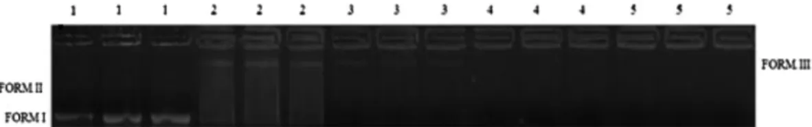
the visualization of the DNA. The DNA samples treated with dis-tilled water were used as the negative control. The plasmid DNA test was assessed by verifying the presence or absence of the relaxed-circular conformation (form III), the helical conformation (form II) and the strained superhelical conformation (form I). A comparison between the DNA bands of the samples and the positive- and negative-control DNA bands was used to diagnose any possible DNA-damaging action of EOO.
2.7. Data analysis
The data on the quantification of total phenolics, tannins and flavonoids, as well as those of the antioxidant, antibacterial and antimutagenic activities were analyzed by the Shapiro-Wilk and Bartlett tests (
a
= 0.05) to confirm the presence of a normal distri-bution and homoscedasticity, respectively. Antioxidant activity data were subjected to a one-way ANOVA test, followed by a Tukey test (a
= 0.05) to separate the means. Antibacterial activity data, which did not meet the assumptions of ANOVA, were submitted to a Kruskal-Wallis test followed by a Dunn’s test (a
= 0.05). The antimutagenic activity data were subjected to a three-way ANOVA (a
= 0.05) to test the interaction among the effects of strains, the metabolic activation and the concentrations of EOO. If an effect was detected, the means were compared by the Tukey test (a
= 0.05). Principal component analysis (PCA) using XLSTAT 2014.5.03 software with Pearson correlation model was applied to clarify the relationship between phenolic compounds and the biological activities of the EOO. All statistical analyses were per-formed using SigmaPlot 12.5 (Systat Software, San Jose, CA, USA).3. Results and discussion
3.1. Chemical analyses
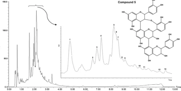
The phytochemical screening of secondary metabolic groups by TLC detected the presence of saponins, flavonoids and tannins. A UPLC–DAD chromatogram of the EOO monitored at 210 nm is shown in Fig. 1. This fingerprinting corroborated the results obtained from the phytochemical prospection, and peaks 1–13 cor-respond to identified phenolic constituents, including condensed tannins (proanthocyanidins) and flavonoid glycosides. Analysis of
first-order MS spectra recorded for each peak, together with MS-MS experiments in positive and negative ESI modes, literature UV comparisons and retention times (RT), allowed the identifica-tion of the extract phytochemicals (Table S1 and Fig. S1). A major peak was detected at 1.61 (compound 1), 2.00 (compound 4) and 2.16 min (compound 5). The use of UPLC coupled to tandem mass spectrometry (MS/MS) can provide abundant information for the structural elucidation of a wide range of compounds (Regueiro et al., 2014).
For fragmentation, the aglycone ion was not observed in the presence of flavone C-glycosides due to the strength of the CAC
connection between the sugar and aglycone moieties. This behav-ior was observed in first-order MS of compounds 8, 9 and 13, sug-gesting that these compounds are flavone C-glycosides. The MS-MS spectra obtained by focusing on each [M H] ion of compounds 8, 9 and 13 exhibited the same pattern of fragmentation ([(M H) – 18 Da] , [(M H) – 90 Da] , [(M H) – 120 Da] , [A+113 Da] , and [A+83 Da] ), which is typical of di-C-glycosylflavones. Positions 6 and 8 appeared to be substituted in each case since the maximum of band II was located at 270 nm or higher. The presence of the [(M H) – 60] fragment in these MS-MS spectra, which is usually generated by the fragmentation of pentose derivatives, along with the presence of the [(M H) – 120 Da] (base peak) and [(M H) – 90 Da] peaks, suggested that the sugar substituents were pen-toses and hexoses (Gattuso et al., 2006). The m/z of the [A +113 Da] and [A+83 Da] ions correspond to the aglycone-bearing sugar fragments, and they are particularly important for aglycone identification. For positive identification and characteri-zation of flavan-3-ol monomers, dimers, trimers and tetramers, the following points were considered: a UV spectrum with a peak near 280 nm (kmax) and molecular ion peaks in positive- and negative-ion ESI modes of MS. The fragmentation pathway hetero-cyclic ring fission (HRF) andretro-Diels–Alder (RDA) fragmentation give information about the hydroxylation of the B-rings and the bonds between two monomeric unit; quinonemethide (QM) frag-mentation defines the two monomeric units and especially the base unit (Jaiswal, Jayasinghe, & Kuhnert, 2012).
Compound 1:The UV spectrum of compound 1 was compatible with a flavan-3-ol type (epi)catechin. One peak was detected atm/z 1153 in positive ESI mode and assigned to a proanthocyanidin tet-ramer [(epi)catechin-(4,8/2,7)-(epi)catechinan-(4,8)-(epi )catechin-(4,8)-(epi)catechin]. This compound produced the MS-MS base
peak in positive ESI mode atm/z865 that derived from the QM cleavage; a secondary peak atm/z1001 [(M H) – 152 Da] origi-nates from an RDA fragment. Another fragment was generated at m/z 983 by the loss of an RDA fragment (152 Da), followed by the loss of a water molecule (18 Da).
Compounds 2, 10 and 12:Three isomers were detected atm/z 577 in the negative ESI mode and were tentatively assigned as (epi)catechin-(epi)catechin dimers with B-type linkages. Com-pounds 2 and 10 produced the MS-MS base peak at m/z 407 [(M H) – 170 Da] by losing an RDA fragment (152 Da), followed by the loss of a water molecule (18 Da); compound 2 showed sec-ondary peaks at m/z 289 ([(epi)catechin-H+]), which originates from a QM fragment) and at 425 [(M-H)+-152 Da], which origi-nates from an RDA fragment. Compound 12 produced the MS-MS base peak atm/z289 [(M H) – 288 Da], which originates from a QM fragment, and secondary peaks atm/z407, caused by the loss of an RDA fragment (152 Da) and the subsequent loss of a water molecule (18 Da), and atm/z299, caused by the loss of an RDA fragment (152 Da) followed by the loss of an HRF fragment (126 Da).
Compound 3:The UV spectrum of compound 3 is compatible with an apigenin aglycone. The negative ESI mode MS-MS experi-ment focused on them/z577 ion showed the presence of a 308-Da disaccharide on an apigenin skeleton, which is composed of a rhamnose unit ([(M H) – 146 Da] ) connected to a glucose unit ([(M H) – 162 Da] ). Since the disaccharide can be considered to be linked to position 7, compound 3 has been identified as apigenin 7-O-rutinoside.
Compounds 4 and 13:Two isomers were detected atm/z 575 while running in negative ESI mode and were assigned to an A-type (epi)catechin dimer. Compound 4 produced the MS-MS base peak atm/z 151 (RDA) and secondary peaks atm/z289 and 285 (QMs) and 449 (HFR). Compound 13 produced the MS-MS base peak at m/z 289 (QM) and secondary peaks at m/z 151 (RDA), 285 (QM), and 449 (HFR).
Compound 5:One peak was detected atm/z863 while running in negative ESI mode and it was assigned to a proanthocyanidin tri-mer, [(epi)catechin-(4,8/2,7)-(epi)catechinan-(4,8)-(epi)catechin]. Compound 5 produced the MS-MS base peak in negative ESI mode atm/z 573 with a neutral loss of an (epi)catechin unit; the sec-ondary peak atm/z711 [(M-H) -152 Da] originates from an RDA fragment. Other important fragments were observed atm/z 451 and 411, which are present from the fragmentation of the (epi )cat-echin trimer ((epi)catechin?A?(epi)catechin?(epi)catechin) connection.
Compounds 6 and 7:The first-order MS spectra of compounds 6 and 7 showed the same protonated species [M+H]+m/z 291. In both MS-MS spectra, the [(M H) – 152 Da] ions were formed by the loss of an RDA fragment. The base peak in both compounds at [M+H]+m/z147 refer to the loss of an H
2O molecule [(M+H) – 18 Da]+, followed by an HFR fragment ( 126 Da). Thus, compounds 6 and 7 are identified as monomeric (epi)catechin.
Compounds 8 and 9:Compound 8 and 9 are isomers with glu-cose in the 6 and 8 positions, respectively. The positive-mode MS-MS spectrum focused onm/z565.32 and 565.45 ([M+]+) ions was not particularly useful for structural identification; only peaks derived from the fragmentation of the sugar units were observed. The presence of the [A+113 Da] and [A+83 Da] peaks in the MS-MS spectrum focused on them/z563.23 and 563.29 ([M H] ) ions, indicating that the aglycone has a molecular weight of 270. The UV spectrum showed typical absorptions for a disubstituted trihydroxyflavone. The loss of 60, 90 and 120 mass units from the deprotonated species [M H] (as discussed previously), in addition to the shift observed for band II in the UV spectrum, con-firmed that the sugar substituents at positions 6 and 8 are pentoses and hexoses. The retention time, UV, and positive- and
negative-mode ESI MS-MS spectra led to the identification of compounds 8 and 9 as apigenin 6-C-pentoside-8-C-hexoside and/or apigenin 6-C-hexoside-8-C-pentoside.
Compound 11:The UV spectra showed absorption bands cen-tered at 272 and 334 nm, which can be attributed to bands II and I, respectively, of a flavone structure. The first-order MS spectrum of compound 11 showed the deprotonated species [M H] atm/z 593.35. In the MS-MS spectra, the [(M H) – 90 Da] and [(M H) – 120] ions suggested the presence of hexoses as substituents at the 6 and 8 positions. Compound 11 was identified as apigenin 6,8-di-C-glucoside (vicenin-2).
3.2. Quantification of phenolic compounds
Quantification of the phenolic compounds in EOO determined the content of total polyphenolics (46.81 ± 3.43mg TAE/mg of EOO), tannins (29.73 ± 2.23mg TAE/mg of EOO) and flavonoids (9.61 ± 0.41mg RE/mg of EOO). Many of these phenolic compounds that have antioxidant activity are used in food products and a number of medical treatments (Li, Hao, Wang, Huang, & Li, 2009).
3.3. Antioxidant activity
The values found for EOO in the DPPH radical scavenging test, the co-oxidation system ofb-carotene/linoleic acid and the lipid peroxidation test show the antioxidant activity measured by each of the different methods (Table 1). The correlation analysis among the total polyphenolic, tannin, and flavonoid concentrations and the antioxidant activity of EOO showed that only the lipid peroxi-dation test directly relates to the content of total polyphenols and total tannins (the angle between the axis of total polyphenol and total tannin parameters and the axis of the lipid peroxidation parameters is nearly 0°). For the system ofb-carotene/linoleic acid, there is a weak correlation with the tannin content. For the DPPH radical-scavenging test, there was no correlation with any of the levels of polyphenols evaluated (angles between the axes are nearly 90°). The flavonoid content had a negative correlation with the lipid peroxidation test, since the axis vectors for the two parameters are in opposite directions (the angle between the axes is 180°) (Fig. 2A). Due to the complexity of the antioxidant pro-cesses, it is important to use different methodologies to assess this activity.
The mechanism of antioxidant action can include suppressing ROS formation, either by inhibition of enzymes or by chelating trace elements involved in free-radical production, scavenging
Table 1
Antioxidant activity of leaf aqueous extract fromOcotea odorifera(EOO).
Treatments/ statistic1
Antioxidant activity IC50of DPPH5
(mg/mL)
b-Carotene/linoleic acid (%)
Lipid
peroxidation (%) NC2 – 0.0 ± 0.0 c 0.0 ± 0.0 c
BHT3 2.6 ± 0.2 b 79.3 ± 3.8 a 96.2 ± 1.3 a
EGb 7614 2.2 ± 0.0 c 0.9 ± 1.8 c 63.5 ± 6.7 b
EOO 4.2 ± 0.2 a 32.5 ± 3.6 b 52.8 ± 6.9 b
F 125.7 541.6 119.2
df 2,6 3,8 3,8
P <0.001 <0.001 <0.001
1One-way ANOVA (a= 0.05). Means (±SD, n = 3) followed by different letters
were significantly different within columns (Tukey test’s,a= 0.05). – dashes indi-cate no data.
2NC – negative control = without extract. 3BHT – butylated hydroxytoluene.
4EGb 761 – standardizedGinkgo bilobaextract.
5Concentration of extract necessary to decrease the initial concentration of DPPH
reactive species, and upregulating or protecting antioxidant defenses (van Acker et al., 1996). Unlike the scavenging of free radicals by DPPH, which is based on electron transfer from an antioxidant compound to an oxidant, the co-oxidation of b-carotene/linoleic acid and lipid peroxidation determine the activity of a compound in protecting a substrate from lipid oxida-tion by neutralizing free radicals formed in the system (Gursoy et al., 2009). Studies claim that the antioxidant activity of phenolic compounds is mainly due to their reducing properties and chemi-cal structure, which play an important role in the neutralizing or sequestering of free radicals and the chelation of transition metals (Kristinova, Mozuraityte, Storro, & Rustad, 2009).
3.4. Antibacterial action
The results of antibacterial activity showed a hole inhibition of 13.5 ± 1.4 and 11.5 ± 1.2 mm for the gram-positive strainsS. aureus 3993 andS. aureus4125, respectively (Table 2). The presence of the
lipopolysaccharide membrane in gram-negative bacteria, such as E. coli24, strongly controls the penetration of antibacterial agents and explains why this strain was not susceptible to the antibacte-rial action of the EOO (Nikaido, 2003). For antibacterial diffusion tests in agar MIC, there is no consensus on the inhibition level acceptable to natural products, when compared with antibiotics standards. However, studies indicate a positive result of antibacte-rial action for an inhibition hole of greater than 7 mm (Nascimento, Locatelli, Freitas, & Silva, 2000) and a MIC of less than 500mg (Aligianis, Kalpoutzakis, Mitaku, & Chinou, 2001). Thus, this value of MIC (40 < MIC < 60mg) classifies the EOO as having strong antibacterial action.
Studies point to phenolic compounds as important promoters and metabolites of antibacterial activity (Mingo, Silván, & Martinez-Rodriguez, 2016). Several modes of action of phenolic compounds have been suggested to cause antibacterial inhibition. These mechanisms include damaging the bacterial cell membrane (Lacombe, Tadepalli, Hwang, & Wu, 2013), the inhibition of
Fig. 2.Ordination diagrams of the principal component analysis (PCA) based on the phenolics compounds and biological activities of aqueous leaf extract fromOcotea odorifera(EOO). (A) Antioxidant activity by methods DPPH radical scavenging,b-carotene/linoleic acid and lipid peroxidation and phenolics compounds. (B) Antibacterial activity and phenolics compounds. (C) Mutagenic inhibition inSalmonella typhimurium/microsome strains, with (+S9) and without ( S9) metabolic activation and phenolics compounds. (D) Antioxidant activity and mutagenic inhibition inSalmonella typhimurium/microsome strains, with (+S9) and without ( S9) metabolic activation.
Table 2
Antibacterial action (hole inhibition ± SD (mm)) and Minimum Inhibitory Concentration (MIC) of leaf aqueous extract fromOcotea odorifera(EOO).
Treatments/statistic1 Concentration Antibacterial action4(%)
S. aureus3993 S. aureus4125 E. coli24
AMP2 150
mg/disk 100 a (43.0 ± 1.0) 100 a (34.7 ± 0.6) 100 (29.7 ± 0.6)
DMSO3 30
mL/disk 0.0 ± 0.0 c (0.0 ± 0.0) 0.0 ± 0.0 c (0.0 ± 0.0) 0.0 ± 0.0 (0.0 ± 0.0)
EOO 1500mg/test 31.4 ± 3.2 b (13.5 ± 1.4) 33.2 ± 3.5 b (11.5 ± 1.2) 0.0 ± 0.0*(0.0 ± 0.0)
H 16.4 16.4 –
df 2 2 –
P <0.001 <0.001 –
MIC 40 < MIC < 60mg 40 < MIC < 60mg –
1 Kruskal-Wallis test’s (a= 0.05). Means (±SD, n = 6) followed by different letters were significantly different within columns (Dunn’s test’s,a= 0.05). – dashes indicate no
data analysis.
2 Ampicillin – positive control. 3 Dimethyl sulfoxide – negative control.
4 Antibacterial action = [(inhibition zone (mm) of treatment/inhibition zone (mm) of positive control)100].
extracellular enzymes, a direct effect on antibacterial metabolism (Scalbert, 1991) and DNA degradation (Brudzynski, Abubaker, & Miotto, 2012). Thus, Fig. 2B shows high statistical correlation between the flavonoid content and its antibacterial activity against the S. aureus 3993 strain (very close axes). Furthermore, the arrangement between the axes of the tannins and polyphenols and axis of the inhibition of the growth of theS. aureus3993 strain shows a negative correlation (axes approximately 180°), showing that the antibacterial activity against this strain is only related to flavonoid content. For theS. aureus4125 strain, the antibacterial activity is not related to any measured polyphenol content (axes arranged near 90°).
3.5. Antimutagenic and antigenotoxic activities
We observed that for the TA97 strain, the antimutagenic activ-ity is observed for 1125.0, 2250.0 and 3375.0mg of EOO in the absence of S9 metabolism, and mutagenic inhibition is observed for these three statistically similar concentrations (Table 3). According to Caillet, Lessard, Lamoureux, and Lacroix (2011), antimutagenic action greater than 70% is considered strong, between 40% and 70% is considered moderate, and below 40% is considered neutral. Note also that the antimutagenic action of the EOO to the TA97 strain in the absence of S9 metabolism was higher than that found in the presence of S9. For the TA98 strain of EOO, there is a moderate antimutagenic response to EOO in the absence of S9 metabolism, and strong activity for 3375.0 and 4500.0mg of EOO in the presence of S9 metabolism. The TA100
and TA102 strains already showed no antimutagenic response to EOO in the absence of S9 metabolism; however, increased antimu-tagenic activity after S9 metabolism was observed (moderate activity against strain TA100 and high antimutagenic activity against the TA102 strain) (Table 3). Thus, we emphasize that the highest antimutagenic activity was observed for the TA97 and TA98 strains, compared with strains TA100 and TA102, in the absence of S9 metabolism. No significant variation among the four different strains was observed in the presence of S9 metabolism. Differences in the mechanism of action of the studied strains are directly related to the obtained results. For TA97 and TA98 strains, the mechanism of action occurs by frameshifts (geneshisD6610 TA97 andhisD3052 TA98), while for the TA100 and TA102 strains (geneshisG46 TA100 andhisG428 TA102), the mechanism occurs by base-pair substitution (Maron & Ames, 1983). The homogenate S9 fraction of the constituent enzymes promotes oxidation reac-tions catalyzed by the cytochrome P450 enzymes (Basheer & Kerem, 2015). Thus, since there is a known interaction between the constituent CYP450 enzymes (Rodeiro et al., 2008), it is believed that these oxidative enzymes have promoted the oxida-tion of polyphenolic compounds present in the EOO, causing decreased antimutagenic activity in the different strains ofS. typhi-muriumthat were evaluated.
The mechanisms of action of the antimutagenic activity of phe-nolic compounds include interference with cytochrome P450-mediated metabolism of mutagens, interaction with active muta-genic metabolites, DNA protection against mutagens presenting electrophilic properties, scavenging of the electrophilic mutagens,
Table 3
Number of colonies and mutagenic inhibition inSalmonella typhimuriumstrains by leaf aqueous extract fromOcotea odorifera(EOO), without ( S9) and with (+S9) metabolic activation.
Strain Concentration (mg/plate)
S9 +S9
N°. of colonies Statistic comparations3 N°. of colonies Statistic comparations
Inhibition (%) 1st 2nd 3rd Inhibition (%) 1st 2nd 3rd
TA97 NC1 317.3 ± 6.1 – 474.0 ± 10.0 –
562.5 438.7 ± 8.3 71.2 ± 2.0 b a a 842.7 ± 30.0 43.0 ± 4.6 a b a 1125.0 333.0 ± 26.3 96.3 ± 6.2 a a a 796.0 ± 20.0 50.3 ± 3.1 a b ab 2250.0 275.0 ± 18.1 110.1 ± 4.3 a a a 804.0 ± 6.9 49.0 ± 1.1 a b a 3375.0 320.0 ± 20.0 99.4 ± 4.8 a a a 825.3 ± 24.1 45.7 ± 3.7 a b b 4500.0 539 ± 26.2 39.8 ± 9.5 c a b 774.7 ± 14.0 46.0 ± 13.4 a a b PC2 738.3 ± 15.2 0.0 ± 0.0 d 1121.3 ± 61.5 0.0 ± 0.0 b
TA98 NC 60.7 ± 10.1 – 40.3 ± 0.6 –
562.5 126.3 ± 1.5 35.8 ± 1.5 bc a b 1094.7 ± 61.2 25.9 ± 4.3 b a a 1125.0 114.7 ± 12.2 47.2 ± 11.9 ab a b 1009.3 ± 115.5 31.9 ± 8.1 b b b 2250.0 115.7 ± 10.1 46.3 ± 9.8 ab a b 830.7 ± 30.3 44.5 ± 2.1 b a a 3375.0 143.0 ± 14.7 19.5 ± 14.4 cd b c 385.3 ± 36.1 75.8 ± 2.5 a a a 4500.0 86.3 ± 20.1 65.9 ± 26.4 a a a 392.0 ± 32.7 74.6 ± 3.4 a a a PC 163.0 ± 8.0 0.0 ± 0.0 d 1464.0 ± 55.0 0.0 ± 0.0 c
TA100 NC 448.0 ± 10.6 – 204.0 ± 48.0 –
562.5 852.3 ± 4.5 12.7 ± 1.0 b b c 1098.0 ± 20.3 28.1 ± 1.6 b a a 1125.0 879.0 ± 11.0 7.0 ± 2.4 b b c 958.7 ± 37.3 39.3 ± 3.0 ab a ab 2250.0 854.7 ± 10.1 12.2 ± 2.2 b b c 890.0 ± 38.0 44.8 ± 3.1 ab a a 3375.0 725.3 ± 16.2 40.1 ± 3.5 a a b 994.0 ± 34.0 36.5 ± 2.7 ab a b 4500.0 712.7 ± 10.3 36.3 ± 13.6 a a b 790.0 ± 36.2 50.5 ± 6.8 a a b PC 911.3 ± 7.6 0.0 ± 0.0 b 1447.7 ± 116.4 0.0 ± 0.0 c
TA102 NC 196.0 ± 8.0 – 210.0 ± 14.0 –
562.5 921.7 ± 24.2 16.0 ± 2.8 ab a c 376.0 ± 14.4 27.5 ± 6.3 c a a 1125.0 925.0 ± 24.0 15.6 ± 2.8 ab b c 318.7 ± 39.2 52.5 ± 17.1 b a a 2250.0 829.3 ± 40.1 26.7 ± 4.6 a b c 326.7 ± 18.5 49.1 ± 8.1 b a a 3375.0 858.3 ± 32.0 23.3 ± 3.7 a b bc 278.7 ± 28.9 70.0 ± 12.6 ab a a 4500.0 856.3 ± 26.1 22.3 ± 5.1 a b b 240.0 ± 42.3 76.4 ± 36.5 a a a
PC 1060.0 ± 4.0 0.0 ± 0.0 b 439.0 ± 3.6 0.0 ± 0.0 d
– dashes indicate no data.
1 Negative control = distilled water (100.0
mg/plate).
2 Positive control = 5.0
mg/plate of 4-Nitro-o-phenylenediamine (TA97-S9 and TA98-S9); 5.0mg/plate of sodium azide (TA100-S9) and 1.0mg/plate of mitomycin-C
(TA102-S9); 5.0mg/plate of 2-aminoanthracene (TA97+S9; TA98+S9; TA100+S9 and TA102+S9).
3 Three-way ANOVA (interaction strain x metabolic activation x concentration:F= 11.0;df= 15,96;P< 0.001). Means (± SD, n = 3) followed by different letters were
and binding or insertion into the outer membrane transporters, which would lead to the blockage of a mutagen that was trans-ferred into the cytosol (Słoczyn´ska, Powroz´nik, Pe˛kala, & Waszkielewicz, 2014). Thus, the results of statistical correlation between polyphenol content and antimutagenic action for the dif-ferent ofS. typhimuriumstrains in the absence and presence of S9 metabolism shows high correlation between the content of total polyphenols and tannins and the antimutagenic action across the TA97 (-S9), TA98 (+S9) and TA98 (-S9) strains (Fig. 2C). As observed for the frameshift mechanism of these strains, it is believed that the phenolic constituents of the EOO are more likely to act by pre-venting a frameshift. For the other strains, with and without S9 metabolism, no correlation was observed between the phenolic content and the antioxidant activity, confirming the lower muta-genicity inhibition values shown inTable 3. There is already an association between antioxidant activity and antimutagenic action; the results inFig. 2D show a high correlation among the results obtained for the antioxidant activity in the lipid peroxida-tion test and the antimutagenic acperoxida-tion of EOO against TA97 ( S9) and TA98 strains ( S9 and +S9). There is also a high correla-tion between the antimutagenic activity against TA98 strains ( S9) and the antioxidant activity in the co-oxidation of b-carotene/ linoleic acid. Due to the similarity of the antioxidant mechanism in these two tests, it is believed that they can be used as pretests for the identification of natural products with potential antimuta-genic action. For strains with base-pair substitution mechanisms (TA100 and TA102), there was a low correlation between the antimutagenic results with and without S9 metabolism and the lipid peroxidation test. However, the DPPH test for scavenging free radicals showed poor correlation with antimutagenic results in dif-ferentS. typhimuriumstrains.
Finally, the antigenotoxic activity of EOO was not observed in any of the test concentrations in the presence of the genotoxic agent SnCl2(Fig. 3). Thus, it is noted that the appearance of a linear arrangement of the plasmid (form III) in lane 3 (lowest concentra-tion EOO) is similar to the electrophoretic profile seen in lane 2 (positive control), in which we notice the absence of the helical conformations and tensioned superhelices, forms II and I, respec-tively, as were observed in lane 1 (negative control). For the two highest concentrations tested, it was not possible to see the pres-ence of the plasmid DNA in agarose gel. Although the EOO has a high phenolic content, the antioxidant activity of the extract was not able to inhibit the redox reactions promoted by the genotoxic agent SnCl2that destabilize DNA.
4. Conclusion
The results obtained allowed the identification of 13 phenolic compounds in the aqueous extract ofOcotea odoriferaleaves; these compounds were identified as tetramers, trimers and dimers of (epi)catechin, in addition to other flavonoids and tannins. More-over, highin vitroantioxidant activity was observed in the different tests, with a high correlation between the content of total phenolic compounds and the antioxidant activity in the lipid peroxidation test. The results showed antibacterial action againstStaphylococcus aureusstrains that highly correlated with the flavonoid content.
Finally, we obtained high antimutagenic action of the EOO against TA97 and TA98Salmonella typhimurium/microsome strains in the absence of S9 metabolism; there is also a correlation among total phenolic content, antimutagenic activity and antioxidant action. The aqueous extract ofO. odoriferaleaves proved to possess inter-esting properties, emerging from both its chemical composition and from the evaluation of its in vitrobiological activities. It is believed, therefore, that the observed therapeutic activities can add value to the use ofO. odoriferaas a food condiment. In fact, food products withO. odoriferaaqueous extract could be an inter-esting possibility, with accentuated therapeutic activities in addi-tion to those of the essential oil (without the recognized toxicity of the safrole compound).
Conflict of interest statement
There are no conflicts of interest involved in this study.
Acknowledgements
We are grateful to the Coordenação de Aperfeiçoamento de Pes-soal de Nível Superior (CAPES), the Conselho Nacional Desenvolvi-mento Científico e Tecnológico (CNPq) and the Fundação de Amparo à Pesquisa do Estado de Minas Gerais (FAPEMIG) for finan-cial supports.
Appendix A. Supplementary data
Supplementary data associated with this article can be found, in the online version, at http://dx.doi.org/10.1016/j.foodchem.2017. 03.087.
References
Abdel-Hameed, E. S. S. (2009). Total phenolic contents and free radical scavenging activity of certain EgyptianFicusspecies leaf samples.Food Chemistry, 114, 1271–1277.
Aligianis, N., Kalpoutzakis, E., Mitaku, S., & Chinou, I. B. (2001). Composition and antimicrobial activity of the essential oil of twoOriganumspecies.Journal of Agriculture and Food Chemistry, 49, 4168–4170.
Basheer, L., & Kerem, Z. (2015). Interactions between CYP3A4 and dietary polyphenols.Oxidative Medicine and Cellular Longevity, 1–15.
Bolsaris, A. S. (2007). Plants used traditionally to treat malaria in Brazil: the archives of Flora Medicinal. Journal of Ethnobiology and Ethnomedicine, 3.
Brudzynski, K., Abubaker, K., & Miotto, D. (2012). Unraveling a mechanism of honey antibacterial action: Polyphenol/H2O2-induced oxidative effect on bacterial cell
growth and on DNA degradation.Food Chemistry, 133, 329–336.
Bruni, R., Medici, A., Andreotti, E., Fantin, C., Muzzoli, M., Dehesa, M., ... Sacchetti, G. (2004). Chemical composition and biological activities of Ishpingo essential oil, a tradicional Ecuadorian spice fromOcotea quixos(Lam.) Kosterm. (Lauraceae) flower calices.Food Chemistry, 85, 415–421.
Caillet, S., Lessard, S., Lamoureux, G., & Lacroix, M. (2011). Umu test applied for screening natural antimutagenic agents.Food Chemistry, 124, 1699–1707.
Castro, R. D., & Lima, E. O. (2011). Antifungal activity of Brazilian sassafrás (Ocotea odoriferaVell.) and Rosemary (Rosmarinus officinalisL.) essential oils against genusCandida.Brazilian Journal of Medicinal Plants, 13, 203–208.
Conforti, F., Statti, G. A., Tundis, R., Menichini, F., & Houghton, P. J. (2002). Antioxidant activity of methanolic extract ofHypericum triquetrifolium Turra
aerial part.Fitoterapia, 73, 479–483.
De Mattos, J. C. P., Dantas, F. J. S., Bezerra, R. J. A. C., Bernardo-Filho, M., Cabral-Neto, J. B., Lage, C., ... Caldeira-de-Araújo, A. (2000). Damage induced by chloride in plasmid DNA.Toxicology Letters, 116, 159–163.
Fig. 3.Antigenotoxic action of leaf aqueous extract fromOcotea odorifera(EOO). Note: 1: negative control (75.5lg DNA + 100.0lL H2O), 2: positive control (75.5lg DNA
Gattuso, G., Caristi, C., Gargiulli, C., Bellocco, E., Toscano, G., & Leuzzi, U. (2006). Flavonoid glycosides in bergamot juice (Citrus bergamia Risso). Journal of Agricultural and Food Chemistry, 54, 3924–3935.
Gursoy, N., Sarikurkcu, C., Cengiz, M., & Solak, M. H. (2009). Antioxidant activities, metal contents, total phenolics and flavonoids of sevenMorchellaspecies.Food and Chemical Toxicology, 47, 2381–2388.
Jaiswal, R., Jayasinghe, L., & Kuhnert, N. (2012). Identification and characterization of proanthocyanidins of 16 members ofRhododendrongenus (Ericaceae) by tandem LC-MS.Journal of Mass Spectrometryß47, 502–515.
Kristinova, V., Mozuraityte, R., Storro, I., & Rustad, T. (2009). Antioxidant activity of phenolic acids in lipid oxidation catalyzed by different prooxidants.Journal of Agricultural and Food Chemistry, 57, 10377–10385.
Lacombe, A., Tadepalli, S., Hwang, C. A., & Wu, V. C. H. (2013). Phytochemicals in Lowbush Wild Blueberry inactivateEscherichia coliO157:H7 by damaging its cell membrane.Foodborne Pathogens & Disease, 10, 944–950.
Lamaison, J. L. C., & Carnet, A. (1990). Teneurs en principaux flavonoides des fleurs deCrataegus monogynaJacq et deCrataegus laevigata(Poiret D. C) en fonction de la vegetation.Pharmaceutica Acta Helvetiae, 65, 315–320.
Li, H., Hao, Z., Wang, X., Huang, L., & Li, J. (2009). Antioxidant activities of extracts and fractions fromLysimachia foenum-graecumHance.Bioresource Technology, 100, 970–974.
Lordello, A. L. L., Cavalheiro, A. J., Yoshida, M., & Gottlieb, O. R. (2000). Phenylpropanoids, sterols and sesquiterpene from Wood ofOcotea odorifera
(Lauraceae).Revista Latinoamericana de Química, 28, 35–39.
Maron, D. M., & Ames, B. N. (1983). Revised methods for the Salmonella
mutagenicity test.Mutation Research, 113, 173–215.
Martinelli, G., & Moraes, M. A. (2013). Livro Vermelho da Flora do Brasil. 1. ed. – Rio de Janeiro: Andrea Jakobsson: Instituto de Pesquisas Jardim Botânico do Rio de Janeiro, 1100 p.
Miller, J. A., & Miller, E. C. (1983). The metabolic activation and nucleic acid adducts of naturally-occurring carcinogens: recent results with ethyl carbamate and the spice flavors safrole and estragole.British Journal of Cancer, 48, 1–15. Mingo, E., Silván, J. M., Martinez-Rodriguez, & Adolfo J. (2016). Selective
antibacterial effect onCampylobacterof a winemaking waste extract (WWE) as a source of active phenolic compounds.LWT – Food Scinceand Technology, 68, 418-424.
Nascimento, G. G. F., Locatelli, J., Freitas, P. C., & Silva, G. L. (2000). Antibacterial activity of plant extracts and phytochemicals on antibiotic-resistant bacteria.
Brazilian Journal of Microbiology, 31, 247–256.
National Committee for Clinical Laboratory Standards, NCCLS (2003).Performance standards of antimicrobial disk susceptibility test. USA: Atlanta.
Nikaido, H. (2003). Molecular basis of bacterial outer membrane permeability revisited.Microbiology and Molecular Biology Reviews, 67, 593–656.
Oh, H. T., Kim, S. H., Choi, H. J., Chung, M. J., & Ham, S. S. (2008). Antioxidative and antimutagenic activities of 70% ethanol extract from masou salmon (Oncorhynchus masou).Toxicology In Vitro, 22, 1484–1488.
Oliveira, B. V., Yamada, L. T., Fagg, C. W., & Brandão, M. G. L. (2012). Native foods from Brazilian biodiversity as a source of bioactive compounds.Food Research International, 48, 170–179.
Oltramari, A. C., Wood, K. V., Bonham, C., Verpoorte, R., Caro, M. S. B., Viana, A. M., ... Maraschin, M. (2004). Safrole analysis by GS-MS of Prototrophic (Ocotea odorifera(Vell.) Rohwer) cell cultures.Plant Cell, Tissue and Organ Culture, 78, 231–235.
Pinto, A. R., Carvalho, R. I. N., Netto, S. P., Weber, S. H., Souza, E., & Uriatti, R. S. (2010). Bioactivity of essential oils of Brazilian sassafrás and eucalyptus against lesser mealworm.Ciência Rural, 40, 637–643.
Regueiro, J., Sánchez-Gonzalez, C., Vallverdú-Queralt, A., Simal-Gándara, J., Lamuela-Raventós, R., & Izquierdo-Pulido, M. (2014). Comprehensive identification of walnut polyphenols by liquid chromatography coupled to linear ion trap–Orbitrap mass spectrometry.Food Chemistry, 152, 340–348.
Rodeiro, I., Donato, M. T., Lahoz, A., Garrido, G., Delgado, R., & Gómez-Lechón, M. J. (2008). Interactions of polyphenols with the P450 system: possible implications on human therapeutics.Mini-Reviews in Medicinal Chemistry, 8, 97–106.
Scalbert, A. (1991). Antimicrobial properties of tannins. Phytochemistry, 30, 3875–3883.
Słoczyn´ska, K., Powroz´nik, B., Pe˛kala, E., & Waszkielewicz, A. M. (2014). Antimutagenic compounds and their possible mechanisms of action.Journal of Applied Genetics, 55, 273–285.
Trabelsi, N., Oueslat, S., Henry-Vitrac, C., Waffo-Téguo, P., Medini, F., Mérillon, J. M., ... Ksouri, R. (2013). Phenolic contents and biological activities ofLimoniastrum guyonianum fractions obtained by Centrifugal Partition Chromatography.
Industrial Crops and Products, 49, 740–746.
Tribess, B., Pintarelli, G. M., Bini, L. A., Camargo, A., Funez, L. A., & Zeni, A. L. B. (2015). Ethnobotanical study of plants used for therapeutic purposes in the Atlantic Forest region, Southern Brazil.Journal of Ethnopharmacology, 164, 136–146.
van Acker, S. A. B. E., van den Berg, D. J., Tromp, M. N. J. L., Griffioen, D. H., van Bennekom, W. P., van der Vijgh, W. J. F., & Bast, A. (1996). Structural aspects of antioxidant activity of flavonoids. Free Radical and Biological Medicine, 20, 331–432.
Verza, S. G., Kreinecker, M. T., Reis, V., Henriques, A. T., & Ortega, G. G. (2007). Evaluation of analytical variables of the Folin-Ciocalteu method for the quantitation of the total tannins content using a Psidium guajavaL. leaves aqueous extract as a model.Química Nova, 30, 815–820.
Wagner, H., & Bladt, S. (1996). Plant drug analysis: a thin layer chromatography atlas. Berlim Heidelberg: Springer Verlag, 2ªed., 384p.
Yamaguchi, M. U., Garcia, F. P., Cortez, D. A. G., Ueda-Nakamura, T., Filho, B. P. D., & Nakamura, C. V. (2011). Antifungal effects of Ellagitannin isolated from leaves of
Ocotea odorifera(Lauraceae).Antonie van Leeuwenhoek, 99, 507–514.
Yu, H. H., Liu, X. G., Xing, R. E., Liu, S., Guo, Z. Y., & Wang, P. B. (2006).In vitro