SUPERVISOR João Manuel Cunha Rodrigues
Xupeng Zhang
MASTER IN NANOCHEMISTRY AND NANOMATERIALS
Dendrimers - Synthesis, Characterization
and
in Vitro
Biological Activity
Low Generation Triazine-based
Dendrimers - Synthesis, Characterization
and
in Vitro
Biological Activity
Dissertation submitted to the University of Madeira in fulfillment of the requirements
for the degree of Master in Nanochemistry and Nanomaterials
by Xupeng Zhang
Work developed under the supervision of
Professor Dr. João Manuel Cunha Rodrigues and
co-supervised by Professor Dr. Helena Maria Pires Gaspar Tomás
Faculdade de Ciências Exatas e de Engenharia,
Centro de Química da Madeira,
Campus Universitário da Penteada,
Funchal – Portugal
i
DECLARATION
I hereby declare that this thesis is the result of my own work, is original and was
written by me. I also declare that its reproduction and publication by Madeira
University will not break any third party rights and that I have not previously (in its
entirety or in part) submitted it elsewhere for obtaining any qualification or degree.
Furthermore, I certify that all the sources of information used in the thesis were
properly cited.
Funchal, 14 of September 2015
ACKNOWLEDGEMENTS
ii
ACKNOWLEDGEMENTS
I would like to express my gratitude to all those who helped me during the
writing of this thesis.
My deepest gratitude goes first and foremost to Professor João Rodrigues, my
supervisor, for his constant encouragement and guidance. He has walked me through all
the stages of my research and writing of this thesis. Without his consistent and
illuminating instruction, this thesis could not have reached its present form.
I would like to express gratitude to Professor Helena Tomás, who led me into
the world of cells. She gave me all the support to needed in the cell tests.
I also owe special gratitude to Cláudia Camacho, who gave me strong support
during all the work that I have done. Thank you for your patience with me who did not
know anything in the beginning. Moreover, I learn from you what responsibility is.
I would like to thank Ana Olival, who gave me all the help in the cell tests.
Thanks to Dina Maciel and Manuel Jardim, who gave me much advice during
my lab work. Also thanks to Nilsa Oliveira, who always helped me to analyze the NMR
spectra.
Thanks to Rosa Perestrelo, who helped me to do the mass spectra.
I would like to thank all members of the Molecular Materials Research Group
(MMRG) of Centro de Química da Madeira (CQM) for all the support and friendship.
This master project was supported by Fundação para a Ciência e a Tecnologia
(FCT) through the CQM Strategic Project PEst-OE/QUI/UI0674/2014, and the NMR
iii
CONFERENCE CONTRIBUTIONS
January/February 2015 – Oral Presentation:
Xupeng Zhang, Cláudia Camacho, Helena Tomás, João Rodrigues, Triazine based
dendrimers: Synthesis and characterization, presented at the 2nd CQM Annual
Meeting/10th Materials Group Meeting – 30th-31st of January 2015 - University of
ABSTRACT
iv
ABSTRACT
In the present study, two low generation triazine-based dendrimers, G1.0(Cl)4
dendrimer and G1.5(OH)8 dendrimer, were synthesized and their cytotoxicity were
tested by using the NIH 3T3 and the A2780 cell lines. In the synthesis process of the
G1.0(Cl)4 dendrimer, cyanuric chloride (CAC) which has high reactivity chlorine atom
was connected to the terminal of triethylene glycol (TEG) via nucleophilic substitution
by controlling temperature. The prepared G1.0(Cl)4 dendrimer was purified by silica
gel column chromatography. Then the four chlorine atoms in the G1.0(Cl)4 dendrimer
were substituted by diethanolamine (DEA) to give dendrimer with the hydroxyl
terminal group G1.5(OH)8.
The starting materials, CAC, G1.0(Cl)4 dendrimer and G1.5(OH)8 dendrimer
were analyzed by one-dimensional NMR, FTIR and MS techniques. The two
dendrimers, G1.0(Cl)4 and G1.5(OH)8, showed perfect stability in the air environment
at room temperature. However, G1.0(Cl)4 is not soluble in water while the G1.5(OH)8
dendrimer is a water soluble compound. Furthermore, cell biological evaluation at the
studied concentrations showed that the CAC, as well as the prepared G1.0(Cl)4 and
G1.5(OH)8 dendrimers, have no cytotoxicity towards the NIH 3T3 and A2780 cell
lines.
v
RESUMO
No presente estudo, dois dendrímeros baseados em triazinas de baixa geração,
G1.0(Cl)4 e G1.5(OH)8 foram sintetizados e a sua citotoxicidade foi testada através da
utilização das linhas celulares NIH 3T3 e A2780.
No processo de síntese do dendrímero G1.0(Cl)4, o cloreto cianúrico, que
possui uma elevada reactividade no átomo de cloro, foi ligado aos grupos terminais do
trietilenoglicol através da substituição nucleofílica por controlo da temperatura. O
dendrímero G1.0(Cl)4 preparado foi purificado por coluna cromatográfica em sílica gel.
Posteriormente, os quatro átomos de cloro do dendrímero G1.0(Cl)4 foram
substituídos por dietanolamina, de modo a obter-se o dendrímero G1.5(OH)8 com
grupos hidroxilo terminais.
O material de partida cloreto cianúrico e os dendrímeros G1.0(Cl)4 e G1.5(OH)8
foram analisados pelas técnicas de RMN unidimensional, FTIR e MS. Verificou-se que
os dois dendrímeros G1.0(Cl)4 e G1.5(OH)8 são estáveis ao ar, à temperatura ambiente.
Contudo, o dendrímero G1.0(Cl)4 não é solúvel em água, enquanto o dendrímero
G1.5(OH)8 é solúvel em água. Além disso, a avaliação biológica celular nas
concentrações estudadas, evidenciaram que o cloreto cianúrico bem como os
dendrímeros preparados G1.0(Cl)4 e G1.5(OH)8 não apresentam citotoxicidade para as
linhas celulares NIH 3T3 e A2780.
CONTENTS
vi
CONTENTS
DECLARATION ... i
ACKNOWLEDGEMENTS ... ii
CONFERENCE CONTRIBUTIONS ... iii
ABSTRACT ... iv
RESUMO ... v
CONTENTS... vi
LIST OF FIGURES ... viii
LIST OF TABLES ... x
LIST OF ABBREVIATIONS ... xi
CHAPTER 1-INTRODUCTION ... 1
1.1 Dendrimers ... 1
1.1.1 The concept and the structural characteristics of dendrimers ... 1
1.1.2 The properties of dendrimers and their unique characteristics ... 3
1.1.3 Toxicity and safety of dendrimers ... 3
1.2 Dendrimers for biomedical applications ... 4
1.2.1 As antimicrobials ... 4
1.2.2 Dendrimers for drug delivery applications ... 5
1.2.3 Dendrimers for gene delivery applications ... 6
1.3 Triazine-based dendrimers ... 8
vii
1.3.2 The applications of triazine derivatives ... 10
1.3.3 Synthesis of triazine-based dendrimers ... 10
1.3.4. The properties and applications of triazine-based dendrimers ... 17
1.4 Objectives and General Strategy of the Project ... 19
CHAPTER 2
–
MATERIALS AND METHODS ... 21
2.1 Reagents and equipment ... 21
2.2 Synthesis of the G1.0(Cl)4 dendrimer ... 22
2.3 Synthesis of the G1.5(OH)8 dendrimer ... 22
2.4 Cell Biological Evaluation ... 23
CHAPTER 3
–
RESULTS AND DISCUSSION ... 25
3.1 Synthesis and characterization of G1.0(Cl)4 dendrimer... 25
3.2 Synthesis and characterization of the G1.5(OH)8 dendrimer ... 27
3.3 The MS spectrum of G1.5(OH)8 dendrimer ... 29
3.4 The FTIR spectrum of CAC, G1.0(Cl)4 dendrimer and G1.5(OH)8 dendrimer ... 30
3.5 In Vitro Cytotoxicity of CAC, G1.0(Cl)4 dendrimer and G1.5(OH)8 dendrimer ... 32
GENERAL CONCLUSIONS ... 36
REFERENCES ... 37
LIST OF FIGURES
viii
LIST OF FIGURES
Figure 1 Outline showing the general structure of dendrimers. 2
Figure 2 Example of a PPI dendrimer with antimicrobial properties 5
Figure 3 Structure of a dendritic molecule as a drug delivery system. 6 Figure 4 The process of using dendrimers to transport nucleic acid into the
cells.
7
Figure 5 Sites of triazine/melamine derivatives that can act as donor or acceptor.
9
Figure 6 Complementary hydrogen bond formation between cyanuric acid and melamine.
9
Figure 7 Chemoselective reactivity of CAC. 10
Figure 8 Triazine derivatives with antitumor properties. 10
Figure 9 The convergent method used in the preparation of triazine-based dendrimers.
12
Figure 10 AB2 monomer in the preparation of triazine-based dendrimers. 12 Figure 11 Synthesis of low generation triazine dendrimer, C1, with the help
of microwave.
13
Figure 12 Synthesis of low generation triazine dendrimer, C2, with the help of microwave.
13
Figure 13 The macromonomer used in which BOC- piperazine was used to protect the –NH2 groups.
14
Figure 14 The divergent method was used to synthesize a triazine-based dendrimer.
15
Figure 15 The divergent method for the preparation of a PEG triazine-based dendrimer.
16
Figure 16 The solid-phase method. 17
Figure 17 The triazine compound 1 and the pyrimidine analog 2. 18
Figure 18 Synthesis process of G(OH)8 dendrimer. 20
Figure 19 1H NMR spectrum of G1.0(Cl)
4 in CDCl3. 25
Figure 20 13C NMR of G1.0(Cl)
4, TEG and CAC in CDCl3. 27
Figure 21 Synthesis of G1.5(OH)8. 28
Figure 22 1H NMR spectrum of G1.5(OH)
8 in DMSO. 29
Figure 23 The mass spectrum of G1.5(OH)8 in MeOH. 30
Figure 24 The FTIR spectrum of CAC (a), G1.0(Cl)4 dendrimer (b) and G1.5(OH)8 dendrimer (c).
ix Figure 25 Cytotoxicity evaluation of CAC, G1.0(Cl)4 dendrimer and
G1.5(OH)8 dendrimer at different concentrations for 48 h and for NIH 3T3 cells using resazurin reduction assay.
33
Figure 26 Cytotoxicity evaluation of CAC, G1.0(Cl)4 dendrimer and G1.5(OH)8 dendrimer at different concentrations for 48 h and for A2780 cells using resazurin reduction assay.
33
Figure 27 Comparison of the cytotoxicity of G1.0(Cl)4 dendrimer at different concentrations between NIH 3T3 cells and A2780 cells after 48h.
34
Figure 28 Comparison of the cytotoxicity of G1.5(OH)8 dendrimer at different concentrations between NIH 3T3 cells and A2780 cells after 48h.
34
Figure A1 The way to transfer one solution to another one under nitrogen condition.
44
Figure A2 1H NMR spectrum of hexane in CDCl
3. 44
Figure A3 1H NMR spectrum of ethyl acetate in CDCl
3. 44 Figure A4 1H NMR spectrum of TEG in CDCl
LIST OF TABLES
x
LIST OF TABLES
Table 1: Most relevant families of dendrimers with their repeated units. 2
Table 2: The solubility of G1.5(OH)8 in each organic solvent or mixture 28
of solvents at room temperature.
Table 3: The comparison of IR spectrum between database and the 30
xi
LIST OF ABBREVIATIONS
AA Antibiotic-Antimycotic
APCI Atmospheric Pressure Chemical Ionization
CAC Cyanuric Chloride
DEA Diethanolamine
DIPEA Diisopropylethylamine
DMEM Dulbecco’s Modified Eagle Medium
DMF Dimethyl Formamide
DMSO Dimethyl Sulfoxide
ED Ethylenediamine
EDTA Ethylene Diamine Tetraacetic Acid
EPR Enhanced Permeability and Retention
ESI Electrospray Ionization
FBS Fetal Bovine Serum
FTIR Fourier Transform Infrared Spectroscopy
HIV Human Immunodeficiency Virus
MS Mass Spectrometry
NMR Nuclear Magnetic Resonance
PAMAM Polyamidoamine Dendrimer
PEG Poly(ethylene glycol)
PETIM Ploy Propyl Ether Imine
PGA Penicillin-G-Amidase
PLL Poly-L-Lysine
PPI Polypropylenimine Dendrimer
RPMI-1640 Roswell Park Memorial Institute
RSV Respiratory System Virus
LIST OF ABBREVIATIONS
xii
TEG Triethylene glycol
TFA Trifluoroacetic Acid
1
CHAPTER 1-INTRODUCTION
1.1 Dendrimers
1.1.1 The concept and the structural characteristics of dendrimers
Nowadays, nanotechnology shows a very rich attraction to the scientists and
researchers, because materials associated with this technology present completely
different properties. Several research groups working in the field of nanotechnology are
interested in dendrimers, a particular type of molecule with regular high structure (1).
Dendrimers have a regular and symmetrical structure that is obtained by
progressively repeated reactions and are the three-dimensional monodisperse type of
macromolecules (2, 3). In 1978, Vogtle and coworkers were the first to try the use of
“cascade” method in the preparation of dendrimers (4). Tomalia and coworkers were the first to synthesize a significantly active polyamidoamine (PAMAM) dendrimer
family (5) which was coined as “starburst polymers”. Dendrimers have showed a wide
range of potential applications such as in photoelectric sensors (6, 7), liquid crystals,
catalysts (8, 9), pharmaceutical formulations (10, 11), gene delivery (12), surfactants,
nano composite materials (13) and as film materials (14).
In general, dendrimers mean that the macromolecule has a perfect dendritic
spherical structure (Figure 1). (i) A molecule that has more than two identical chemical
functions is used as a core; (ii) Starting from the core, branches are grown, consisting of
repeated units, including at least one point of junction (this repetition follows some
rules that result in different and radially concentric layers called “generations”);
Nowadays the generation of dendrimers normally reach around 1-10 generations, and
the molecular weight may correspond to thousands or even millions of daltons; and (iii)
Different types of terminal groups, usually located on the surface of dendrimers; these
CHAPTER 1 - INTRODUCTION
2
Figure 1: Outline showing the general structure of dendrimers (15).
Until now, hundreds of different types of dendrimers are published in the literature,
but only a few of them are commercially available. In Table 1, some most frequent
dendrimers used in research are presented.
Table 1: Most relevant families of dendrimers with their repeated units.
Name Repeated units Ref.;
Cationic PAMAM
dendrimer
(5,16)
Polypropylenimine (PPI)
dendrimer
(4,17)
Triazine dendrimer (18, 19)
Carbosilane dendrimer (20, 21)
Poly(PropylEtherImine)
(PETIM) dendrimer
3
Phosphorus dendrimer (24, 25)
Poly-L-Lysine (PLL)
dendrimer
(26, 27)
Viologen dendrimer (28, 29)
1.1.2 The properties of dendrimers and their unique characteristics
Dendrimers have many features such as regular structure, high symmetry, high
density of terminal groups, controlled relative molecular mass, intramolecular
adjustable cavities, good hydromechanical properties, and low melt viscosity (30).
These properties give to this kind of molecules a series of unusual characteristics such
as a reactive behavior (their surface is easily modifiable to endow the dendrimer with
multi-functionality), an easy film formation, and good mechanical properties (30).
Dendrimers also are widely used for drug delivery, bioimaging, and as antiviral and
transfecting agents (31, 32).
1.1.3 Toxicity and safety of dendrimers
To be used in biomedical applications, dendrimers should present low toxicity,
be non-immunogenic (33) and also preferably biodegradable (34). For example,
cationic dendrimers can cause cell lysis when they interact with the negative charges of
biological membranes (35). Because the dendrimers are nanosized, they can easily
interact with the cellular components (36).
The cytotoxicity of dendrimers is related to the functional groups located on the
core, branches and surface. However, the most strong influence on the cytotoxicity of
the dendrimers comes from the terminal groups (34). According with several studies,
CHAPTER 1 - INTRODUCTION
4 phosphonate were more cytotoxic than the dendrimers with anionic groups (37). For
that reason, PAMAM dendrimers modified with –OH groups, showed lower
cytotoxicity than PAMAM-NH2 because the –OH groups are neutral at the
physiological pH (38). Another way to reduce the cytotoxicity of cationic dendrimers is
by modifying the surface of cationic dendrimers with poly(ethylene glycol) (PEG) or
pyrrolidone (33).
1.2 Dendrimers for biomedical applications
As we know, PAMAM dendrimers are interesting in the biomedical area (39).
PAMAMs have been reported to allow several cell types for high-efficiency
transfection (40). Additionally, the use of PEG chains in dendrimer surface is good for
dendrimers’ biocompatibility (12, 41). The work of Calabretta et al. shows that PAMAM dendrimers partially modified with PEG present high cytotoxicity against the
Gram-negative bacteria Pseudomonas aeruginosa, while it is not cytotoxic to human
corneal epithelial cells at the same concentration (42).
1.2.1 As antimicrobials
Nowadays, there is a huge market demand for new antimicrobials. Dendrimers
with cationic and amphiphilic properties can disrupt the membrane of mammalian cells,
bacteria, and fungi, thus providing new directions in the search for new antimicrobial
agents (43).
Chen et al. have reported a family of PPI dendrimers (Figure 2) whose
antibacterial properties were analyzed by using a bioluminescence method. The results
showed that the antibacterial properties are related to the size of the dendrimers, and the
5
Figure 2: Example of a PPI dendrimer with antimicrobial properties synthesized by Chen et al. (44).
1.2.2 Dendrimers for drug delivery applications
Dendrimers have a branched structure that offers internal cavities, which can
help in the solubilization of medicines (45). Once most of the chemotherapeutic drugs
present hydrophobicity, their delivery by using intravenous or intraperitoneal injections
is limited. Also, when free drugs exist in the blood and do not enter cells, the kidneys
can filter and remove them quickly, implying the need for multiple treatments sessions.
Dendrimers are expected to help to overcome some of the obstacles presented before.
Firstly, dendrimers can work like unimolecular micelles to encapsulate and solubilize
drugs inside of their cavities (45). Secondly, if a dendrimer that carries a drug is larger
than 5nm, the carrier’s size exceeds the renal threshold for clearance and then is less
likely to be removed by the kidneys. So the drug can stay in the patient’s body longer
(45). Finally, because in the normal tissues the microvascular endothelial gap is
compact, and its structure is intact, it is not easy for the dendrimers to pass through the
blood vessel walls when they have a size larger than 5 nm (45). In the tumor tissues,
CHAPTER 1 - INTRODUCTION
6 lymphatic drainage is lacking. All these properties endow the tumor tissues with high
selective permeability and retention for macromolecular substances. This phenomenon
is called the high permeability and retention effect of solid tumor tissues referred to as
the enhanced permeability and retention (EPR) effect. Therefore, the use of dendrimers
is a passive form to target the tumor tissues and deliver chemotherapeutic drugs.
Researchers such as Fox et al. (46) describe how dendrimer structure is related to the
EPR effect in a very interesting review.
There is an interesting bio-application of dendrimers called enzymatic activation
of low generation dendritic prodrugs. These dendrimers can disassemble triggered by
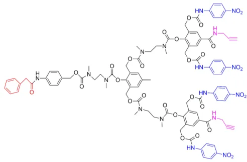
special enzymatic conditions (47). As illustrated in Figure 3, the red part is
penicillin-G-amidase (PGA) (which release is enzymatically triggered), the blue part is
4-nitroaniline, and the purple part are two acetylene functional groups which were used
to connect various PEG-azide units. When this dendrimer was modified using PEG tails,
the aqueous solubility of this dendrimer increased.
Figure 3: Structure of a dendritic molecule as a drug delivery system (47).
1.2.3 Dendrimers for gene delivery applications
A wide variety of viral gene delivery systems has been presented to deliver
7 limitations that consist in high carcinogenicity and immunogenicity in vivo (48).
Non-viral vectors, in general, show lower efficiency, but they offer safety and
flexibility (49, 50).
Because of their monodispersity, a large number of terminal groups, and regular
structure, dendrimers systems can be used with advantage for gene delivery (51, 52).
For instance, the capacity of PAMAM dendrimers as gene delivery systems was
demonstrated because they possess a huge number of cationic amines at the surface (40,
53, 54). The positive charge of these dendrimers allow them to bind to negatively
charged nucleic acids and, also, favor the interactions with the cell membrane (50, 55).
Also, transfection efficiency can be improved by modifying the dendrimers with
different groups such as alkyl chains, and membrane targeting peptides (56, 57).
Figure 4 schematically illustrates the cells uptake of dendriplexes. In order to
transfer the nucleic acid to cells, the dendrimer is first mixed with the nucleic acid
resulting in charge neutralization and compaction of the DNA. After, the dendriplex
interacts with the cell membrane that is negatively charged. Then an endosome is
formed by cell endocytosis. Finally, the endosome suffers lysis (that is helped by the
proton sponge effect exerted by the dendrimer inside the endosome), and the DNA is
released in the cell interior (48).
CHAPTER 1 - INTRODUCTION
8
1.3 Triazine-based dendrimers
The dendrimers which use 1,3,5-triazine ring as a branching unit or core are
called triazine-based dendrimers. The triazine unit has good electron affinity, and may
perform as an electron transport component (58). The triazine dendrimer branches
start from the triazine ring that is usually obtained from the commercially available
cyanuric chloride (CAC) compound. Different types of diamines are used to connect
triazines. Due to diverse characteristics such as the length and the flexibility of
diamines, triazine dendrimers may exhibit multiple properties (59).
Triazine dendrimers show a wide compositional variety and can be
synthesized to exhibit orthogonally functional surfaces appropriated for drug delivery
applications (60). The process of triazine dendrimer synthesis can be designed (using
reactions that are fast, chemoselectivity and with high yields) to improve the
solubility of dendrimers and to obtain the adequate structure-activity relation (61).
1.3.1 The properties of triazine ring as starting material in the preparation of
dendrimers
Triazine ring is the six-membered heterocyclic ring containing nitrogen, is
chemically very stable, and can be decomposed by heating in concentrated sulfuric acid
at 150 ºC (62). Due to its good stability (at a range of pH and thermal conditions), it can
pass through a variety of harsh reaction conditions without being destroyed/
decomposed. Triazine ring (especially melamine) can be used in the supramolecular
area by recognizing molecules through hydrogen bonding interaction (Figure 5). One
system based on the interaction between melamine and CAC showed significance in
the self-assembly of linear and cyclic hydrogen-bonded assemblies (Figure 6) (63).
Between atoms N to N, N to O, O to O, different types of hydrogen bond were formed.
Due to this property, it is possible to increase the water solubility of triazine dendrimers
9
Figure 5: Sites of triazine/melamine derivatives that can act as donor or acceptor (62).
Figure 6: Complementary hydrogen bond formation between cyanuric acid and melamine (63).
The way to synthesize triazine dendrimer relies on the nucleophilic substitution
reaction of trichlorotriazine (60). The main materials to supply the triazine ring are
CAC, melamine, and cyanuric acid. In terms of industrial production, CAC is an
inexpensive reagent. In the structure of CAC, three carbons connect respectively, three
nitrogen atoms. Due to the effect of the C=N unsaturated bond, the reactivity of
chlorine is high, and it is easy to occur nucleophilic substitution, but the active extent of
three chlorines is different (Figure 7). The chlorine can be substituted by functional
groups like OH, NH2, SH, NHR and form new materials with different characteristics
and applications. When we only need to replace one chloride, the temperature of
reaction should be controlled at temperatures of -15 to 5ºC and the reaction should be
under the attendance of an acid-binding agent; for the removal of the second chlorine,
the temperature should be increased to 40-60ºC and the acid-binding agent is necessary;
CHAPTER 1 - INTRODUCTION
10 temperature 90-100ºC and the acid-binding agent presence (64).
Figure 7: Chemoselective reactivity of CAC (60).
1.3.2 The applications of triazine derivatives
Triazine derivatives refer to the chemical compounds containing triazine
structures. These derivatives are antibacterial, antitumor and have good optical
properties. They are widely used in pesticides, medicine, fluorescent whitening agents,
antioxidants, lubricants, paper treatment agents, rubber and in textile auxiliaries areas
(37, 65).
Kukla et al. reported the ability of triazine against the human
immunodeficiency virus (HIV) (66). Menicagli et al. were among the first to show the
potential of the antitumor properties of triazine derivatives, by studying the in vitro
antitumor properties of a series of 2-alkyl-(alk-1′-ynyl,aryl)-4,6-dialkoxy-1,3,5
-triazines derivatives (65) (Figure 8). Their findings showed that the presence of
saturated or unsaturated moiety bonded to the heterocyclic ring via a C-C bond, as well
as the type of the structure of the carbon-carbon bonded residue, seem to play a
significant role in the observed cytotoxic activity.
Figure 8: Triazine derivatives with antitumor properties synthesized by Menicagli et al. (65).
1.3.3 Synthesis of triazine-based dendrimers
Nowadays, dendrimers are usually synthesized by two methods called divergent
11 core. More procedures are needed to achieve successive generations. In the convergent
method, the branches/dendrons are synthesized separately and then connected to the
core. Accordingly, the theoretical output is related to the amount of branches that are
initially used (67). Also, other methods are used to synthesize triazine dendrimers such
as solid phase synthesis (68).
(1) Convergent method
The convergent method for triazine-based dendrimers generally presents three
steps: firstly, utilize the selectivity of CAC to get AB2 monomer; secondly, repeat the
first step to growth AB2 and arrive to dendron; finally, connect the dendron to a
compatible core to obtain the triazine-based dendrimer.
The nucleophilic aromatic substitution reaction of the chloride (which are on
the triazine rings) with phenolates was used to synthesize triazine dendrimers. To
improve this substitution reaction, a poor electron group such as heterocyclic or the
groups, which are connected on the heterocyclic structure are needed. Specially, it is
more efficient to substitute the chlorine that is on the heterocyclic structure (like CAC)
(69).
Verheyde et al. (70) used the convergent method to synthesize a triazine-based
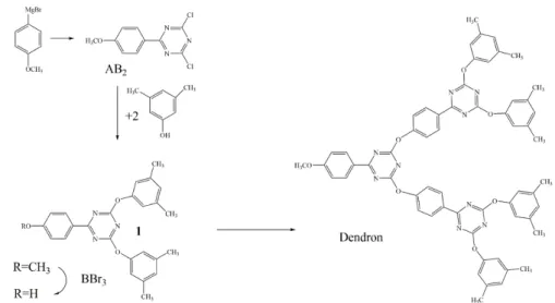
dendrimer containing multiple triazine rings (Figure 9). First, through the Grignard
reaction the AB2 monomer was obtained. The two chlorine atoms of the AB2 monomer
reacted with two 3,5-bis-t-butylphenol that is inexpensive. Then the –CH3 of 1 (Figure 9) was converted to –H with the presence of boron tribromide. After, two groups of 1 were connected to AB2 (Figure 9) and the dendron was obtained. After repeating the
above reaction to getting branches, the triazine-based dendrimer was obtained by
CHAPTER 1 - INTRODUCTION
12
Figure 9: The convergent method used by Verheyde et al. in the preparation of triazine-based dendrimer
(70).
Takagi et al. used CAC and p-nitrophenol as raw materials, and got the AB2
monomer (Figure 10). Then compound 1 was prepared for the terminal groups of the dendrimer. To connect the –NO2 of compound 1, a reduction step was necessary. Under
the presence of Fe/FeSO4·7H2O, the nitro group was reduced to amino-group (Figure
10). Then the amino-group reacted easily with the chlorine atom of the triazine core by
removal of HCl. N-generation of dendron was obtained by repeating the reaction.
Finally, the triazine-based dendrimer was got using the CAC as the core (71).
Figure 10: AB2 monomer in the preparation of triazine-based dendrimers synthesized by Takagi et al.
(71).
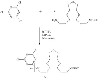
Recently, Enciso et al. synthesized a low generation triazine dendrimer in high
yields by using microwave assisted reactions (72). The main advantage is that the
13 assistance of microwave, when compared to the traditional reaction approach.
In the first reaction, to ensure the complete conversion of CAC, more than 2
equivalents of diamine were used (Figure 11). When this reaction was finished,
monosubstitution compounds were not discovered. The whole reaction was under the
presence of microwave at 60 °C, and only 10 min was needed to finish this reaction.
Finally, a clear oil product was obtained with 95% yield (72).
For the synthesis of C2, harsher conditions were needed. 10 equivalents of
diamine were used to reduce the product of the undesired dimer (Figure 12). This
reaction needed more time (30 min) than the synthesis reaction of C1, and a high
temperature (95 °C) was also needed. Finally, a clear oil product was obtained with 87%
yield (72).
Figure 11: Synthesis of low generation triazine dendrimer, C1, with the help of microwave (72).
CHAPTER 1 - INTRODUCTION
14 For the high generation of triazine dendrimers, an expected yield is not obtained,
and traditional thermal conditions are still required. However, the microwave-assisted
synthesis method can reduce the time for preparing low generation dendrimers and
provides more time for the studies of high generation dendrimers (72). Depending on
the synthesis steps, the same authors synthesized the generation 9 of this family of
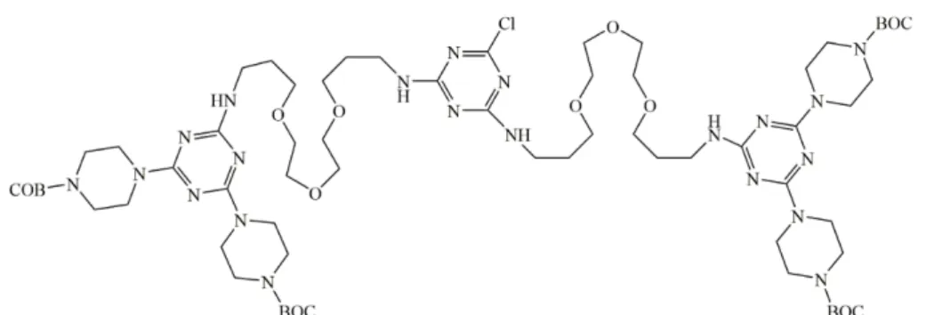
dendrimers using a macromonomer (Figure 13) (73).
Figure 13: The macromonomer was used by Enciso et al. in which the BOC-piperazine is used to
protect the –NH2 groups (73).
(2) Divergent method
In the divergent method, one step is needed to protect the functional group.
However, this approach revealed to be experimentally too complicated. CAC was
widely used as a core to synthesize triazine dendrimers due to its selective chemical
property of three chlorine atoms (60).
Takagi et al. (71) used CAC and p-nitroaniline as starting material to synthesize
15
Figure 14: The divergent method used by Takagi to synthesize a triazine-based dendrimer (71).
Namazi and Adeli (74, 75) reported the use of PEG as the core and CAC as the
main material to synthesize triazine-based dendrimers (G1.0, G1.5,G2.0) through the
divergent method. The synthetic route is showed in Figure 15. In the reaction i, the PEG
and CAC was mixed in dichloromethane, using as the base NaOH. The yield of the
product G1.0 is 100%. In the reaction ii, excess diethanolamine (DEA) and G1.0 was
mixed at room temperature. After refluxing for 4 h, the product G1.5 was obtained with
100% yield. In step iii, the CAC reacted first with phenol. Then the product 4 of this
reaction was mixed with G1.5 in dried dichloromethane under argon, using NaOH as
CHAPTER 1 - INTRODUCTION
16
Figure 15: The divergent method used by Namazi and Adeli (75) for the preparation of a PEG
triazine-based dendrimer.
(3) Solid phase synthesis
Dilly et al. (68) introduced a new way to prepare triazine-based dendrimers
using solid phase synthesis method. These kinds of dendrimers were separated from
solid phase under the action of trifluoroacetic acid (TFA). The synthesis route is
showed in Figure 16. First, diisopropylethylamine (DIPEA) was used as a base. The
product 1 was obtained by the reaction of CAC with the supporting material. Then the
product 1 reacted with ethylenediamine (ED) at 80-100ºC under the action of DIPEA,
and the product 2 was obtained. Product 3 was achieved by the reaction of product 2
and CAC. Yoo et al. also used solid phase approach and divergent method to synthesize
17
Figure 16: The solid-phase method used by Dilly et al. (68).
1.3.4. The properties and applications of triazine-based dendrimers
In general, the solubility of the triazine-based dendrimers is poor, but it can be
improved by modifying the terminal groups or linkers of the dendrimers. The
triazine-based dendrimers normally have good thermal oxidative stability. Kraus and
Louw (77) used a convergent method to synthesize the triazine-based dendrimers by
using the trimeric acid (1,3,5-benzenetricarboxylic acid) as the core and the silicon
phthalocyanine as the functional group. The obtained triazine-based dendrimers were
stable in the external environment and, according to the authors, can be useful for
applications in photosynthetic materials (77).
Due to the unique biological activity of the triazine-based dendrimers, they can
be used in the research of antibiotics, anticancer drugs and in the drug delivery field.
Biphenyl drugs are effective inhibitors of respiratory system virus (RSV). Nikitenko et
al. (78) reported a kind of biphenyl compound which contains triazine ring (compound
CHAPTER 1 - INTRODUCTION
18 compound that contains a pyrimidine analog (compound 2 in Figure 17).
Figure 17: The triazine compound 1 and the pyrimidine analog 2 (78).
Simanek’s group studies (79) showed that the triazine-based dendrimers can
reduce the toxicity of drugs when they are used as drug carriers. They synthesized a
triazine-based dendrimer molecule that can decrease the toxicity of anticancer drugs
(methylamine purine, 6-mercaptopurine) using the injected peritoneal injection method
(80). By mixing these drugs with triazine-based dendrimers, a good increase on the
solubility of these drugs was observed. The mice study showed that the use of
triazine-based dendrimers can reduce the toxicity of anticancer drugs and allow the use
of increased doses (79, 81, 82).
A series of aryl- triazine-based dendrimers that have conjugated systems was
19 Compared with the other families of dendrimers, triazine-based dendrimers are
less studied and, despite the obtained achievements, is a promising field of study,
particularly for the preparation of metallodendrimers. The stability and biological and
optical activities of triazine-based dendrimers provide them with a broad prospect of
applications.
1.4 Objectives and General Strategy of the Project
The objective of this project was to synthesize low generation triazine-based
dendrimers by using, as starting materials, CAC and triethylene glycol (TEG), and test
the cytotoxicity of the prepared compounds. Our interest in the triazine dendrimers is
related with our previous experience in the field of the preparation of
metallodendrimers for non-linear applications (85) and on the use of PAMAM (57) and
PPI dendrimers as scaffolds for the preparation of low-generation ruthenium based
metallodendrimers (86) for drug and gene delivery. The abundant possibilities to
synthesize triazine-based dendrimers with expected solubility in aqueous environments
and low cytotoxicity made us start a preliminary study on the preparation of these type
of dendrimers for biological applications (e.g. drug delivery).
As showed in Figure 18, the main synthesis process depends on the
chemoselectivity of three different chloride atoms from the starting aromatic material,
CAC. This molecule was connected to the core, the TEG, by a nucleophilic reaction to
get G1.0(Cl)4. The product was purified by silica chromatography. Then the DEA was
used to modify the G1.0(Cl)4 to get G1.5(OH)8. The products were characterized by
nuclear magnetic resonance (NMR) techniques, Mass Spectrometry (MS), Fourier
Transform Infrared Spectroscopy (FTIR). Finally, the cytotoxicity of these compounds
was investigated in vitro using the NIH 3T3 fibroblast cell line of mouse (used as a
model of normal cells) and the A2780 human ovarian carcinoma cell line (used as a
CHAPTER 1 - INTRODUCTION
20
21
CHAPTER 2
–
MATERIALS AND METHODS
2.1 Reagents and equipment
CAC (purity > 99%) was purchased from ACROS ORGANICS (New Jersey,
USA). Sodium hydroxide, chloroform, n-hexane, tetrahydrofuran (THF), 1,4-dioxane,
TEG were bought from Thermo Fisher Scientific (MA, USA). Ethyl acetate was
obtained from VWR (PA, USA). DEA was purchased from Sigma-Aldrich (MO,
USA). Deuterated chloroform, deuterated dimethyl sulfoxide (DMSO), and
diisopropylamine were bought from MERCK (Darmstadt, Germany).
In the cytotoxicity studies, two types of cell lines were used, NIH 3T3 and
A2780. The Dulbecco’s Modified Eagle Medium (DMEM) was acquired from
Sigma-Aldrich (MO, USA) and to this was added 10% fetal bovine serum (FBS) and
1% antibiotic-antimycotic (AA) and this medium was used for the NIH 3T3 cells. The
Roswell Park Memorial Institute media (RPMI-1640) was acquired from
Sigma-Aldrich (MO, USA) and was also used with 10% FBS and 1% AA and this
medium was used for the A2780 cells. These media were used to culture the cells. The
trypsin-EDTA solution was used to detach the cells from the bottom of the plates.
NMR Spectroscopy
Acquisition of 1H and 13C spectra was made on an NMR Spectrometer of 400
MHz from Bruker (UltraShieldTM 400 Plus ULTRA LONG HOLD) Console
AVANCE 400 II+.
MS Spectroscopy
A Mass Spectrometer type Ion Trap Multipolar LC-MSMS (Bruker Esquire
6000 Mass Spectrometer) with Electrospray Ionization (ESI) and Atmospheric
Pressure Chemical Ionization (APCI) sources and MSn capacity was used in the mass
CHAPTER 2 – MATERIALS AND METHODS
22
FTIR spectroscopy
The FTIR spectra were acquired using KBr pellets on a Perkin Elmer
Spectrum Two spectrometer.
2.2 Synthesis of the G1.0(Cl)
4dendrimer
Under nitrogen atmosphere, a solution of CAC (17.08g, 92.6mmol) in 50ml of
dry THF (Solution 1) was prepared and a solution of TEG (6.95g,46.3mmol) with
N,N-DIPEA (16.1ml, 46.3mmol) in 6ml of dry THF at 0 °C (a water solution with salt
and ice was used to control the temperature) (Solution 2). Then, these two solutions
were mixed as showed in Figure A1 in the ANNEX, at 0°C, under a nitrogen
atmosphere. After being stirred for 27 h at 0°C, the mixture was filtered, and the
obtained white solid was washed with 20 ml of THF. The excess solvent was
evaporated using a rotary evaporator system. The white solid was purified by silica gel
column chromatography using a mixture of hexane and ethyl acetate with a ratio of 4:1.
Finally, the product (which was from silica gel column) was washed with hexane to
remove the ethyl acetate. Finally 5.5g of G1.0(Cl)4 was obtained as a white solid (yield:
26.6%). 1H NMR (CDCl
3, 400 MHz) [δ: 4.627, 4.634, 4.638, 4.642, 4.650(m, 4H); 3.843, 3.851, 3.854, 3.858, 3.866 (m, 4H); 3.683 (s, 4H)].13C NMR (CDCl
3, 400 MHz)
[δ: 60.55, 68.69, 69.46, 71.02, 172.721]. IR (KBr) ν: 2920 (-CH2), 1384 (-C-Cl) cm-1.
2.3 Synthesis of the
G1.5(OH)
8dendrimer
Because 1,4-dioxane combines with atmospheric oxygen upon prolonged
exposure to air, forming potentially explosive peroxides, this reaction was processed in
a three neck flask (250mL) under nitrogen.
In a flask, 25 mL of 1, 4-dioxane was added to 2.5g of G1.0 (Cl)4 (5.6 mmol) under
stirring. Then, the solution was moved under a nitrogen atmosphere to a three neck
23 We added DEA(2.7mL,28mmol)to the solution of G1.0(Cl)4 drop wise. Then
sodium hydroxide solution (0,9g, 22.4mmol NaOH in 1 ml water) was added as a base
(its molar ratio to G1.0(Cl)4 was 4:1). This mixture was stirred for 0.5h at room
temperature, then the temperature was increased to 80 °C and refluxed for 4.0h. At the
end of the reaction, the mixture was filtered by using a vacuum filter funnel. The excess
of 1,4-dioxane was removed using a rotary evaporator (under 17mbar, at 37 °C
conditions), the filtrate was dispersed and washed with THF and acetone, filtered again,
and the filter cake was dried using the rotary evaporator. Finally, the product was
washed with acetone to remove the impurities:1.24 g of G1.5(OH)8 was obtained as a
white oil (yield: 33.3%).1H NMR (DMSO-d6, 400 MHz) [δ:4.683, 4.694, 4.706, 4.724,
4.736, 4.749 (m,8H); 4.272, 4.284, 4.295 (t,4H); 3.672, 3.661, 3.684 (tri, 4H); 3.568(s,
36H)]. IR (KBr) ν: 3382 (-OH), 2948 (-CH2), 2879 (-CH2), 1583 (-C=N-), 1520
(-C=N-), 1342 (-CH2) cm-1. MS (ESI) [M+H] + m/z: calcd. for C28N10O12H52, 720.77;
found, 721.4.
2.4 Cell Biological Evaluation
The NIH 3T3 and A2780 cell lines were used to evaluate the cytotoxicity of
CAC, G1.0(Cl)4 and G1.5(OH)8. First, NIH 3T3 fibroblasts and A2780 cells were
plated in 96-well plates. After 24h, the old media were removed and complete medium
and the test solutions (10μL of DMSO, CAC-DMSO, G1.0(Cl)4-DMSO and
G1.5(OH)8-DMSO solutions ) were added to the cell culture wells and then incubated
for 48 h, at 37 °C. Medium and DMSO were used as controls.
The resazurin reduction assay was chosen to measure the cellular metabolic
activity to conclude about the cell viability. Then the medium was removed; the wells
were filled with fresh medium containing 10% of resazurin, and the cells were kept in
the incubator for 3 h. After 3 h, the medium with resazurin was transferred to 96-well
white opaque plates and the resorufin fluorescence (λex=530nm, λem=590 nm) was read
CHAPTER 2 – MATERIALS AND METHODS
24 One-way ANOVA statistical analyzes with Tukey’s test was chosen to evaluate
the statistical significance of experimental data. All results are reported as mean ± SD.
0.05 was selected as the significance level, and the data was indicated with (**) for p <
25
CHAPTER 3
–
RESULTS AND DISCUSSION
3.1 Synthesis and characterization of G1.0(Cl)
4dendrimer
Due to the chemoselectivity and by controlling temperature, CAC is used to
synthesize a large number of triazine-based dendrimers.
In this work, two equivalents of CAC were connected to TEG as a core. The
oxygen atoms in the chain of TEG can improve the polarity of the final dendrimer and
form H-bonding with H2O, which can increase the solubility of the final dendrimer in
aqueous solution.
The 1H NMR spectrum of G1.0(Cl)
4 in CDCl3 is shown in Figure 19. The peak
shift at 7.260 ppm is from CDCl3. The solvent impurities originating from column
chromatography are depicted as (d) and (e) in Figure 19.
Figure 19: 1H NMR spectrum of G1.0(Cl)
CHAPTER 3 – RESULTS AND DISCUSSION
26 In Figure A2 in the ANNEX, we have the 1H NMR of hexane prepared in
CDCl3 acquired from the Spectral Database for Organic Compounds (SDBS). In the 1H
NMR spectrum of G1.0(Cl)4 in Figure 19, signals at δ: 0.890, 1.271, 1.280 correspond
to hexane. Compared with the 1H NMR of ethyl acetate in CDCl
3 obtained from the
SDBS (see ANNEX, Figure A3), the signals indicated at (e) in the 1H NMR spectrum of
G1.0(Cl)4 were from ethyl acetate.
In the 1H NMR spectrum of TEG in CDCl
3 (see ANNEX, Figure A4) there
are peaks at 3.721, 3.711 and 3.700 ppm (these peaks are labeled as A in the Figure);
3.652 ppm (this peak is labeled as C in the Figure); 3.599, 3.587 and 3.577 ppm (these
peaks are labeled as B in the Figure). In Figure 19, a single peak at C which has a
chemical shift at 3.683 ppm (s, 4H) is related with the single peak at 3.652 ppm (s,4H)
observed in the SDBS spectrum for the TEG starting material. Due to the position of the
carbon in the middle of G1.0(Cl)4, which is indicated as C in the chemical structure in
Figure 19, the chemical shift of the proton at this carbon C was not influenced by the
atoms from the G1.0(Cl)4 termini.
When the oxygen atom from the -OH group of TEG was connected with CAC,
the protons at carbon A in the G1.0(Cl)4 chemical structure (see Figure 19) were less
shielded. So the signal of the protons at carbon A in G1.0(Cl)4 moved to the left. The
signals of the protons at carbon A should be [δ: 4.627, 4.634, 4.638, 4.642 and 4.650
ppm (m, 4H)] and the peaks of the protons at carbon B [δ: 3.843, 3.851, 3.854, 3.858
and 3.866 ppm (m, 4H)].
The 13C NMR spectrum of ethyl acetate has four signals at 171.080, 60.440,
21.000 and 14.280 ppm (SDBS). Looking at the 13C NMR spectrum of G1.0(Cl) 4, in
Figure 20, the observed peaks at 14.348, 20.299, 21.182, and 171.233 ppm correspond
to ethyl acetate.
The 13C NMR spectra in Figure 20 also revealed a relevant peak with a
chemical shift at 172.721 ppm for both CAC and G1.0(Cl)4. This means that there is a
27
Figure 20: 13C NMR of G1.0(Cl)
4, TEG and CAC in CDCl3.
In the 13C NMR spectrum of TEG, there are 3 signals at 72.791, 70.431, and
61.675ppm. The 13C NMR spectrum of G1.0(Cl)
4 presents peaks at 71.016, 69.461,
68.689 and 60.546 ppm that belong to the TEG core, confirming that the connection
between TEG and CAC was successful.
The 1H NMR spectrum of the G1.0(Cl)
4 compound acquired at room
temperature after two weeks confirmed its stability.
3.2 Synthesis and characterization of the
G1.5(OH)8 dendrimerAs presented in Figure 21, DEA was used to increase the generation and the
CHAPTER 3 – RESULTS AND DISCUSSION
28
Figure 21: Synthesis of G1.5(OH)8.
The results of the solubility test for G1.5(OH)8 in solvents with different
polarity are presented in Table 2. Except DMSO, water, dimethyl formamide (DMF)
and methanol, the other pure solvents cannot dissolve the product even at 60 °C. The
table shows that only solvents with higher polarity (except acetonitrile) and a mixture
of dioxane:methanol (9:3) can dissolve G1.5(OH)8 totally.
Table 2: Solubility of G1.5(OH)8 in selected organic solvents or mixture of solvents at room
temperature.
Solvent
Polarity* Solubility atRoom
Temperature
Methylene chloride 3.10 No obvious dissolution
Tetrahydrofuran 4.00 No obvious dissolution
Chloroform 4.10 No obvious dissolution
Ethyl acetate 4.40 No obvious dissolution
Dioxane 4.80 No obvious dissolution
Acetone 5.10 No obvious dissolution
Methanol 5.10 Well dissolved
Acetonitrile 5.80 No obvious dissolution
DMF 6.40 Well dissolved
DMSO 7.20 Well dissolved
Water 10.20 Well dissolved
Dioxane:Methanol=9:3 Well dissolved
29 Comparing the 1H NMR spectra of G1.5(OH)
8 and G1.0(Cl)4, the most relevant
difference corresponds to peak D in Figure 22, which can be attributed to the proton of
the –OH groups from the G1.5(OH)8 termini. As the spectrum showed, the shape of this
signal is two triplets instead of the expected wide single peak of the –OH. Probably, the
main reason for the observed effect on the proton peaks (8H) comes from the fact that
DMSO may be bound to one or two –OH groups depending on the concentration
(87).As to peaks A, B and C of G1.5(OH)8, it is similar to G1.0(Cl)4 according to
compare Figure 19 with Figure 22.
Figure 22: 1H NMR spectrum of G1.5(OH)
8 in DMSO.
3.3 The MS spectrum of G1.5(OH)
8dendrimer
The mass spectrum of the G1.5(OH)8 dendrimer in MeOH (Figure 23)
presents two special fragments. One fragment was the molecular peak M=721.4 that is
in accordance with the molecular weight of G1.5(OH)8 dendrimer and the other one at
M=436.1 corresponds to the half of G1.5(OH)8 dendrimer. The peak 743.4 is probably
CHAPTER 3 – RESULTS AND DISCUSSION
30
Figure 23: The mass spectrum of G1.5(OH)8 in MeOH.
3.4 The FTIR spectrum of CAC, G1.0(Cl)
4dendrimer and G1.5(OH)
8dendrimer
The characteristic vibrations of the chemical bonds in the functional groups of
any prepared compound will display special bands in the FTIR spectrum (88). So we
can use FTIR spectroscopy to compare different molecules to find the relationship
between different compounds. In Table 3 the IR spectrum vibrational bands obtained
for the materials prepared in this work are compared with those obtained from
Bio-Rad database.
Table 3: Comparison of IR spectrum stretching vibrations of the materials in this work and those obtained from the database.
Bonds Data from this work Stretching vibration*
C-Cl 1270, 1384 cm-1 1200-1000 cm-1
C=N 1498, 1583, 1520 cm-1 1500 cm-1
-OH 3463, 3382 cm-1 3650-3320 cm-1
CH2 2920, 2948, 2879, 1342 cm-1 2936-2843 cm-1
1485-1445 cm-1
*Stretching vibration values obtained from the Bio-Rad data base (http://www.bio-rad.com/)
31 band also appeared in the spectrum of Figure 24c, which wavenumbers are as = 1583
cm-1 and
as = 1520 cm-1, respectively. In the case of spectrum 24b, the stretching
vibration of –C=N- is unclear. The peaks at as = 1270 cm-1 in Figure 24a and as =
1384 cm-1 in Figure 24b are from –C-Cl of CAC. In Figure 24b and 24c, the
maximum at as = 1342 cm-1, as = 2879 cm-1, as = 2920 cm-1 and as = 3382 cm-1 are
attributed to the –CH2 bonds. In Figure 24c, the stretching vibration –OH correspond
to as = 3382 cm-1. In Figure 24b, there is a wide peak (as = 3463 cm-1). Considering
that KBr was used to prepare the FTIR sample, maybe the type and amount of sample
in the KBr, the preparation of the FTIR sample and the quantity of moisture in the
KBr matrix affect the resolution of the IR analysis. Comparing the achieved results
with the NMR and MS data (in the case of G1.5(OH)8), we can confirm the successful
CHAPTER 3 – RESULTS AND DISCUSSION
32
Figure 24: The FTIR spectrum of CAC (a), G1.0(Cl)4 dendrimer (b) and G1.5(OH)8 dendrimer (c).
3.5
In Vitro
Cytotoxicity of CAC, G1.0(Cl)
4dendrimer and G1.5(OH)
8dendrimer
The application of triazine-based dendrimers in the biological field depends on
their inherent toxicities, which can hinder or limit their biological applications. The in
vitro cytotoxicity of CAC , G1.0(Cl)4 dendrimer and G1.5(OH)8 dendrimer were
tested by using NIH 3T3 and A2780 cell lines. The resazurin reduction assay showed
the results of cell viability (see Figures 25 and 26). The wells that only had medium
were used as a control. A second control used a mixture of medium (either DMEM or
33 As shown in Figures 25 and 26, the viability of cells in the presence of CAC, G1.0(Cl)4
dendrimer and G1.5(OH)8 dendrimer exceeded 80% in the concentration range 1 µM to
100 µM for both cell lines. As such, the compounds present very low toxicity within the
selected concentration range.
Figure 25: Cytotoxicity evaluation of CAC, G1.0(Cl)4 dendrimer and G1.5(OH)8 dendrimer at
different concentrations for 48 h and for NIH 3T3 cells using resazurin reduction assay. All results were expressed as the mean ±SD, n =3. One-way ANOVA with Tukey’s test was used to assess the statistical difference between the group means (**p <0.01, *** p <0.001).
Figure 26: Cytotoxicity evaluation of CAC, G1.0(Cl)4 dendrimer and G1.5(OH)8 dendrimer at
CHAPTER 3 – RESULTS AND DISCUSSION
34 Figures 27 and 28 make a comparison between the cytotoxicity of G1.0(Cl)4
dendrimer and G1.5(OH)8 dendrimer for increasing compound concentrations and for
NIH 3T3 cells and A2780 cells. G1.0(Cl)4 and G1.5(OH)8 have respectively
significant difference on the toxicity of these two kinds of cells.
Figure 27: Comparison of the cytotoxicity of G1.0(Cl)4 dendrimer at different concentrations
between NIH 3T3 cells and A2780 cells after 48h. All results were expressed as the mean ±SD, n =3. One-way ANOVA with Tukey’s test was used to assess the statistical difference between the group means (**p <0.01, *** p <0.001).
Figure 28: Comparison of the cytotoxicity of G1.5(OH)8 dendrimer at different concentrations
35 The inherent toxicity of dendrimers hinder their application in pharmaceutical
field (89) and, compared with the interior groups, the surface groups is much more
active to influence the toxicity of dendrimers (60). In general, when the surface charge
of dendrimers is cationic, the dendrimers show high toxicity. The main reason for that
behavior is related with the fact that cationic charges can interact with the biological
membranes that are negatively charged. As a result of this interaction, the cell
membrane can be disrupted due to the creation of nanoholes (89). The terminal atoms
of the dendrimer G1.0(Cl)4 are chloride atoms that are very electronegative (90). So,
the surface of G1.0(Cl)4 is rich in electrons and presents a negative character. Oxygen is
also an electronegative atom. Once the dendrimer G1.5(OH)8 has 8 –OH terminal
groups, its surface also has a strong negative character. For these reasons the toxicity of
GENERAL CONCLUSIONS
36
GENERAL CONCLUSIONS
In summary, the G1.0(Cl)4 dendrimer and the G1.5(OH)8 dendrimer were
synthesized and characterized by using common synthesis techniques. These two
dendrimers showed good stability on the atmospheric air at room temperature.
Importantly, once the G1.5(OH)8 dendrimer displays a good solubility in water (and
DMSO), it is possible to foresee their use in biological applications. CAC, G1.0(Cl)4
and G1.5(OH)8 dendrimer do not have cytotoxicity according to the results of the
resazurin reduction assay with 0-100 µM concentrations.
During this work, special techniques of organic chemistry were used, as well
as common and advanced synthesis techniques, to confirm the preparation of the
reported triazine-based dendrimers. Despite the small number of results obtained in
the time available, it is expected that they can open the way for the preparation of
higher dendrimer generations for biological applications, particularly for drug
37
REFERENCES
1. Dong, R. J.; Zhou, Y. F.; Zhu, X. Y., Supramolecular Dendritic Polymers: From Synthesis to Applications. Acc. Chem. Res. 2014, 47, 2006-2016.
2. Tomalia, D. A.; Naylor, A. M.; Goddard, W. A., Starburst Dendrimers: Molecular-Level Control of Size, Shape, Surface Chemistry, Topology, and Flexibility from Atoms to Macroscopic Matter. Angew. Chem. Int. Ed. Engl.
1990, 29, 138-175.
3. Bosman, A. W.; Janssen, H. M.; Meijer, E. W., About Dendrimers: Structure, Physical Properties, and Applications. Chem. Rev. 1999, 99, 1665-1688.
4. Buhleier, E.; Wehner, W.; Vögtle, F., " Cascade"-and" Nonskid-Chain-like" Syntheses of Molecular Cavity Topologies. Synth. Stuttgart 1978, 1978,
155-158.
5. Tomalia, D. A.; Baker, H.; Dewald, J.; Hall, M.; Kallos, G.; Martin, S.; Roeck, J.; Ryder, J.; Smith, P., A new class of polymers: starburst-dendritic macromolecules. Polym. J. 1985, 17, 117-132.
6. Matsui, M.; Wang, M.; Funabiki, K.; Hayakawa, Y.; Kitaguchi, T., Properties of Novel Perylene-3,4 : 9,10-tetfacarboxidiimide-Centred Dendrimers and Their Application as Emitters in Organic Electroluminescence Devices. Dyes Pigm.
2007, 74, 169-175.
7. Junge, D. M.; McGrath, D. V., Photoresponsive dendrimers. Chem. Commun.
1997, 9, 857-858.
8. Esposito, A.; Delort, E.; Lagnoux, D.; Djojo, F.; Reymond, J. L., Catalytic Peptide Dendrimers. Angew. Chem. Int. Ed. 2003, 42, 1381-1383.
9. Kofoed, J.; Reymond, J. L., Dendrimers as Artificial Enzymes. Curr. Opin. Chem. Biol. 2005, 9, 656-664.
10. Wu, X. Y.; Huang, S. W.; Zhang, J. T.; Zhuo, R. X., Preparation and Characterization of Novel Physically Cross-linked Hydrogels Composed of Poly(vinyl alcohol) and Amine-terminated Polyamidoamine Dendrimer. Macromol. Biosci. 2004, 4, 71-75.
11. Razinkov, V.; Gazumyan, A.; Nikitenko, A.; Ellestad, G.; Krishnamurthy, G., RFI-641 Inhibits Entry of Respiratory Syncytial Virus Via Interactions With Fusion Protein. Chem. Biol. 2001, 8, 645-659.
12. Luo, D.; Haverstick, K.; Belcheva, N.; Han, E.; Saltzman, W. M., Poly(ethylene glycol)-Conjugated PAMAM Dendrimer for Biocompatible, High-Efficiency DNA Delivery. Macromolecules 2002, 35, 3456-3462.
13. Jiang, G.; Wang, L.; Chen, W., Studies on the Preparation and Characterization of Gold Nanoparticles Protected by Dendrons. Mater. Lett. 2007, 61, 278-283. 14. Kouketsu, T.; Duan, S.; Kai, T.; Kazama, S.; Yamada, K., PAMAM dendrimer
composite membrane for CO2 separation: Formation of a chitosan gutter layer. J.
Membr. Sci. 2007, 287, 51-59.
REFERENCES
38
Prog. Polym. Sci. 2014, 39, 268-307.
16. Conti, D. S.; Brewer, D.; Grashik, J.; Avasarala, S.; da Rocha, S. R. P., Poly (amidoamine) dendrimer nanocarriers and their aerosol formulations for siRNA delivery to the lung epithelium. Mol. Pharmaceutics 2014, 11, 1808-1822. 17. Chisholm, E. J.; Vassaux, G.; Martin-Duque, P.; Chevre, R.; Lambert, O.; Pitard,
B.; Merron, A.; Weeks, M.; Burnet, J.; Peerlinck, I., Cancer-specific transgene expression mediated by systemic injection of nanoparticles. Cancer Res. 2009,
69, 2655-2662.
18. Steffensen, M. B.; Hollink, E.; Kuschel, F.; Bauer, M.; Simanek, E. E., Dendrimers based on [1, 3, 5]‐triazines. J. Polym. Sci., Part A: Polym. Chem.
2006, 44, 3411-3433.
19. Merkel, O. M.; Mintzer, M. A.; Librizzi, D.; Samsonova, O.; Dicke, T.; Sproat, B.; Garn, H.; Barth, P. J.; Simanek, E. E.; Kissel, T., Triazine dendrimers as nonviral vectors for in vitro and in vivo RNAi: the effects of peripheral groups and core structure on biological activity. Mol. Pharmaceutics 2010, 7, 969-983. 20. Oosterom, G. E.; van Haaren, R. J.; Reek, J. N. H.; Kamer, P. C. J.; van Leeuwen,
P. W. N. M., Catalysis in the core of a carbosilane dendrimer. Chem. Commun.
1999, 1999, 1119-1120.
21. Perisé-Barrios, A. J.; Jiménez, J. L.; Domínguez-Soto, A.; de la Mata, F. J.; Corbí, A. L.; Gomez, R.; Muñoz-Fernandez, M. Á., Carbosilane dendrimers as gene delivery agents for the treatment of HIV infection. J. Controlled Release
2014, 184, 51-57.
22. Lakshminarayanan, A.; Ravi, V. K.; Tatineni, R.; Rajesh, Y. B. R. D.; Maingi, V.; Vasu, K. S.; Madhusudhan, N.; Maiti, P. K.; Sood, A. K.; Das, S., Efficient Dendrimer–DNA Complexation and Gene Delivery Vector Properties of Nitrogen-Core Poly (propyl ether imine) Dendrimer in Mammalian Cells. Bioconjugate Chem. 2013, 24, 1612-1623.
23. Ullas, P. T.; Madhusudana, S. N.; Desai, A.; Sagar, B. K. C.; Jayamurugan, G.; Rajesh, Y. B. R. D.; Jayaraman, N., Enhancement of immunogenicity and efficacy of a plasmid DNA rabies vaccine by nanoformulation with a fourth-generation amine-terminated poly (ether imine) dendrimer. Int. J. Nanomed. 2014, 9, 627-634.
24. Gissibl, A.; Padié, C.; Hager, M.; Jaroschik, F.; Rasappan, R.; Cuevas-Yañez, E.; Turrin, C. O.; Caminade, A. M.; Majoral, J. P.; Reiser, O., Synthesis and application of phosphorus dendrimer immobilized azabis (oxazolines). Org. Lett.
2007, 9, 2895-2898.
25. Ferenc, M.; Pedziwiatr-Werbicka, E.; Nowak, K. E.; Klajnert, B.; Majoral, j. P.; Bryszewska, M., Phosphorus Dendrimers as Carriers of siRNA— Characterisation of Dendriplexes. Molecules 2013, 18, 4451-4466.
26. Denkewalter, R. G.; Kolc, J.; Lukasavage, W. J., Surface modifying agents, metal chelating agents, substrates for drugs. US Patent 4289872, 1981.
39
dendritic poly (L-lysine) for loss-of-function analysis. J. Controlled Release
2008, 126, 59-66.
28. Kathiresan, M.; Walder, L.; Ye, F.; Reuter, H., Viologen-based benzylic dendrimers: selective synthesis of 3,5-bis(hydroxymethyl)benzylbromide and conformational analysis of the corresponding viologen dendrimer subunit. Tetrahedron Lett. 2010, 51, 2188-2192.
29. Li, J.; Lepadatu, A. M.; Zhu, Y.; Ciobanu, M.; Wang, Y.; Asaftei, S. C.; Oupický,
D., Examination of Structure–Activity Relationship of Viologen-Based Dendrimers as CXCR4 Antagonists and Gene Carriers. Bioconjugate Chem.
2014, 25, 907-917.
30. Gajjar, D.; Patel, R.; Patel, H.; Patel, P. M., Designing of Triazine based dendrimers and its application in removal of heavy metal ions from water. Chem. Sci. Trans. 2014, 3, 897-908.
31. Ciolkowski, M.; Rozanek, M.; Bryszewska, M.; Klajnert, B., The influence of PAMAM dendrimers surface groups on their interaction with porcine pepsin. Biochim. Biophys. Acta, Proteins Proteomics 2013, 1834, 1982-1987.
32. Luis Jimenez, J.; Pion, M.; Javier de la Mata, F.; Gomez, R.; Munoz, E.; Leal, M.; Angeles Munoz-Fernandez, M., Dendrimers as topical microbicides with activity against HIV. New J. Chem. 2012, 36, 299-309.
33. Sevenson, S.; Tomalia, D. A., Dendrimers in biomedical applications-reflections on the field. Adv. Drug Delivery Rev. 2012, 57, 2106-2129.
34. Duncan, R.; Izzo, L., Dendrimer biocompatibility and toxicity. Adv. Drug Delivery Rev. 2005, 57, 2215-2237.
35. Fischer, D.; Li, Y. X.; Ahlemeyer, B.; Krieglstein, J.; Kissel, T., In vitro cytotoxicity testing of polycations: influence of polymer structure on cell viability and hemolysis. Biomaterials 2003, 24, 1121-1131.
36. Mecke, A.; Majoros, I. J.; Patri, A. K.; Baker, J. R.; Holl, M. M. B.; Orr, B. G., Lipid bilayer disruption by polycationic polymers: The roles of size and chemical functional group. Langmuir 2005, 21, 10348-10354.
37. Chen, H. T.; Neerman, M. F.; Parrish, A. R.; Simanek, E. E., Cytotoxicity, hemolysis, and acute in vivo toxicity of dendrimers based on melamine, candidate vehicles for drug delivery. J. Am. Chem. Soc. 2004, 126,
10044-10048.
38. Lee, J. H.; Lim, Y. B.; Choi, J. S.; Lee, Y.; Kim, T. I.; Kim, H. J.; Yoon, J. K.; Kim, K.; Park, J. S., Polyplexes assembled with internally quaternized PAMAM-OH dendrimer and plasmid DNA have a neutral surface and gene delivery potency. Bioconjugate Chem. 2003, 14, 1214-1221.
39. Nowacka, O.; Milowska, K.; Bryszewska, M., Interaction of PAMAM dendrimers with bovine insulin depends on nanoparticle end-groups. J. Lumin.
2015, 162, 87-91.
40. Haensler, J.; Szoka, F. C., Polyamidoamine cascade polymers mediate efficient transfection of cells in culture. Bioconjugate Chem. 1993, 4, 372-379.