ISSN 0104-6632 Printed in Brazil
www.abeq.org.br/bjche
Vol. 32, No. 02, pp. 509 - 518, April - June, 2015 dx.doi.org/10.1590/0104-6632.20150322s00003066
Brazilian Journal
of Chemical
Engineering
SYNTHESIS AND CHARACTERIZATION OF
COPOLYMERIC AND TERPOLYMERIC
HYDROGEL-SILVER NANOCOMPOSITES BASED
ON ACRYLIC ACID, ACRYLAMIDE AND
ITACONIC ACID: INVESTIGATION OF THEIR
ANTIBACTERIAL ACTIVITY AGAINST
GRAM-NEGATIVE BACTERIA
A. Bal
1*, F. E. Çepni
2, Ö. Çakir
2, I. Acar
1and G. Güçlü
11
Istanbul University, Faculty of Engineering, Chemical Engineering Department, Avcılar-Istanbul,34320, Turkey.
Phone: + 90 212 473 70 70, Fax: +90 212 473 71 80 E-mail: [email protected]
2
Istanbul University, Faculty of Science, Molecular Biology and Genetics Department, Vezneciler - Istanbul, 34118, Turkey.
(Submitted: October 26, 2013 ; Revised: September 23, 2014 ; Accepted: October 13, 2014)
Abstract - In this study, copolymeric and terpolymeric hydrogel-silver nanocomposites based on poly(acrylamide-co-itaconic acid), poly(acrylic acid-poly(acrylamide-co-itaconic acid) and poly(acrylic acid-co-acrylamide-poly(acrylamide-co-itaconic acid) were synthesized by free-radical polymerization. These nanocomposites were characterized by Fourier Transform Infrared Spectroscopy (FTIR), Scanning Electron Microscopy (SEM), UV-Visible Spectrophotometry (UV-Vis) and X-Ray Diffraction (XRD) analysis, as well as their swelling behaviors. In addition, antibacterial
properties of these hydrogel-silver nanocomposites were investigated against Pseudomonas aeruginosa.
Acrylic-based hydrogel-silver nanocomposites demonstrated antibacterial activity against Gram-negative bacteria. These hydrogel-silver nanocomposites can be used as antibacterial material in the medical field.
Keywords: Antibacterial; Gram-negative bacteria; Hydrogel; Nanocomposite; Silver nanoparticle.
INTRODUCTION
Hydrogels are three-dimensionally crosslinked polymer networks composed of hydrophilic homo or hetero copolymers, and they have the ability to absorb significant amounts of water (Byrne et al., 2002). Hydrogels have been used in agricultural applica-tions, the food industry, water treatment processes, and biotechnological and medical fields (Karadağ et
al., 1997; Karadağ and Üzüm, 2012; Ju et al., 2009; Özkahraman et al., 2011). Today, hydrogel-silver nanocomposites have also been widely used in bio-medical applications (Kim et al., 2004; Varaprasad et al., 2010; Yiamsawas et al., 2008; Gils et al., 2010; Guzman et al., 2012).
2007). In recent years, silver nanoparticles have been preferred because of their antimicrobial effect to fight against infection and diseases (Ravindra et al., 2012). Silver exhibits strong cytotoxicity for various microorganisms and it has been extensively used to control infections since ancient times (Travan et al., 2009; Pinto et al., 2009; Zhou et al., 2012). Metallic and ionic forms of silver have a bacteriostatic (growth inhibition) or a bactericidal (antibacterial) im-pact (Pinto et al., 2009). In addition, low concentra-tions of silver nanoparticles are non-toxic to human cells and silver is known to be an environmentally friendly antibacterial material (Murthy et al., 2008; Thatiparti et al., 2009). Therefore, silver has been extensively preferred for synthesis of medical prod-ucts. Silver-based medical products can be used as ointments and bandages which have retarding and preventing properties for bacterial infections (Travan et al., 2009). Hydrogels play an important role in the stabilization of silver nanoparticles (Khan et al., 2011). Because of this, silver nanoparticles are em-bedded in hydrogel networks for preparation of anti-bacterial hydrogel-silver nanocomposites(Murthy et al., 2008). Today, there has been growing interest in the development of antibacterial materials combining the antibacterial properties of silver with the original performance of the polymer matrix (Travan et al., 2009) and there are numerous research studies on hydrogel/silver nanocomposites.
Poly(acrylic acid-co-acrylamide) (Yiamsawas et al., 2008; Thomas et al., 2007; Buikliskii et al., 2012; Mohan et al., 2010; Aggor et al., 2010), poly(acrylamide)/poly(vinyl pyrrolidone) (Murthy et al., 2008), poly(acrylamide)/poly(vinyl alcohol) (Varaprasad et al., 2010), poly(2-hydroxyethyl methacrylate-co-acrylic acid) (Gils et al., 2010), poly(ethylene glycol dimethacrylate-co-acrylonitrile) (Kim et al., 2004), poly(N-isopropylacrylamide)-co-sodium acrylate)(Mohan et al., 2007; Mohan et al., 2006); poly(N-isopropylacrylamide-co-acrylic acid-co-butylmethacrylate)(Thatiparti et al., 2009), poly (acrylamide-co-2-acrylamido-2-methylpropanesulfonic acid) (Ravindra et al., 2012), poly(2-hydroxyethyl methacrylate-(poly(ethylene glycol) methyl ether methacrylate-methacrylic acid) (Xiang and Chen, 2007), poly(N,N-dimethylacrylamide)-g-poly(vinyl alcohol)(Luo et al., 2009) and poly(2-hydroxylethyl acrylate)epoly(ethylenimine)(Ho et al., 2004) based hydrogel-silver nanocomposites have been previously synthesized.
In the above mentioned studies, the antibacterial activities of hydrogel-silver nanocomposites were in-vestigated and the use of these nanocomposite hydrogels in pharmaceutical and biomedical fields
foreseen. In addition, there are various studies on the use of hydrogel-silver nanocomposites as wound dressings (Hong, 2007; Rujitanaroj et al., 2008), for biologic labeling (Xu et al., 2012) and drug delivery (Xiang and Chen, 2007), and as biosensors (Endo et al., 2008) and photonic crystals (Xu et al., 2003). There are also different articles about polymeric mem-branes containing silver salts (Pollo et al., 2012) and, MgO nanoparticles as antibacterial agent (Tang et al., 2012; Tang et al., 2014.)
A literature survey has not yielded any research about terpolymeric hydrogel-silver nanocomposites based on poly(acrylic acid-co-acrylamide-co-itaconic acid). There were no detailed data about the investi-gation of antibacterial activity against gram-negative bacteria of this terpolymer in the literature.
In this study, poly(acrylamide-co-itaconic acid) [p(AAm-co-IA)], poly(acrylic acid-co- itaconic acid) [p(AA-co-IA)] and poly(acrylic acid-co-acrylamide-co-itaconic acid) [p(AA-co-AAm-co-IA)] based hydrogel-silver nanocomposites were synthesized by free-radical polymerization. These nanocomposites were characterized by Fourier Transform Infrared Spectroscopy (FTIR), Scanning Electron Microscopy (SEM), UV-Visible Spectrophotometry (UV-Vis) and X-Ray Diffraction (XRD) analysis. Their swelling behaviors were also investigated in detail. In addi-tion, antibacterial properties of these hydrogel-silver nanocomposites were investigated against the Gram-negative bacterium, Pseudomonas aeruginosa.
EXPERIMENTAL
Materials
Acrylamide (AAm), acrylic acid (AA), polyethyl-ene glycol (PEG) with an average molecular weight of 10.000, silver nitrate (AgNO3) and sodium boro-hydride (NaBH4), were purchased from Merck (Ger-many). Ammonium persulphate (APS), itaconic acid (IA) and N,N-methylenebisacrylamide (NMBA) were obtained from ABCR (Germany), Fluka (USA) and Aldrich (USA), respectively. Other reagents were chemically pure grade, and all solutions and stan-dards were prepared with distilled water.
Synthesis of Hydrogels
hydrogels were performed as follows.
Firstly, monomers were dissolved in deionized water at the desired mole ratios in cylindrical glass tubes and PEG (5% w/w of total monomer weight) was added to this aqueous monomer solution. Then, initiator (1% w/w of total monomer weight) and crosslinking agent (5% w/w of total monomer weight) were also directly added. After sealing the mouth of these tubes with rubber caps, the solution was purged with nitrogen gas for 30 min and the polymerization reaction was performed at 80 °C for 3 h. At the end of the reaction, the glass tubes were carefully broken and hydrogels were cut into discs 10 mm in length. These hydrogel discs were im-mersed in deionized water at room temperature for 72 h. During this time, the water was replaced once a day with fresh distilled water in order to remove residual monomer. Afterwards, hydrogels were dried in an oven at 50 °C. Dried pure hydrogels were used for preparation of hydrogel-silver nanocomposites. Feed compositions and symbols of the pure hy-drogels are given in Table 1.
Table 1: Feed compositions of the hydrogels.
AAm AA IA APS NMBA PEG
Sample
(molar ratio) (weight %)*
p(AAm-co-IA) 2 - 1 1 5 5
p(AA-co-IA) - 2 1 1 5 5
p(AA-co-AAm-co-IA) 1 1 1 1 5 5
* based on total monomer amount
Preparation of Hydrogel-Silver Nanocomposites
Hydrogel-silver nanocomposites (HSNC) were prepared according to a procedure given in the litera-ture(Mohan et al., 2010). This procedure was real-ized as follows.
Firstly, dry pure hydrogel (H) discs (50 mg) were completely swollen in distilled water for 2 days and then the freshly swollen hydrogels were equilibrated in 30 mL of aqueous AgNO3 solution (2g/L, 0.012 mol/L) for 24 hours. After removing the excess of AgNO3 solution from the surface of the swollen drogels with filter paper, the silver salt loaded hy-drogels (HS) were immersed in 50 mL of NaBH4 solution (2g/L, 0.053 mol/L) for 24 hours to reduce the absorbed silver ion (Ag+) in the hydrogel struc-ture to metallic silver nanoparticles (Ago). The for-mation of the silver nanoparticles in the hydrogel structure was observed by the appearance of a brown color. Symbols for the pure hydrogels, silver loaded hydrogels and hydrogel-silver nanocomposites are given in Table 2.
Table 2: Symbols of the hydrogels, silver-loaded hydrogels and hydrogel-silver nanocomposites.
Symbols
Composition Hydrogels Silver-loaded
hydrogels
Hydrogel-silver nanocomposites
p(AAm-co-IA) H1 HS1 HSNC1
p(AA-co-IA) H2 HS2 HSNC2
p(AA-co-AAm-co-IA) H3 HS3 HSNC3
Swelling Studies
The swelling properties of each one of the H, HS, and HSNC samples were investigated gravimetri-cally in distilled water at room temperature accord-ing to the well-known tea-bag method (Yazdani-Pedram et al., 2000; Al et al., 2008; Dalaran et al., 2009; Dalaran et al., 2011; Çöle et al., 2013). To apply this method, the tea-bag made of 200 mesh nylon screen containing dried sample was immersed in water until the equilibrium swelling ratio (Qe) was reached. Then the tea-bag was taken out and the excess water removed with filter paper. The tea-bag containing the swollen hydrogel was weighed. The equilibrium swelling ratio (swelling capacity) of samples was calculated using to following equation:
wet dry
e
dry
W W
Q
W − =
Qe is the equilibrium swelling ratio in grams of water per gram of sample. Wwet and Wdry are the weights of the swollen sample and the dry sample in grams, respectively.
FTIR Analyses
The infrared spectra of the H and HS samples were obtained with a “Perkin-Elmer Spectrum One” FTIR Spectrophotometer in the range 400-4000 cm-1. In order to prepare the samples in pellet form for FTIR analysis, dried samples were diluted with IR grade Merck potassium bromide powder (KBr) (sam-ples/ KBr: 1/200 (w/w)) and compressed into a disc under a pressure. Then these discs were subjected to FTIR analysis.
XRD Analyses
analysis, and these samples were placed in the meas-urement chamber of diffractometer and subjected to XRD analysis.
UV-Vis Spectroscopic Analyses
UV-Vis spectroscopic analyses of the HSNC sam-ples were carried out with a “Shimadzu UV-160A UV-Visible Spectrophotometer” with a scan range of 200-600 nm. For UV analysis, hydrogel-silver nano-composites were allowed to swell with deionized water, and then UV-Vis spectra were recorded using deionized water for background correction. At this stage, silver nanoparticles were extracted from the swollen hydrogel-silver nanocomposite samples into the deionized water sample. Then a certain amount of sample was taken from this medium to measure of absorption spectra.
UV -Vis. analyses were applied to selected sam-ples. Sample selection for UV-Vis. analysis was de-termined based on the swelling experiment results. In the swelling studies, the equilibrium swelling ratio values of HSNC2 and HSNC3 were very close to each other. Therefore, two samples (HSNC1 and HSNC3) that had different equilibrium swelling ratio values were selected.
SEM Analyses
The scanning electron microscopy (SEM) images of the H and HSNC samples were obtained using a “Quanta FEG 450 Scanning Electron Microscope”. In these analyses, the samples were coated with gold before SEM measurements. This coating is required to obtain a clear image of an insulating material, but is so thin (200 A°) that it does not hinder the identifi-cation of specific minerals (Welton, 2003). At the end of the SEM analysis, EDAX spectra and SEM micrographs were obtained.
The SEM micrographs of H samples were very similar to each other. Similarly, the SEM micro-graphs of HSNC samples were also very similar to each other. Since the surfaces of all H and HSNC samples were almost the same, only the SEM micro-graph of H3 and HSNC3 samples are given in the manuscript.
Antibacterial Activity
The antibacterial activities of the HSNC samples were determined by the modified disc diffusion method (Bauer et al., 1966) against Pseudomonas aeruginosa ATCC 9027 (Gram negative bacterium). The overnight bacterial culture was diluted with tryptic soy broth (TSB) medium up to 0.5 McFarland
standards to obtain a bacterial cell density around 108 CFU/mL and 100 mL of this culture was spread homogenously on the tryptic soy agar (TSA) plate. The HSNC samples (5mg/mL) were deposited on the plate after drying and incubated for 24 h at 37 °C. Geneticin (10 µg, Oxoid) and the hydrogels without Ag were used as positive and negative controls, re-spectively. The tests were carried out in triplicate. Antibacterial activity was evaluated by measuring the zone of inhibition against the test organism. These values were calculated with Graph Pad Prism software and reported as mean ±SD.
Antibacterial activity tests were applied to se-lected samples. Sample selection for the antibacterial tests was determined based on the swelling experi-ment results. Because the equilibrium swelling ratio values of HSNC2 and HSNC3 were very close to each other, two samples (HSNC1 and HSNC3) with dif-ferent equilibrium swelling ratio values were selected.
RESULTS AND DISCUSSION
This study deals with both the synthesis of anti-bacterial hydrogel-silver nanocomposites based on acrylic acid, acrylamide, itaconic acid and the inves-tigation of the antibacterial activities of these hy-drogel-silver nanaocomposites against the Gram-negative bacterium, Pseudomonas aeruginosa. For this purpose, three types of hydrogels were synthe-sized and hydrogel-silver nanocomposites were pre-pared using AgNO3-NaBH4. These HSNC samples were characterized by FTIR, UV-Vis. spectroscopy, XRD and SEM techniques. In addition, the swelling properties and antibacterial activities of HSNC sam-ples were also investigated.
Structural analysis of the samples by FTIR indi-cated that the copolymeric and terpolymeric hy-drogels were successfully synthesized. FTIR analysis also indicated that the interaction between silver and the polymer network occurred. This interaction is evidence for the presence of silver ions in the silver-loaded hydrogel structure. The results of XRD and UV-Vis. analyses confirmed the presence of silver nanoparticles in the hydrogel structure. Morphologi-cal analysis of samples by SEM clearly indicated the formation of silver nanoparticles in the hydrogel network. The EDAX spectrum also confirmed this result. In swelling studies, the order of the swelling capacity of the gels was found to be in the following order:
pure hydrogel hydrogel silver nanocomposite
silver loaded hydrogel.
> − >
In the antibacterial activity studies, the hydrogel-silver nanocomposites exhibited antibacterial activity against the Gram-negative bacterium, Pseudomonas aeruginosa. All analyses are examined in detail in the separate headings below.
FTIR Analyses
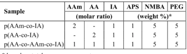
The FTIR spectra of H1, H2, H3, HS1, HS2 and HS3 are given in Figures 1-3. The characteristic peaks of acrylic-based copolymers were found at about 1400 cm-1 (C-O symmetric stretching) and 1450 cm-1 attributed to the.-CH2 group(Silverstein and Bassler, 1966). As expected, these peaks were observed in all spectra.
In the case of the AAm-IA based copolymers H1, the double peaks observed at about 1668 and 1720 cm-1 correspond to the amide and carboxyl groups (Silverstein and Bassler, 1966), respectively. In the case of the AA-IA based copolymer H2, a single peak was observed at 1721 cm-1 corresponding to the carboxyl groups of acids. In parallel, in the case of the AA-AAm-IA based terpolymer H3, peaks were observed at 1660 and 1727 cm-1 for the amide (shoul-der) and carboxyl groups (sharp peak), respectively.
Figure 1: The FTIR spectra of H1 and HS1.
Figure 2: The FTIR spectra of H2 and HS2.
Figure 3: The FTIR spectra of H3 and HS3.
For the silver loaded hydrogels (H-Ag+) HS1, HS2, HS3 a new peak was observed at about 1550 cm-1 attributed to the ionized carboxylate ion (Gils et al., 2010; Silverstein and Bassler, 1966). This indicates ionization of carboxyl groups and complexation be-tween the COO_ groups and Ag+ ions (Xiang and Chen, 2007; Khan et al., 2011). This provides evi-dence of the presence of silver ions in the hydrogel structure and of interaction between silver nanoparti-cles and the polymer backbone.
XRD Analyses
The XRD pattern of H and HSNC samples was used to determine the nanoparticle formation in the gel networks. XRD patterns of H1, H2, H3, HSCN1, HSCN2 and HSCN3 are demonstrated in Figures 4-6. Pure hydrogel samples did not exhibit any sharp peaks in XRD patterns, with only a broad peak at
∼20 θ attributed to the polymer networks (Varapsad
et al., 2009). In the case of HSNC, four sharp peaks were observed. These four diffraction peaks in the XRD patterns of HSCN samples, at angles 2θ=38.06o, 44.09o, 64.27o, 77.45o are ascribed to Bragg reflections through (111), (200), (220) and (311) planes of the face-centered cubic (fcc) packing of silver nanoparti-cles, respectively (Gils et al., 2010; Murthy et al., 2008). However, these peaks due to the formation of metallic silver nanoparticles in the gel networks (Varapsad et al., 2009) were not observed in XRD patterns of H1, H2 and H3 hydrogels, as expected. These results confirm that all HSCN samples contain silver nanoparticles.
UV-Vis Spectroscopic Analyses
are given in Figure 7. For this study, 150 mg of HSNC was dispersed in 15 mL of deionized water for 2 days to extract all silver nanoparticles into the aqueous phase and absorption spectra recorded with a scan range of 200–600 nm for these solutions. As shown in Figure 7, the spectra of HSNC samples have a characteristic absorption peak at around 405-410 nm due to the surface-plasmon resonance band of the silver nano-particles (Yiamsawas et al., 2008; Mohan et al., 2007), indicating the presence of silver nanoparticles (Varapsad et al., 2010) in the hydrogel structure.
Figure 4: XRD patterns of H1 and HSCN1.
Figure 5: XRD patterns of H2 and HSCN2.
Figure 6: XRD patterns of H3 and HSCN3.
Figure 7: UV-Vis. spectra of HSNC1 and HSNC3.
SEM Analyses
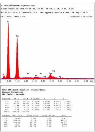
Surface morphologies of the hydrogels and hydrogel-silver nanocomposites were investigated by SEM and EDAX analysis. SEM micrographs of H3, HSNC3 samples and the EDAX spectrum of the HSNC3 sample are illustrated in Figures 8, 9 and 10, respectively. The SEM micrographs were taken at 80.000 magnifications.
Figure 8: SEM micrograph of H3.
Figure 10: EDAX spectrum of HSNC3
The SEM micrograph of pure hydrogel (H, with-out nanoparticles) showed a clear network and smooth surface throughout the structure, while the SEM micrograph of HSNC clearly showed the formation of silver nanoparticles in the network. The SEM micrographs of HSNC showed that the silver nano-particles were dispersed and embedded in the poly-mer matrix in all micrographs. The EDAX spectrum of the HSNC sample in Figure 10 also confirmed this result.
Swelling Studies
The results of swelling studies are presented to-gether in Figure 11. As seen from the figure, the swelling capacities of pure hydrogels are higher than those of hydrogel-silver nanocomposites and silver-loaded hydrogels.
After the hydrogels were treated with AgNO3 so-lution and silver ions loaded into the hydrogel net-work, the presence of silver ions in the hydrogel network caused a decrease of the swelling degree of silver-loaded hydrogels. This diminution originates from complexation of the Ag+ ion with carboxyl groups of the polymer backbone. As seen from the
figure, after the addition of Ag+, a significant amount of shrinkage was observed in the hydrogel network. When the silver-loaded hydrogels were treated with NaBH4 solution, silver nanoparticles were found in the hydrogel network. Thus, the swell-ing ratio of hydrogel-silver nanocomposites im-proved upon formation of Ago nanoparticles by the reduction reaction. The improvement of the swelling capacity in the hydrogel-silver nanocomposites can be attributed to the presence of silver nanoparticles of different sizes and different surface charges in the hydrogel network (Gils et al., 2010; Murthyet al., 2008; Vimalaet al., 2009, Yiamsawas et al., 2008). As a result, the order of the swelling capacity of the gels is the following: H > HSNC > HS.
Figure 11: Equilibrium swelling ratio values of drogels (H), silver-loaded hydrogels (HS) and hy-drogel-silver nanocomposites (HSNC).
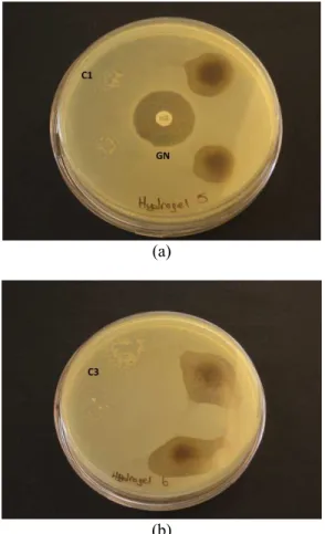
Antibacterial Activity
(a)
(b)
Figure 12: Antibacterial activity of (a) HSNC1 and (b) HSNC3 against Pseudomonas aeruginosa (ATCC 9027), C1: Hydrogel 1 without silver and C3: Hydrogel 3 without silver as negative control, GN: Geneticin as positive control.
Table 3: Antibacterial activity of HSNC samples.
Samples Gram-negative
bacteria HSNC1 HSNC3
GN (Control)
Pseudomonas aeruginosa (ATCC 9027)
1.8±0.17 2.27±0.24 2.4±0
Inhibition zone diameters were expressed as mean ±SD (mm). GN: Geneticin (10 µg)
CONCLUSION
In this study, copolymeric and terpolymeric hy-drogel-silver nanocomposites based on acrylic acid, acrylamide and itaconic acid were synthesized for investigation of antibacterial activities against Pseu-domonas aeruginosa. These hydrogel-silver nano-composites were characterized by FTIR, UV-Vis. spectroscopy, XRD and SEM techniques as well as their swelling properties. The following conclusions can be drawn from the results obtained:
FTIR spectra of HS samples showed the presence of silver ions in the hydrogel structure, and interac-tion between silver nanoparticles and the polymer backbone.
XRD patterns of HSNC samples confirmed the presence of silver nanoparticles in hydrogel structure.
UV-Vis. spectra of HSNC indicated the formation of silver nanoparticles.
SEM micrographs of HSNC showed the distribu-tion of silver nanoparticles in the polymer network. In addition, silver nanoparticles embedded in the poly-mer matrix were also observed in the EDAX spectrum.
In swelling studies, a significant amount of shrink-age of the gel network was observed after the addi-tion of Ag+ ion. Nevertheless, when Ago nanoparti-cles were formed by reduction, the swelling capacity of hydrogel-silver nanocomposites improved. This situation reflects the surface charge of the nanoparti-cles and an expansion in hydrogel network. The or-der of the swelling capacity of the gels was the fol-lowing: Hydrogel > Hydrogel-Ago > Hydrogel-Ag+.
Acrylic-based hydrogel-silver nanocomposites ex-hibited antibacterial activity against Pseudomonas aeruginosa according to antibacterial test results.
In conclusion, the silver nanoparticles were suc-cessfully produced within hydrogel networks. Syn-thesized acrylic-based hydrogel-silver nanocompo-sites demonstrated antibacterial activity against the Gram-negative bacterium, Pseudomonas aeruginosa. The hydrogel-silver nanocomposites obtained in this study are suitable for antibacterial applications in the medical field.
REFERENCES
Al, E., Güçlü, G., İyim, T. B., Emik, S. and Özgümüş, S., Synthesis and properties of starch-graft-acrylic acid/na-montmorillonite superabsorbent nano-composite hydrogels. J. Appl. Polym. Sci., 109, 16-22 (2008).
Aggor, F. S., Ahmed, E. M., El-Aref, A. T. and Asem, M. A., Synthesis and characterization of poly(Acrylamide-co-Acrylic acid) hydrogel con-taining silver nanoparticles for antimicrobial ap-plications. J. Am. Sci., 6(12), 648-656 (2010). Bauer, A. W., Kirby, W. M. M., Sherris, J. C. and
Turck, M., Antibiotic susceptibility testing by a standardized single disk method. Am. J. Clin. Pathol., 45, 493-496 (1966).
Byrne, M. E., Park, K. and Peppas, N. A., Molecular imprinting within hydrogels. Adv. Drug. Deliv. Rev., 54, 149-161 (2002).
Çöle, G., Gök, M. K. and Güçlü, G., Removal of basic dye from aqueous solutions using a novel nano-composite hydrogel: N-vinyl 2-pyrrolidone/itaconic acid/organo clay. Water Air Soil Poll., 224(1760), 1-16 (2013).
Dalaran, M., Emik, S., Güçlü, G., İyim, T. B. and Özgümüş, S., Removal of acidic dye from aque-ous solutions using poly (DMAEMA-AMPS-HEMA) terpolymer/MMT nanocomposite hydro-gels. Polym. Bull., 63, 159-171 (2009).
Dalaran, M., Emik, S., Güçlü, G., İyim, T. B. and Özgümüş, S., Study on a novel polyampholyte nanocomposite superabsorbent hydrogels: Syn-thesis, characterization and investigation of re-moval of indigo carmine from aqueous solution. Desalination, 279, 170-182 (2011).
Endo, T., Ikeda, R., Yanagida, Y. and Hatsuzawa, T., Stimuli-responsive hydrogel-silver nanoparticles composite for development of localized surface plasmon resonance-based optical biosensor. Anal. Chim. Acta, 611, 205-221 (2008).
Gils, P. S., Ray, D. and Sahoo, P. K., Designing of silver nanoparticles in gum arabic based semi-IPN hydrogel. Int. J. Biol. Macromol., 46(2), 237-244 (2010).
Guzman, M., Dille, J. and Godet, S., Synthesis and antibacterial activity of silver nanoparticles against gram-positive and gram-negative bacteria. Nanomed.-Nanotechnol., 8(1), 37-45 (2012). Ho, C. H., Tobis, J., Sprich, C., Thomann, R., Tiller
and J. C., Nanoseparated polymeric networks with multiple antimicrobial properties. Adv. Ma-ter., 16(2), 957-961 (2004).
Hong, K. H., Preparation and properties of electro-spun poly (vinyl alcohol)/silver fiber web as wound dressings. Polym. Eng. Sci., 47, 43-49 (2007). Ju, X. J., Zhang, S. B., Zhou, M. Y., Xie, R., Yang, L.
and Chu, L. Y., Novel heavy-metal adsorption material: Ion-recognition P(NIPAM-co-BCAm) hydrogels for removal of lead(II) ions. J. Hazard. Mater., 167, 114-118 (2009).
Karadağ, E., Saraydın, D. and Güven, O., Cationic dye adsorption by acrylamide/itaconic acid hy-drogels in aqueous solutions. Polym. Adv. Tech., 8(9), 574-578 (1997).
Karadağ, E. and Üzüm, Ö. B., A study on water and dye sorption capacities of novel ternary acryla-mide/sodium acrylate/PEG semi IPN hydrogels. Polym. Bull., 68, 1357-1368 (2012).
Khan, A., El-Toni, A. M., Alrokayan, S., Alsalhi, M., Alhoshan, M. and Aldwayyan, A. S.,
Microwave-assisted synthesis of silver nanoparticles using poly-N-isopropylacrylamide/acrylic acid microgel particles. Colloid Surface A: Physicochem. Eng. Aspects, 377(1-3), 356-360 (2011).
Kim, J. W., Lee, J. E., Kim, S. J., Lee, J. S., Ryu, J. H., Kim, J., Han, S. H., Chang, I. S. and Suh, K. D., Synthesis of silver/polymer colloidal compos-ites from surface-functional porous polymer mi-crospheres. Polymer, 45(14), 4741-4747 (2004). Luo, Y. L., Wei, Q. B., Xu, F., Chen, Y. S., Fan, L. H.
and Zhang, C. H., Assembly, characterization and swelling kinetics of Ag nanoparticles in PDMAA-g-PVA hydrogel networks. Mater. Chem. Phys., 118(2-3), 329-336 (2009).
Mohan, Y. M., Premkumar, T., Lee, K. and Geckeler, K. E., Fabrication of silver nanoparticles in hy-drogel networks. Macromol. Rapid. Commun., 27(16), 1346-1354 (2006).
Mohan, Y. M., Lee, K., Premkumar, T. and Geckeler, K. E., Hydrogel networks as nanoreactors: A novel approach to silver nanoparticles for anti-bacterial applications. Polymer, 48(1), 158-164 (2007).
Mohan, Y. M., Vimala, K., Thomas, V., Varaprasad, K., Sreedhar, B., Bajpai, S. K. and Raju, K. M., Controlling of silver nanoparticles structure by hydrogel networks. J. Colloid Inter. Sci., 342(1), 73-82 (2010).
Murthy, P. S. K., Mohan, Y. M., Varaprasad, K., Sreedhar, B. and Raju, K. M., First successful de-sign of semi-IPN hydrogel–silver nanocompo-sites: A facile approach for antibacterial applica-tion. J. Colloid Inter. Sci., 318, 217-224 (2008). Özkahraman, B., Acar, I. and Emik, S., Removal of
cationic dyes from aqueous solutions with poly (N-isopropylacrylamide-co-itaconic acid) hydrogels. Polym. Bull., 66, 551-570 (2011).
Pinto, R. J., Marques, P. A., Neto, C. P., Trindade, T., Daina, S. and Sadocco, P., Antibacterial activity of nanocomposites of silver and bacterial or vegetable cellulosic fibers. Acta Biomaterialia, 5(6), 2279-2289 (2009).
Pollo, L. D., Duarte, L. T., Anacleto, M., Habert, A. C. and Borges, C. P., Polymeric membranes con-taining silver salts for propylene/propane sepa-ration. Brazilian J. Chem. Eng., 29(2), 307-314 (2012).
Rujitanaroj, P., Pimpha, N. and Supaphol, P., Wound-dressing materials with antibacterial activity from electrospun gelatin fiber mats containing silver nanoparticles. Polymer, 49, 4723-4732 (2008). Silverstein, R. M. and Bassler, G. C., Spectrometric
Identification of Organic Compounds. 4th Ed., John Wiley, New York (1966).
Tang, Z.-X., Fang, X.-J., Zhang, Z.-L., Zhou, T., Zhang, X.-Y. and Shi. L-E., Nanosize MgO as an-tibacterial agent: Preparation and characteristics. Braz. J. Chem. Eng., 29(4), 775-781 (2012). Tang, Z.-X., and Lv, B.-F., MgO nanoparticles as
antibacterial agent: Preparation and activity. Braz. J. Chem. Eng., 31(3), 591-601 (2014).
Thatiparti, T. R., Kano, A., Maruyama, A. and Takahara, A., Novel silver-loaded semi-interpene-trating polymer network gel films with antibacte-rial activity. J. Polym. Sci., Part A: Polym. Chem., 47(19), 4950-4962 (2009).
Thomas, V., Mohan, Y. M., Sreedhar, B. and Bajpai, S. K., A versatile strategy to fabricate hydrogel– silver nanocomposites and investigation of their antimicrobial activity. J. Colloid Interf. Sci., 315(1), 389-395 (2007).
Travan, A., Pelillo, C., Donati, I., Marsich, E., Benincasa, M., Scarpa, T., Semeraro, S., Turco, G., Gennaro, R. and Paoletti, S., Non-cytotoxic silver nanoparticle-polysaccharide nanocomposites with antimicrobial activity. Biomacromol., 10(6), 1429-1435 (2009).
Varaprasad, K., Mohan, Y. M., Ravindra, S., Reddy, N. N., Vimala, K., Monika, K., Sreedhar, B. and Raju, K. M., Hydrogel-silver nanoparticle com-posites: A new generation of antimicrobials. J. Appl. Polym. Sci., 115(2), 1199-1207 (2010).
Vimala, K., Sivudu, K. S., Mohan, Y. M., Sreedhar, B. and Raju, K. M., Controlled silver nano-particles synthesis in semi-hydrogel networks of poly(acrylamide) and carbohydrates: A rational methodology for antibacterial application. Carbo-hyd. Polym., 75(3), 463-471 (2009).
Welton, J. E., SEM Petrology Atlas, Chevron Oil Field Research Company, Methods in Explora-tion Series No. 4. The American AssociaExplora-tion of Petroleum Geologists, Oklahoma, U.S.A. (2003). Xiang, Y. and Chen, D., Preparation of a novel
pH-responsive silver nanoparticle /poly(HEMA-PEGMA-MAA) composite hydrogel. Eur. Polym. J., 43(10), 4178-4187 (2007).
Xu, L., Li, X., Takemura, T., Hanagata, N., Wu, G., and Chou, L. L., Genotoxicity and molecular re-sponse of silver nanoparticle (NP)-based hydrogel. J. Nanobiotech., 10(16), 1-11 (2012).
Xu, S., Zhang, J., Paquet, C., Lin, Y. and Kumacheva, E., From hybrid hydrogels to photonic crystals. Adv. Funct. Mater., 13(6), 468-472 (2003). Yazdani-Pedram, M., Retuert, J. and Quijada, R.,
Hydrogels based on modified chitosan, 1 Synthe-sis and swelling behavior of poly(acrylic acid) grafted chitosan. Macromol. Chem. Phys., 201(9), 923-930 (2000).
Yiamsawas, D., Boonpavanitchakul, K., Sangsiri-mongkolying, R. and Kangwansupamonkon, W., ICONN2008, International Conference on Nano-science and Nanotechnology. Proceeding Book of ICONN 2008, 90-93 (2008).