Squamous cell carcinoma of the tongue:
clinical and morphological analysis of 57 cases
and correlation with prognosis
Carcinoma de células escamosas de língua: análise clínica e morfológica
de 57 casos e correlação com o prognóstico
Marcelo Gadelha Vasconcelos1; Rodrigo Porpino Mafra2; Rodrigo Gadelha Vasconcelos3; Ana Miryam Costa de Medeiros4; Lélia Maria Guedes Queiroz5
First submission on 23/04/14; last submission on 16/08/14; accepted for publication on 02/09/14; published on 20/10/14
1. Doctorate in Oral Pathology from Universidade Federal do Rio Grande no Norte (UFRN); professor at the Dentistry Department of Universidade Estadual da Paraíba (UEPB). 2. Dental surgeon; master’s student of Oral Pathology at UFRN.
3. Doctoral student of Oral Pathology at UFRN; professor at the Dentistry Department of UEPB.
4. Doctorate in Oral Pathology from UFRN; professor at the Post-Graduate Program in Oral Pathology of UFRN.
5. Doctorate in Oral Pathology from Universidade de São Paulo (USP); professor at the Post-Graduate Program in Oral Pathology of UFRN.
ABSTRACT
Introduction: Oral squamous cell carcinoma (OSCC), which represents more than 90% of head and neck malignant neoplasms, has a
poor prognosis due to its high frequency of lymph node metastasis and local invasion. Previous studies have investigated parameters related to the biological behavior of OSCC and its correlation with disease outcome (DO). Objective: To evaluate clinical and morphological data in cases of tongue squamous cell carcinoma (TSCC), correlating these indings with prognosis. Material and methods: Fifty-seven specimens of TSCC were obtained from patients undergoing surgical excision at a referral hospital in Natal, Brazil. Clinical data, such as tumor-node-metastasis (TNM) stage and DO, were collected from medical records. Hematoxylin and eosin-stained sections were analyzed regarding histological grade of malignancy (HGM), based on the system proposed by Bryne (1998). Results: The majority of patients (38.6%) were diagnosed as TNM stage III, and 57.9% developed metastases. Remission of the tumor occurred in 77.2% of the cases. The parameter “metastasis” exhibited a signiicant association with DO (p = 0) and TNM stage (p = 0.001), thus constituting a good indicator of tumor progression. Correlation of HGM and TNM stage with DO was not evidenced. Nevertheless, statistical analysis showed a signiicant association between HGM and TNM stage (p = 0.006). Conclusion: TNM clinical staging and HGM, evaluated in association, may be
useful to estimate the prognosis of TSCC.
Key words: squamous cell carcinoma; oral pathology; prognosis.
INTRODUCTION
Oral squamous cell carcinoma (OSCC) is the most common head and neck cancer, accounting for more than 90% of all tumors
in this region(1). The tongue is the most commonly affected site of
the oral cavity and has a poor prognosis, probably because of the high frequency of lymph node metastasis(17, 19) and local invasion(1).
Despite recent advances in treatment for this malignancy, the overall ive-year survival rate of patients is usually below 50%(17).
OSCCs are treated by primary surgical excision, alone or in combination with radiotherapy or chemotherapy in cases
of advanced stages of the disease(11, 15, 20). Improved prognostic
markers would be clinically useful to determine the biological aggressiveness of these tumors and to enable speciic appropriate therapies to be applied to each tumor(17, 19). Regional lymph
node metastasis is one of the most important prognostic factors of OSCC(1, 11, 13, 17, 19, 20) and may reduce the cure rate by
50%(7), but anatomic site(1, 22), histological grading systems
of malignancy(1, 4, 5, 9, 11-13, 17, 18), tumor-node-metastasis (TNM)
classiication of malignant tumors(1, 2, 5, 10, 11, 13, 17, 19, 21) and depth
of invasion(11, 19, 20) have also been cited as prognostic indicators.
Patients with OSCC can die as a result of diagnostic delays, regional lymph node metastases(19) or locoregional recurrences(20). Analysis of
the prognostic factors is important for predicting the outcome and achieving a high survival rate in these patients(21). Several reports have
examined the usefulness of clinical and pathologic factors related to the prognosis of OSCC(1, 2, 5, 9, 10, 17-20, 22). Therefore, the objective of this
study was to analyze the incidence of clinical and morphological data in a series of cases of tongue squamous cell carcinoma (TSCC), as well as their inluence in prognosis.
OBJECTIVE
The aim of this study was to evaluate and correlate clinical and morphological data on 57 TSCC cases, in a Brazilian population, contributing to the knowledge concerning the prognosis of oral cancer.
MATERIAL AND METHODS
The study was approved by the Research Ethics Committee of Universidade Federal do Rio Grande do Norte (protocol number: 029/10). Fifty-seven TSCC specimens were obtained from patients undergoing deinitive surgical resection, at Hospital Dr. Luiz Antônio, located in Natal, Brazil. Patients who had been followed up clinically for at least ive years were preferentially selected to determine the presence or absence of cervical lymph node metastases at the time of diagnosis. Patients who had developed cervical or distant lymph node metastases during treatment or follow-up were excluded. Metastases were demonstrated by imaging exams, such as computed tomography or magnetic resonance. Clinical data, such as age, gender, TNM stage and DO, were obtained from the patients’ medical records.
For histological analysis, 5-µm thick sections were cut from parafin-embedded tumor material and stained with hematoxylin-eosin. Malignancy at the invasion front was evaluated using the histological grading system proposed by Bryne (1998)(7). For each
tumor, degree of keratinization, nuclear pleomorphism, invasion pattern, and inlammatory response were analyzed semiquantitatively and given scores of 1 to 4. Cases with a total score of 4 to 8 were classiied as low-grade malignancy, and cases with a total score higher than 8 were classiied as high-grade malignancy(10).
RESULTS
Profile of the sample
Most patients with TSCC were males (70.2%) and aged from 61 to 70 years (35.1%). Caucasian patients predominated in the sample (n = 32; 56.2%). The combination of smoking
and alcohol consumption was the most frequent habit (42.1%), followed by smoking alone (31.6%). TNM stage III was the most frequent (38.6%), followed by stages I, IV and II. As regards the outcome, tumor remission was observed in 77.2% of the cases, and 57.9% developed metastases (Tables 1and2).
Statistical analysis of the clinical and
morphological aspects
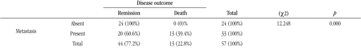
A statistically signiicant association was found between metastasis and DO, according to the chi-square (χ2) test (p =
0.000). Among the 24 cases with no metastasis, 100% achieved remission of the disease. Among the 33 cases with metastasis, less than 40% of patients died (Table 2).
TABLE 1 – Correlation between metastasis and clinical TNM stage. Signiicance level according to chi-square test (χ2) Clinical TNM stage
I II III IV Total (χ2) p
Metastasis
Absent 8 (57.1%) 9 (90)% 6 (27.3%) 1 (9.1%) 24 (100%) 17.613 0.001
Present 6 (42.9%) 1 (10%) 16 (72.7%) 10 (90.9%) 33 (100%)
Total 14 (24.5%) 10 (17.5%) 22 (38.6)% 11 (19.3%) 57 (100%)
TNM: tumor-node-metastasis.
TABLE 2 – Correlation between metastasis and disease outcome. Signiicance level according to chi-square test (χ2)
Disease outcome
Remission Death Total (χ2) p
Metastasis
Absent 24 (100%) 0 (0)% 24 (100%) 12.248 0.000
Present 20 (60.6%) 13 (39.4%) 33 (100%)
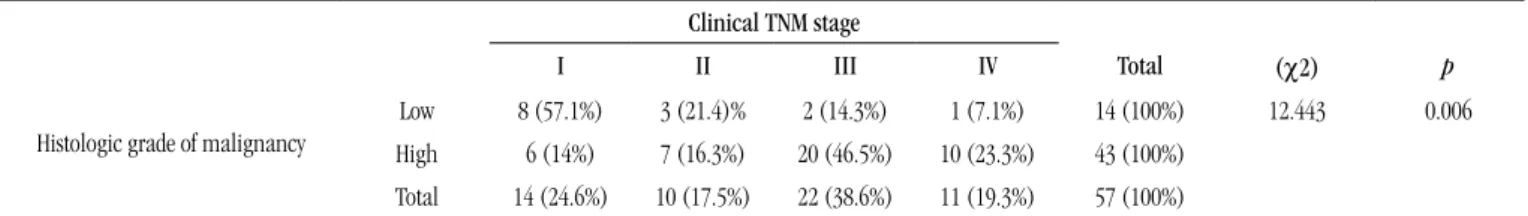
TABLE 3 – Correlation between histological grade and clinical TNM stage. Signiicance level according to chi-square test (χ2)
Clinical TNM stage
I II III IV Total (χ2) p
Histologic grade of malignancy
Low 8 (57.1%) 3 (21.4)% 2 (14.3%) 1 (7.1%) 14 (100%) 12.443 0.006
High 6 (14%) 7 (16.3%) 20 (46.5%) 10 (23.3%) 43 (100%)
Total 14 (24.6%) 10 (17.5%) 22 (38.6%) 11 (19.3%) 57 (100%)
TNM: tumor-node-metastasis.
Considering the relationship between metastasis and clinical TNM stage, there was a greater probability of developing metastasis in more advanced stages (III and IV). In the group with absence of metastasis, most of the sample was at stage II, followed by stage I. The χ2 test showed a statistically signiicant correlation between
the studied variables (p = 0.001) (Table 1).
Analysis of the relationship between histological grade of malignancy (HGM) and clinical TNM stage showed a greater frequency of low-grade malignancy cases in stages I and II, followed by stages III and IV. In relation to the high-grade cases, a higher prevalence of stages III and IV was found, in comparison to stages I and II. The χ2 test showed statistically signiicant
differences among these variables (p = 0.006) (Table 3).
100% and 85.7% of cases that were in stages I and II, respectively, as well as the predominance of deaths in cases classiied in stage IV. In a retrospective study of 130 OSCC cases, patients in stages III and IV had greater death rates, thus showing a statistically signiicant correlation between TNM stage and DO(2). Furthermore, in a recent
study of 202 TSCC cases, a statistically signiicant correlation of TNM stage with overall survival and disease-speciic survival was reported, emphasizing the relevance of TNM stage as an indicator of prognosis and parameter for treatment decision(17).
Considering the relationship between metastasis and TNM stage, according to the χ2 test, a statistically signiicant association
among these variables (p = 0.001) was evidenced, since TSCC
lesions in the most advanced stages (III and IV) were more likely to
DISCUSSION
Some studies point to factors which may inluence prognosis and survival of OSCC patients, namely: size and location of the primary tumor, regional lymph node involvement, presence of distant metastasis, and HGM(20). In a retrospective study, Goldstein
et al.(11) evaluated the association of other clinicopathologic
parameters with the outcome of TSCC. They found a statistically signiicant correlation between the occurrence of relapses and the variables “tumor size”, “invasion depth”, “surgical margin involvement” and “HGM”.
The TNM staging system has been the main indicator of prognosis in OSCC patients(13). This system consists of an
anatomical staging, proposed by the International Union Against Cancer, which describes the local extent of the primary tumor, as well as the involvement of regional lymph nodes and of distant sites. Following quantiication of those prognostic parameters, tumors are classiied in stages ranging from I to IV, and it is well established in literature that the head and neck SCCs classiied as stage I exhibit better prognoses than those in stage IV(1, 2, 15).
In view of these considerations, although TNM classiication is considered a factor associated to prognosis(5, 10), in our study we
could not demonstrate a statistically signiicant relationship between TNM stage and DO. However, we veriied remission of the lesion in
develop metastases (Table 1). We also found a signiicant correlation between metastasis and DO, considering that the 24 cases (100%) with absence of metastasis achieved remission of the disease, although among the 33 cases with metastasis, 39.4% of the patients died (Table 2). Thus, metastasis constitutes a good indicator of tumor progression, directly inluencing regional disease control and survival rate, as highlighted in previous studies(4, 6).
While several clinical parameters are related to the aggressiveness of OSCC, many aspects regarding the progression and biological behavior of this neoplasm are still unclear, particularly morphological characteristics, considered a controversial issue in
the literature(5, 9, 18). Currently, there are several HGM systems for
OSCC described in the literature, as already mentioned, with the purpose of providing morphological subsidies for an interpretation
of tumor aggressiveness(13).
In spite of attempts to improve the accuracy of HGM methods, by using scores based on mathematical data, the analysis of morphological parameters is based on the subjectivity of indings, what may lead to conlicting results. Considering these facts, the use of histological grading systems represents additional information for determining the prognosis of OSCC(1, 4).
Among all HGM methods, we can emphasize the system recommended by Bryne in 1998(7): because the key molecular
molecules, secretion of proteolytic enzymes, increased cell proliferation and initiation of angiogenesis) occur in tumor-host interface (invasion front), the author proposes a grading system which restricts morphological evaluation to the invasion front. This method represents an adaptation of a system proposed by
Bryne et al. in 1989(8). In this new grading, the criterion “number
of mitoses” was removed, while the parameters “degree of keratinization”, “cellular pleomorphism”, “invasion pattern” and “intensity of the inlammatory iniltrate” remained.
Bryne(7) and Noguchi et al.(16) reported that cells from the
invasion front are less differentiated and present a high degree of cell dissociation in comparison to other areas of the tumor. In view of this, one can infer that the invasion front constitutes one of the resources for comprehension of the tumor biological behavior, providing additional information on the choice of therapeutic strategies for OSCC patients, whereas the tool for better understanding of the invasive and metastatic behavior of neoplastic cells consists of the analysis of tumor margins.
In the present study, the HGM system recommended by Bryne(7)
was adopted, considering that it is a quick and easily performed method that presents prognostic value, what was demonstrated in previous researches(1, 3, 4, 7). Similarly to these investigations, we
opted to use the modiication proposed by Miranda(14), in order to
classify the SCC lesions according to the degree of malignancy. The majority of cases were deined as high grade (75.4%), with no signiicant association with the variables “DO” and
“metastasis”. However, it was descriptively demonstrated that most low- and high-grade cases correlated with disease remission and metastasis, respectively. Our results partially support the indings of previous studies, which demonstrated lack of correlation between HGM and prognostic variables, such as overall survival(1, 13, 18).
In contrast, regarding the analysis of the relation between HGM and clinical TNM staging, a greater frequency of low- and high-malignancy cases was evidenced statistically (p = 0.006) in early
(I and II) and advanced stages (III and IV), respectively (Table 3). These considerations are in agreement with previous studies(9, 12),
which reported that histological grading at the invasion front has prognostic value for OSCC, although other TNM system-related parameters may present more statistically signiicant results. Therefore, in the current study, metastasis constituted an indicator of biological behavior in cases of TSCC, since this variable was directly related to DO (death/remission) and clinical TNM stage, whereas HGM showed a signiicant correlation only with clinical stage.
CONCLUSION
Based on our results regarding clinical and morphological parameters, we believe that the TNM system and HGM, associated, may be useful tools for predicting high recurrence rate, poor survival rate and disease-free time of TSCC, thus contributing signiicantly to the understanding of the biological behavior of this neoplasm.
RESUMO
Introdução: O carcinoma de células escamosas oral (CCEO) representa mais de 90% das neoplasias malignas de cabeça e pescoço. Possui prognóstico desfavorável devido à alta frequência de metástase linfonodal e invasão local. Estudos anteriores investigaram parâmetros relacionados com o comportamento biológico do CCEO e sua correlação com o desfecho clínico. Objetivo: Avaliar dados clínicos e morfológicos em casos de carcinoma de células escamosas de língua (CCEL), correlacionando esses achados com o prognóstico. Material e métodos: Cinquenta e sete espécimes de CCEL foram obtidos de pacientes submetidos à excisão cirúrgica em um hospital de referência de Natal, Brasil. Dados clínicos, a exemplo do estágio tumor-linfonodo-metástase (TNM) e do desfecho da doença (DD), foram coletados dos prontuários médicos. Secções histológicas coradas em hematoxilina e eosina foram analisadas quanto à gradação histológica de malignidade (GHM), com base no sistema de Bryne (1998). Resultados:
A maioria dos pacientes (38,6%) foi diagnosticada no estágio TNM III, e 57,9% desenvolveram metástases. A remissão do tumor ocorreu em 77,2% dos casos. O parâmetro metástase apresentou associação significativa com o DD (p = 0) e o estágio TNM (p = 0,001), constituindo, dessa forma, um bom indicador de progressão da neoplasia. Não se observou correlação de GHM e estágio TNM com DD, contudo a análise estatística revelou associação significativa entre GHM e estágio TNM (p = 0,006). Conclusão: O estadiamento clínico TNM e a GHM, avaliados em conjunto, podem ser úteis para determinar o prognóstico do CCEL.
MAILING ADDRESS
Lélia Maria Guedes Queiroz
Departamento de Odontologia; Programa de Pós-Graduação em Patologia Oral; Av. Senador Salgado Filho, 1.787; Lagoa Nova; CEP: 59056-000; Natal-RN, Brazil; e-mail: [email protected].
REFERENCES
1. ALVES, P. M. et al. Signiicance of galectins-1, -3, -4 and -7 in the progression of squamous cell carcinoma of the tongue. Pathol Res Pract, v. 207, n. 4, p. 236-40, 2011.
2. ARAÚJO JÚNIOR, R. F., COSTA, A. L. L., RAMOS, C. F. Parâmetros clínico-patológicos como indicadores de prognóstico em carcinoma epidermoide oral. Pesq Bras Odontoped Clin Integr João Pessoa, v. 6, n. 2, p. 125-30, 2006.
3. BÀNKFALVI, A.; PIFFKÒ, J. Prognostic and predictive factors in oral cancer: the role of the invasive tumour front. J Oral Pathol Med, v. 29, n. 7, p. 291-8, 2000.
4. BARROS, S. S. L. V. Expressão imuno-histoquímica de metaloproteinases em carcinoma epidermóide de lábio inferior e língua. Natal, 2006. Tese (Doutorado em Patologia Oral) – Universidade Federal do Rio Grande do Norte.
5. BELL, R. B. et al. Tongue cancer: is there a difference in survival compared with other subsites in the oral cavity? J Oral Maxillofac Surg, v. 65, n. 2, p. 229-36, 2007.
6. BETTENDORF, O.; PIFFKÒ, J.; BÀNKFALVI, A. Prognostic and predictive factors in oral squamous cell cancer: important tools for planning individual therapy? Oral Oncol, v. 40, n. 2, p. 110-9, 2004.
7. BRYNE, M. Is the invasive front of an oral carcinoma the most important area for prognostication? Oral Dis, v. 4, n. 2, p. 70-7, 1998. 8. BRYNE, M. et al. New malignancy grading is a better prognostic indicator than Broders’ grading in oral squamous cell carcinomas. J Oral Pathol Med, v. 18, n. 8, p. 432-7, 1989.
9. COSTA, A. L. L.; ARAÚJO JÚNIOR, R. F.; RAMOS, C. C. F. Correlation between TNM classiication and malignancy histological feature of oral squamous cell carcinoma. Rev Bras Otorrinolaringol, v. 71, n. 2, p. 181-7, 2005.
10. GARAVELLO, W. et al. Prognostic inluence of gender in patients with oral tongue cancer. Otolaryngol Head Neck Surg, v. 138, n. 6, p. 768-71, 2008.
11. GOLDSTEIN, D. P. et al. Outcomes of squamous cell cancer of the oral tongue managed at the Princess Margaret Hospital. Head Neck, v. 35, n. 5, p. 632-41, 2013.
12. KUROKAWA, H. et al. The high prognostic value of the histologic grade at the deep invasive front of tongue squamous cell carcinoma. J Oral Pathol Med, v. 34, n. 6, p. 329-33, 2005.
13. LINDENBLATT, R. C. R. et al. Oral squamous cell carcinoma grading systems – analysis of the best survival predictor. J Oral Pathol Med, v. 41, n. 1, p. 34-9, 2012.
14. MIRANDA, J. L. Expressão de proteínas da matriz extracelular em carcinoma epidermóide de língua e lábio inferior. Natal, 2002. Tese (Doutorado em Patologia Oral) − Universidade Federal do Rio Grande do Norte. 15. NEVILLE, B. W. et al. Patologia oral & maxilofacial. 3. ed. Rio de Janeiro: Elsevier, 2009.
16. NOGUCHI, M. et al. Invasive front in oral squamous cell carcinoma: image and low cytometric analysis with clinicopathologic correlation. Oral Surg Oral Med Oral Pathol Oral Radiol Endod, v. 93, n. 6, p. 682-7, 2002. 17. RODRIGUES, P. C. et al. Clinicopathological prognostic factors of oral tongue squamous cell carcinoma: a retrospective study of 202 cases. Int J Oral Maxillofac Surg, v. 43, n. 7, p. 795-801, 2014.
18. SILVEIRA, E. J. D. et al. Correlation of clinical, histological and cytokeratin proiles of squamous cell carcinoma of the tongue with prognosis. Int J Surg Pathol, v. 15, n. 4, p. 376-83, 2007.
19. SÜSLÜ, N. et al. Carcinoma of the oral tongue: a case series analysis of prognostic factors and surgical outcomes. J Oral Maxillofac Surg, v. 71, n. 7, p. 1283-90, 2013.
20. THIAGARAJAN, S. et al. Predictors of prognosis for squamous cell carcinoma of oral tongue. J Surg Oncol, v. 109, n. 7, p. 639-44, 2014. 21. WANG, X. et al. Intratumor genomic heterogeneity correlates with histological grade of advanced oral squamous cell carcinoma. Oral Oncol, v. 42, n. 7, p. 740-4, 2006.