Tissue microarray: physical and chemical parameters
involved in the construction of recipient blocks
Microarranjo de tecidos: parâmetros físicos e químicos envolvidos
na construção dos blocos receptores
Marcos P. Santos; Thaiane A. Robeldo; Edwin David Castañeda; Ana Carolina Pagliarone; Karina N. Z. Pinto; Ricardo C. Borra
Universidade Federal de São Carlos (UFSCar), São Paulo, Brazil.
First submission on 14/03/17; last submission on 14/03/17; accepted for publication on 22/05/17; published on 20/08/17
ABSTRACT
Introduction: Tissue microarray (TMA) is considered an innovative method in several ields, with a great diversity of applications and advantages over traditional histomorphometric techniques. The most important advantage that TMA offers is the simultaneous evaluation of a large number of specimens from a limited source of material. However, TMA exhibits a high rate of non-viable samples in the inal stages of the process, which compromise their use in analyzes that can not be repeated. Objective: Considering this disadvantage, the
objective of this study was to optimize the methodology to maximize the viability of the samples, as well as to increase the eficiency of the technique. Material and methods: For this purpose, several variables involved in the construction of the recipient blocks, including parafin composition, diameter, spacing distance, localization and type of the tissue samples in the block were tested in order to establish correlations between the quality of the values and the parameters studied. Results: The results showed that the blocks built with polymer-enriched parafin, subjected to the fusion protocol at 37ºC, associated to a tempering, and constructed with one millimeter diameter samples and 1000 µm spacing between tissues, produced slides whith superior features. Conclusion: The data obtained from the physical
and chemical adjustments of the TMA recipient blocks provided vital information that, when applied in TMA research projects, may reduce the losses associated with the method.
Key words: histological techniques; parafin; quality improvement.
INTRODUCTION
Since it was irst reported in 1998, the tissue microarray (TMA) technique has been essential in hundreds of research projects involving diseases such as cancer, inlammatory and
neurodegenerative diseases(1). The construction of TMA blocks
is based on the extraction of cylindrical punches of parafin-embedded tissues from a diverse set of donor blocks, which are later reintroduced in the same recipient parafin block in speciic positions. This method allows the construction of blocks comprising nearly of 1,000 different samples. After assembling the TMA blocks, histological slides containing all the samples are obtained, enabling the simultaneous analysis of molecular targets in rigid and standardized conditions(2, 3).
TMA represents an important technological innovation in several different ields that use histomorphometric analysis as a research tool, including pathology, histology and immunology(3).
In the ield of oncology, different TMA models embedding optimized tissues of interest have been used to test markers associated with the prevalence, progression or prognosis of malignant neoplasms(4).
The TMA method has also been of great importance for studies of inlammatory diseases and neurodegenerative diseases, such as Alzheimer’s disease. In this case, TMA slides embedded with different samples of the brain were used to determine the number of amyloid plaques present in the affected organ. TMA can also be used for studies of healthy tissues. This approach is vital to allow for understanding of the expression of target genes in normal tissues(5).
TMA presents a range of advantages in comparison with other techniques that use slides derived from parafin blocks, such as the
ampliication of a limited source of material, the simultaneous analysis of a large quantity of specimens, experimental uniformity, and a reduction in the time and costs required for laboratorial procedures(3). The large-scale production of data by this method
is possible due to the number of samples that can be processed simultaneously, a number which, depending on the apparatus used for construction, may reach 1,000 spots per block(6, 7).
In addition, TMA allows cylindrical specimens with a small diameter to be removed from the donor block without causing any signiicant damage to the donor tissues. This prevents the depletion of the original tissues present in the donor blocks, maintaining them interpretable for future morphological and/or molecular analysis(8). Despite its advantages, however, the TMA
method presents a high rate of non-viable samples at the end of the process. This is caused by losses, scratches or folds in the tissue during the stages of the cutting and transferring of the sections from one block to the other(2). Previously reported data estimated
that tissue damage at the end of the TMA process can reach 10% to 30% of the original material used(9-13). This drawback can
compromise the use of the method in the laboratory experiments where repetitions are not possible due to the use of limited or scarce samples(14).
Variables such as type of tissue, parafin characteristics of the recipient block, parafin block tempering protocol of both blocks (donor and recipient), dimensions of the cylinder punch of the sample, spacing and positioning between the punches in the block, may act as determinant factors affecting the quality of the product. Despite their importance, these variables have not been studied in a systematic and controlled way. Therefore, the aim of the present study was to analyze the physical and chemical parameters involved in recipient block construction to optimize the TMA method and to reduce the high rate of non-viable samples produced.
MATERIAL AND METHODS
Embedded parafin blocks containing a variety of healthy mice organs were used to analyze the physical and chemical parameters employed in the construction of the recipient blocks. The experimental protocol followed the ethical principles in animal research indicated by the International Guiding Principles for Biomedical Research Involving Animals(15, 16) and was approved
by the Commitment to the Ethical Use of Animals in Research at the Universidade Federal de São Carlos (CEUA 7669010815).
The variables studied were: block tempering protocol (37ºC, 40ºC and 64ºC), tissue type (dorsal skin, heart, small bowel, lung, liver, spleen, stomach, kidney and tongue), recipient block composition
(pure parafin, pure parafin with increasing percentages of beeswax 5%, 10% and 15%, parafin enriched with Histotec® polymers from
Merck Millipore, Darmstadt, Germany), punch sample diameter (0.6 mm, 1.0 mm and 1.5 mm), spacing between samples in the recipiente block (400 μm, 700 μm and 1000 μm), and positioning in the block (upper right, upper left, center left, center right, lower left and lower right). The punch diameter values and the spacing between them were based on publications on TMA methodology and the options enabled by the TMA Master equipment (3-D Histech®,
Budapest, Hungary). This equipment is a drilling and punching machine for the construction of the TMAs. The operator has a range of possibility to control parameters such as size of punch, layout of distribution and size of the samples.
Animals
The mice used in the experiment were healthy, three-month-old male Swiss mice that had not undergone any type of previous treatment or procedure. The mice were sacriiced with an overdose (150 mg/kg) of Ketamine (Sespo, Paulínia, SP, Brazil)(17). Tissues
such as the dorsal skin, heart, small bowel, lung, liver, spleen, stomach, kidney and tongue were removed and immediately ixed in 10% buffered formalin (Sigma-Aldrich, St. Louis, MO, USA) for 24 hours at 25ºC. After the ixation period, the tissues
were washed in running water and kept in a 70% ethanol solution at 4ºC for 24 hours. Subsequently, the samples were prepared for inclusion in parafin using an automatic tissue processor (Lupetec, São Carlos, Brazil).
Construction of the TMA blocks
Five different reagents were used for the construction of the recipient blocks: pure conventional parafin, a mixture of pure conventional parafin with 5%, 10% or 15% of beeswax, and parafin enriched with polymers – Histosec® (Merck Millipore, Darmstadt,
Germany). Only the blocks constructed with Histosec® did not
suffer scratches after trimming of the surface with a microtome, and for this reason, the parafin enriched with polymers was used for the inal construction of all the TMA blocks employed in the subsequent tests.
Experimental design of the recipient blocks used
for analysis of the block tempering protocols
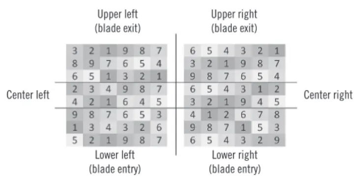
For analysis of the block tempering protocols, the recipient blocks received 1.5 mm diameter cylinders made up of samples from different tissues, with a spacing between cylinders of 1000 μm. The arrangement of the tissue samples was identical among the three recipient blocks, although the samples were distributed to occupy the different regions of the block (Figure 1).
blocks were tempered, and the blocks were then allowed to stand overnight at room temperature (25ºC)(18).
• Protocol 2: the recipient blocks were submitted to a temperature of 37ºC overnight, placing them in the stove with the surface facing downwards, with a clean slide as support. Subsequently, the blocks were submitted to a temperature of -12ºC for 10 minutes, followed by two heating-cooling (37ºC/-12ºC) cycles of one hour each(19).
• Protocol 3: the recipient blocks were submitted to a temperature of 40ºC for 10 minutes, with the surface of the block facing downwards using a clean slide as support.
Preparation of slides for quality analysis – block
tempering protocols
After the qualitative veriication of the integrity of the respective recipient blocks, those submitted to block tempering protocols at temperatures of 37ºC and 40ºC were used for obtaining sample sections of 5 μm thickness, as they did not suffer dimensional distortions. Subsequently, three sections from each block were extended on glass slides and analyzed under light microscopy for quality evaluation. Five μm thickness was the lowest value that allowed the performance of cuts in all the extensions of the block (3 cm × 2 cm) and the attainment of the slides without loss of tissues.
Preparation of the slides for quality analysis –
diameter and spacing
The block tempering protocol that presented the best result was used for the construction of the other nine recipient blocks from the combination of the three diameters of tissue (0.6 mm, 1.0 mm or 1.5 mm) and the three spacing values (400 μm, 700 μm or 1000 μm). The blocks were cut using a microtome loaded with a new blade. Three sections from each block were again extended on slides for later analysis of the quality of the sections through light microscopy.
Establishing the level and quality indicators
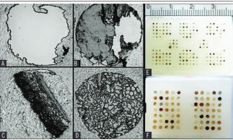
Each tissue sample was analyzed individually, with qualitative value described based on an index of quality comprising the values 0, 1, 2, and 3. Value 0 was attributed for samples that presented complete absence of tissues (Figure 2A) in the section.
Value 1 indicated that the tissue was present (totally or partially) in the section, but presented bends and scratches (Figure 2B). Value 2 meant the tissue was present in a complete form, exhibiting slight spacing between the borders and the parafin of the recipient block
(Figure 2C). Finally, value 3 represented the complete and integral
presence of the tissue without exhibiting any spacing (Figure 2D).
Lower left (blade entry)
Lower right (blade entry)
Center right Center left
Upper right (blade exit) Upper left
(blade exit)
FIGURE 1 − Example of the experimental design of the recipient blocks
The numbers represent the location of each tissue distributed throughout the block: 1) skin; 2) heart; 3) bowel; 4) lung; 5) liver; 6) spleen; 7) stomach; 8) kidney; 9) tongue.
Experimental design of recipient blocks used for
analysis of the remaining variables
The TMA were produced with cylindrical punches of the sample, with three different diameters (0.6 mm, 1.0 mm and 1.5 mm), positioned with a spacing between samples of 400 μm, 700 μm or 1000 μm. The blocks were designed so that each tissue would be distributed uniformly throughout each recipient block. Additionally, the tissues were located at least once in the irst line of the entry and exit point of the blade, receiving the irst and last contact between the blade and the block during the cutting of the histological section (Figure 1).
Performing the block tempering protocols
The block tempering protocol is used to incorporate the tissue sample present in the cylinder obtained from the donor block in the recipient block. Three different block tempering protocols currently described in TMA experiments were tested.
To improve the optimization of the TMA technique, the three quality values of each sample analyzed in triplicate were merged into one value by multiplying them together, so that zero represented the worst quality condition and the value 27 stands for the best possible situation. For the dichotomization of the quality indicators, values equal to or greater than nine were considered acceptable. This type of sample did not receive zero values (worst quality) at any stage but achieved a score of three on two occasions (best quality).
Quality analysis of slides exposed to hematoxylin
and eosin (HE) staining
After the quality analysis, the block with the best result was used to obtain four sections with 6 µm thickness, which were immediately positioned on histological slides. The sections were maintained at 37ºC in a drying stove for 24 hours to keep the tissue dry and to attach it to the slide before the staining process. Subsequently, the sections underwent deparafinization and HE staining based on the standard protocols. The slides were scanned and digital images were obtained using the Panoramic Desk (3D Histech®, Budapest, Hungary). Each tissue was qualitatively
assessed using the quality indicator values created in this study.
Statistical analysis
The experimental variables were analyzed using the Mann-Whitney, Kruskal-Wallis and Dunn Test with an adjusted p-value.
The adjusted p-value was adjusted by Benjamini-Hochberg correction. Statistical tests were performed using the IBM SPSS Statistics (SPSS Inc.®) and R (version 3.2 – www.r-project.org)
software packages, with p ≤0.05 considered signiicant in all tests.
RESULTS
Structure of the recipient blocks
The blocks constructed with pure conventional parafin did not endure the entry of the blade, releasing them from the cassette. They also suffered scratches and breaks along their entire length. The blocks constructed from the mixtures of pure parafin and different percentages of beeswax endured the microtomy phase. However, the cuts performed by the blade were carried out under great friction, resulting in low quality of the sections. These characteristics were found in all mixtures, regardless of the percentage used. The sections constructed with parafin enriched with polymers presented the best results, and so this material was used in all the other steps.
Block tempering protocol
The selection of the block tempering protocol was a determinant for the quality of the experiments. The recipient block submitted to a temperature of 64ºC (protocol #1) did not retain its integrity, melting during the irst heating phase. However, protocols #2 and #3 produced blocks without distortions, however, the quality of the samples from the blocks submitted to protocol #2 with tempering [index of quality (IQ) = 12] were signiicantly better than those undergoing protocol #3 without tempering (IQ = 4) (Table 1; p < 0.01, Mann-Whitney U test). Since tempering protocol #2 was the most eficient, this treatment was used as the basis for testing the others variables.
Diameter of sample inserted in the recipient block
Statistical analysis showed that the quality values of the samples inserted in the recipient blocks varied in relation to the diameter of the punch (p < 0.01, Kruskal-Wallis statistical
test). Samples with a diameter of 1.0 mm presented a median of quality which was statistically higher (IQ = 18, p < 0.01, Dunn’s
test) than the samples with diameters of 1.5 mm (IQ = 8) and 0.6 mm (IQ = 6). However, the samples with a diameter of 1.0 mm had a median value statistically higher (p < 0.01) than those with
a diameter of 0.6 mm (Table 1). There was no statistical difference between the 1.5 mm and 0.6 mm diameter samples.
Spacing between samples inserted in the recipient
block
The quality value results were statistically different depending on the spacing distances (p < 0.01; Kruskal-Wallis statistical test). The data showed that the median of the quality values for the blocks with 1000 μm spacing (IQ = 12) was statistically higher (p < 0.01;
FIGURE 2 − Quality levels established for quantitative analysis of tissue samples
The levels were organized in an increasing order of quality: A) value 0; B) value 1; C) value 2; D) value 3. TMA blocks built with polymer-enriched paraffin, submitted to the 37ºC block tempering protocol, associated with a tempering process and filled by 0.4 (E) and 1.5 mm diameter samples (F) with 1000 µm spacing between the tissues.
TMA: tissue microarray. A
C D
B
E
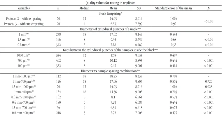
TABLE 1 − Quality of blocks in relation to physical parameters used to construct the TMA
Quality values for testing in triplicate
Variables n Median Mean SD Standard error of the mean p
Block tempering*
Protocol 2 – with tempering 70 12 14.93 8.916 1.066
< 0.01
Protocol 3 – without tempering 70 4 6.53 7.699 0.92
Diameters of cylindrical punches of sample**
1 mm(a) 238 18 17.62 9.145 0.593
-1.5 mm(b) 166 8 9.95 8.756 0.68 < 0.01
0.6 mm(c) 342 6 7.68 6.469 0.35 < 0.01
Gaps between the cylindrical punches of the samples inside the block**
1000 μm(a) 344 12 12.8 9.034 0.487
-700 μm(b) 402 8 10.12 8.895 0.444 < 0.001
400 μm(b) 382 8 9.43 9.001 0.461 < 0.001
Diameter vs. sample spacing combination**
1 mm-1000 μm(a) 112 18 18.25 8.337 0.788
-1 mm-700 μm(a, b) 126 18 17.06 9.807 0.874 0.720
1.5 mm-1000 μm(b) 70 12 14.93 8.916 1.066 0.028
1 mm-400 μm(b) 164 18 14.36 9.006 0.703 < 0.001
0.6 mm-1000 μm(c) 162 8 8.1 6.861 0.539 < 0.001
0.6 mm-700 μm(c) 180 6 7.29 6.087 0.454 < 0.001
1.5 mm-700 μm(c, d) 96 4 6.31 6.618 0.675 < 0.001
0.6 mm-400 μm(d) 218 3 5.72 7.008 0.475 < 0.001
TMA: tissue microarray; SD: standard deviation.
*: Mann-Whitney U test; **: Dunn statistical test with p-value adjusted by the false discovery rate according to the correction proposed by Benjamini-Hochberg; a, b, c and d: show statistically significant differences between groups.
Dunn statistical test) than the blocks with 700 μm spacing (IQ= 8) and 400 μm spacing (IQ = 8). No signiicant difference was found between the blocks with spacing of 700 μm and 400 μm (Table 1).
Correlation among sample spacing and diameter
in relation to quality values
After identifying the existence of an individual correlation between diameter and the spacing between samples in relation to the quality values, analysis was performed to check whether the spacing-diameter combination could inluence quality values
(p < 0.01; Kruskal-Wallis statistical test). The data revealed an
interaction between the two variables. The combinations of 1.0 mm diameter and 1000 μm spacing (IQ = 18); and 1.0 mm diameter and 700 μm spacing (IQ = 18) presented the best quality values in comparison to the other combinations (p < 0.01; Dunn statistical test). The samples with diameter of 0.6 mm regardless of spacing, displayed the worst quality values (1000 μm, IQ = 8; 700 μm, IQ = 6; 400 μm, IQ = 3) (Table 1).
Tissue type
Analyzing the inluence of the tissue type with the quality indicators, the results showed that the values of quality of the lung
(IQ = 12), tongue (IQ = 12), bowel (IQ = 9) and kidney (IQ = 9) were signiicantly superior to the others (p < 0.05; Dunn statistical test). Among these tissues, there were no statistical differences. The tissues: skin (IQ = 4), spleen (IQ = 6) and stomach (IQ = 4) showed the worst results. Liver (IQ = 8) and heart (IQ = 8) showed intermediate values of quality (Table 2).
Samples positioning
Assays were performed to verify the existence of a correlation between the positions of the samples in the recipient blocks and their respective quality values. When results were analyzed, it was veriied that, except for the upper right position (IQ = 6), which was considered the worst, there was no statistical difference in the quality of samples based on the position of samples in the recipient blocks (Table 2).
Correlation between tissue types and sample
diameters and quality indicators
TABLE 2 − Quality of blocks in relation to the type of tissue
used to construct the TMA Quality values for testing in triplicate
Variables n Median Mean SD Standard error of the mean p Type of tissue*
Lung(a) 124 12 14.26 8.756 0.786
-Tongue(a) 122 12 12.35 9.24 0.837 0.4048
Bowel(a) 128 9 11.89 9.368 0.828 0.1794
Kidney(a) 124 9 11.65 8.378 0.752 0.1164
Liver(b) 129 8 11.19 8.319 0.732 0.0382
Heart(b) 126 8 11.04 8.922 0.795 0.0268
Skin(b, c) 127 4 9.02 9.671 0.858 < 0.001 Spleen(c) 124 6 8.07 8.187 0.735 < 0.001 Stomach(c) 124 4 6.84 8.635 0.775 < 0.001
Position of the samples in the block*
Lower left(a) 202 12 12 9.295 0.654
-Center left(a) 157 8 11.5 8.96 0.715 0.981
Center right(a) 165 8 11.25 8.855 0.689 0.896
Lower right(a) 199 9 10.88 9.201 0.652 0.609
Upper left(a, b) 202 8 10.31 8.825 0.621 0.217 Upper right(b) 203 6 8.55 8.959 0.629 < 0.001 TMA: tissue microarray; SD: standard deviation.
*: Dunn statistical test with p-value adjusted by the false discovery rate according to the correction proposed by Benjamini-Hochberg; a, b and c: show statistically significant differences between groups
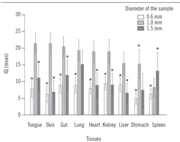
through the combinations of tongue/1 mm (IQ = 27), skin/1 mm (IQ = 27), bowel/1 mm (IQ = 27), lung/1 mm (IQ = 18), lung/1.5 mm (IQ = 15), heart/1 mm (IQ = 18), kidney/1 mm (IQ = 18) and liver/1 mm (IQ = 18). Overall, all the tissues presented the best quality values when the 1 mm diameter was used, with the exception of spleen and lung, which did not exhibit statistical differences between the 1 mm (IQ = 18) and 1.5 mm (IQ = 15) diameters. Regarding the spleen, an improvement in the mean quality value was obtained when the 1.5 mm diameter was utilized (Figure 3).
DISCUSSION
Experiments showed that physical and chemical parameters related to the construction of the receptor block can affect the quality of the histological sections. The structure of the blocks constructed with pure parafin was too rigid so that the recipient blocks were not capable of supporting the action of the blade during the microtomy phase, releasing from the cassettes and suffering fractures in their structures. When 5%, 10% or 15% of beeswax was incorporated into the pure parafin, the process of microtomy was achieved. The beeswax increased the tenacity of the block, allowing the parafin to withstand the shear pressure
FIGURE 3 − Influence of the combination of the type of tissue and the diameter of
cylinders in the IQ of samples inserted in the recipient blocks, considering the size of gaps between them
The values of quality were represented as mean ± confidence interval (95%). Each combination of tissue and diameter was compared with the value from the best sample (tongue – 1.0 mm). The results were analyzed by the Kruskal-Wallis and Dunn statistical tests. The p-value were adjusted using the false discovery rate proposed by Benjamini-Hochberg and the (*) p-value < 0.05 was considered significant.
IQ: index of quality.
Tongue Skin Gut Lung Heart Kidney Liver Stomach Spleen
IQ (mean)
Diameter of the sample 0.6 mm 1.0 mm 1.5 mm 30
25
20
15
10
5
0
Tissues
without breaking. The blocks constructed with parafin enriched with polymers were the only ones that endured cutting with the blade without great resistance or additional problems. In this case it is likely that the mixture of various polymers of different densities enriched the parafin, resulting in a lubricant characteristic and a greater malleability/tenacity, ensuring that the block could receive a great quantity of energy (impact) without suffering fractures.
Regarding the block tempering protocols, the block submitted to a temperature of 64ºC could not withstand such heat, and its structure melted. The other block tempering protocols tested (tempered and not tempered) used temperatures below the point of fusion of the parafin enriched with polymers (37ºC and 40ºC, respectively) and thus, the integrity of the block was preserved.
samples to be properly allocated in the holes of the recipient block, preventing proper adhesion between the tissue and the parafin on the recipient block. For the tempered block, the time of exposure (overnight) at 37ºC of the recipient block was signiicantly higher. In addition, the block underwent two phases of tempering with heating-cooling cycles. This could have allowed more time for the samples to be properly allocated in the recipient block and enabled the embedding of the parafin based on the contraction and expansion cycles of the materials.
Regarding the relationship between sample size and quality, blocks constructed with tissues with 1.0 mm diameter reached better results than those with 1.5 mm or 0.6 mm diameters. The low quality of the group comprised of 0.6 mm diameter samples could be explained by the small area of contact between the tissue and the parafin. Although the recipient blocks constructed with 1.5 mm diameter samples also had a performance worse than those of 1.0 mm, in this case, as the sample diameter increased, the area of the sample submitted to cutting force also increased, impairing the effect produced by the perimeter increase. Therefore, samples with 1.0 mm diameter presumably present the best relation between area and perimeter to resist the forces produced during the path of the blade. Regarding the combinations of diameter and spacing, the construction of the blocks with 1.0 mm diameter with 700 μm or 1000 μm tissues spacing was enough to obtain the resistance required for the histological sections. On the other hand, the inluence of the two types of tissue on the mean quality can be characterized as a function of the intrinsic nature of the tissues, as well as the effect of the structure after the stages of ixation and processing for parafin inclusion. Tissues that present a structural composition with pores or villi, such as the lung, tongue, bowels and kidney could have beneitted during the tages of iniltration and inal insertion in the liquid parafin. In this case, the parafin could have penetrated such cavities and due to a greater stability to the tissue-parafin complex of the inal recipient. In contrast, tissues with a highly dense or fragile structure such as the spleen, heart, or liver tend not to adhere correctly to the parafin of the recipient block, or to withstand the action of the blade during the cutting of the histological sections, resulting in the lower quality of the inal sample. Some tissues such as the skin and stomach mucosa may have had their ine structures modiied greatly by the ixation, dehydration, clariication and iniltration process, which could have negatively inluenced the annealing of the tissue to the parafin, and the microtomy stage.
The fact that the best mean quality values for most of the tissues were obtained with a diameter of 1.0 mm can be explained by an increase in the total perimeter (internal and external) between the tissues and the parafin of the recipient block. For the
spleen, a diameter of 1.5 mm was capable of overcoming, at least partially, the low quality of the tissue samples. The impact suffered by the spleen during the puncturing process of the donor block was probably lessened by the greater area of the tissue. In this case, the puncture pressure was better distributed throughout the section than the punctures performed in smaller areas, due to a greater stability of the spleen structure helping to maintain its integrity.
The position of the samples in the recipient blocks did not inluence the quality values presented, and the entry and exit point of the blade were not correlate to the inal result. The only signiicant difference during the tests involved the upper right position on the recipient block, which corresponds to the exit point of the blade. This result could indicate a potential inluence during the process of microtomy, or related to the construction of the histological slides.
For the slides that underwent HE staining, there was a clear degradation of around 40% of the samples (data not shown). The cause may be due to the conditions they were exposed during the deparafinization process which requires the use of aggressive reagents that decrease tissue quality. This result enforces the importance of the optimization of the parameters to produce high-quality TMA slides which are submitted to staining procedures to limit the failure rate.
CONCLUSION
The physical and chemical adjustments of the recipient blocks of the TMA method provided vital information which, when applied in TMA research projects, can reduce the losses associated with the method. Based on the analysis of the variables tested, the construction of the recipient blocks with polymer enriched parafin, assembled with samples of 1.0 mm diameter, 1000 μm spacing, and submitted to a 37ºC block tempering protocol associated with a tempering process, presented the best quality values. Although highly probable, the optimal block design/spacing speciications obtained from this instrument may be extrapolated to the other instruments that have been designed and marketed for the construction of TMAs.
ACKNOWLEDMENTS
opinions, indings, conclusions or recommendations expressed in this article are those of the authors and do not necessarily relect the views of funding bodies. The funding bodies played no role in the
REFERENCES
1. Lustosa SAS, Viana LS, Affonso, RJ, et al. Expression proiling using a cDNA array and immunohistochemistry for the extracellular matrix genes FN-1, ITGA-3, ITGB-5, MMP-2, and MMP-9 in colorectal carcinoma progression and dissemination. ScientiicWorldJournal. 2014; 2014(8): 1-27.
2. Bubendorf L, Kononen J, Koivisto P, et al. Survey of gene ampliications during prostate cancer progression by high-throughput luorescence in situ hybridization on tissue microarrays. Cancer Res. 1999; 59(4): 803- 7.
3. Chorilli M, Michelin DC, Salgado HRN. Animais de laboratório: o camundongo. Rev Ciênc Farmac Bas Aplic. 2007; 28(1): 11-23. 4. CIOMS. Council for International Organization of Medical Sciences; ICLAS. The International Council for Laboratory Animal Science. International Guiding Principles for Biomedical Research Involving Animal; 2012. Available at: https://cioms.ch/publications/ guidelines/1985_texts_of_guidelines.htm. [accessed on: June 10, 2015]. 5. Gavrielides MA, Conway C, O’Flaherty N, Gallas BD, Hewitt SM. Observer performance in the use of digital and optical microscopy for the interpretation of tissue-based biomarkers. Anal Cell Pathol (Amst). 2014; 2014(8): 1-10.
6. Hoos A, Urist MJ, Stojadinovic A, et al. Validation of tissue microarrays for immunohistochemical proiling of cancer specimens using the example of human ibroblastic tumors. Am J Pathol. 2001; 158(4): 1245-51.
study design; collection, analysis, and interpretation of data; in the writing of the manuscript; and in the decision to submit the manuscript for publication.
RESUMO
Introdução: O microarranjo tecidual (MAT) é considerado um método inovador em vários campos, com uma vasta diversidade de aplicações e vantagens em relação às técnicas histomorfométricas clássicas. A vantagem mais importante que o MAT oferece é a avaliação simultânea de um grande número de espécimes de uma fonte limitada de material. Contudo, ele apresenta uma taxa elevada de amostras não viáveis nos estádios finais do processo, o que compromete sua utilização em análises que não podem
ser repetidas. Objetivos: Considerando essa desvantagem, o objetivo deste estudo foi otimizar a metodologia para maximizar a
viabilidade das amostras, bem como aumentar a eficiência da técnica. Material e métodos: Para tanto, foram testadas várias
variáveis envolvidas na construção dos blocos receptores, como composição da parafina, diâmetro, distância de espaçamento, localização e tipo das amostras de tecido no bloco, a fim de estabelecer correlações entre a qualidade dos valores e os parâmetros
estudados. Resultados: Os resultados mostraram que os blocos construídos com parafina enriquecida em polímero, submetidos
ao protocolo de fusão a 37ºC, acoplados a ciclos de aquecimento e resfriamento e construídos com amostras de um milímetro de
diâmetro e espaçamento entre os tecidos de 1000 µm, produziram lâminas com características superiores. Conclusão: Os dados
obtidos dos ajustes físicos e químicos dos blocos de receptores de MAT forneceram informações vitais que, quando aplicadas em projetos de pesquisa de MAT, podem reduzir as perdas associadas ao método.
Unitermos: técnicas histológicas; parafina; melhoria de qualidade.
7. Hoos A, Cordon-Cardo C. Tissue microarray proiling of cancer specimens and cell lines: opportunities and limitations. Lab Invest. 2001; 81(10): 1331-8.
8. Jawhar NMT. Tissue microarray: a rapidly evolving diagnostic and research tool. Ann Saudi Med. 2009; 29(2): 123-7.
9. Kononen J, Bubendorf L, Kallioniemi A, et al. Tissue microarrays for high throughput molecular proiling of tumor specimens. Nature Med. 1998; 4 (7): 844-7.
10. Mousses S, Kallioniemi A, Kauraniemi P, Elkahloun A, Kallioniemi OP. Clinical and functional target validation using tissue and cell microarrays. Curr Opin Chem Biol. 2002; 6(1): 97-101.
11. Mucci NR, Akdas G, Manely S, Rubin MA. Neuroendocrine expression in metastatic prostate cancer: evaluation of high throughput tissue microarrays to detect heterogeneous protein expression. Hum Pathol. 2000; 31(4): 406-14.
12. Richter J, Wagner U, Kononen J, et al. High throughput tissue microarray analysis of cyclin E gene ampliication and overexpression in urinary bladder cancer. Am J Pathol. 2000; 157 (3): 787-94.
13. Rimm DL, Camp RL, Charette LA, Olsen DA, Provost E. Ampliication of tissue by construction of tissue microarrays. Exp Mol Pathol. 2001; 70(3): 255-64.
conventional method and by a modified technique. J Bras Patol Med Lab. 2008; 44(5): 327-45.
15. SBCAL. Sociedade Brasileira de Ciência em Animais de Laboratórios. Princípios éticos. Available at: http://www.cobea.org.br/conteudo/ view?ID_CONTEUDO=65. [accessed on: June 10, 2015].
16. Schrami P, Kononen J, Bubendorf L, et al. Tissue microarrays for gene ampliication surveys in many different tumor types. Clin Cancer Res. 1999; 5(8): 1966-75.
17. Simon R. Tissue microarrays: methods and protocols. 1 ed. Hamburgo: Humana Press; 2010.
18. Taylor CR, Rudbeck L. Immunohistochemical staining methods. 6 ed. Dinamarca: Dako’s Educational Guidebook; 2013.
19. Yuksel A. Constructing tissue microarrays. The tumour bank – the children’s hospital at Westmead, 2012. Available at: http://abrn.net/ wp-content/uploads/2014/04/CHW-TB-SOP-Constricting-Tissue-Microarrays-05.05.004-.pdf. [accessed on: June 10, 2015].
CORRESPONDING AUTHOR
Ricardo Carneiro Borra