85
Ophthalmomyiasis as a cause of canalicular lesion
Fabio P. Saraiva,1 José B. V. D. Fernandes,2 Vivian O. Tomikawa,1 Patrícia G. Costa,1 Suzana Matayoshi3
0021-7557/05/81-01/85
Jornal de Pediatria
Copyright © 2005 by Sociedade Brasileira de Pediatria
Abstract
Objective: Myiasis is the invasion of human tissues by Diptera larvae. Ocular involvement is uncommon. Trauma is the major cause of lacrimal apparatus lesions. However, it is rarely associated with parasitic infestation. The objective of this paper is to report a case of canalicular laceration caused by Dermatobia hominis larva.
Description: An eight-year-old girl presented preseptal cellulitis that was refractive to antibiotics. A
Dermatobia hominis larva was observed inside the lacrimal sac. Surgical extraction was performed and laceration of the lacrimal drainage system was noted.
Comments: Parasitic infection of the lacrimal apparatus is rare. Surgical extraction is the treatment of choice in such cases. Despite being uncommon, ophthalmomyiasis should be considered as a possible diagnosis when cellulitis is not responsive to antibiotics, especially in endemic areas. This is the first description of lacrimal drainage system injury by Dermatobia hominis larva.
J Pediatr (Rio J). 2005;81(1):85-7: Miasis, cellulitis, parasitic eye infections, lacerations, lacrimal apparatus.
1. Resident physician, Ophthalmologic Clinic, Hospital das Clínicas da Universidade de São Paulo (HC-USP), São Paulo, SP, Brazil. 2. Fellow physician, Division of Eye Plastic Surgery, Ophthalmologic Clinic,
HC-USP, São Paulo, SP, Brazil.
3. Ph.D. Assistant physician, Division of Eye Plastic Surgery, Ophthalmologic Clinic, HC-USP, São Paulo, SP, Brazil.
Manuscript received Mar 02 2004, accepted for publication Aug 11 2004.
Suggested citation: Saraiva FP, Fernandes JB, Tomikawa VO, Costa PG, Matayoshi S. Ophthalmomyiasis as a cause of canalicular lesion. J Pediatr (Rio J). 2005;81:85-7.
Introduction
Myiasis is the invasion of human tissue by the larva of a fly of the order Diptera. There are more than 85,000 Diptera species. Few, however, cause ocular injuries.1 Ocular involvement is responsible for 5% of all cases of myiasis and is denominated ophthalmomyiasis.2 Keyt was the first to describe a case of ophthalmomyiasis in 1900.3 It can be classified as internal or external (depending on whether or not the ocular bulb is penetrated) or orbital.4 When larvae reach the intra-ocular space they can be located in the anterior chamber, vitreous body or the sub-retinal space.5,6
External ocular infestation is more common than internal and the most common etiologic agent is Oestrus ovis.7,8 In these cases larval invasion usually occurs in the conjunctiva.9 Ophthalmomyiasis can also be associated with larvae of
Cuterebra, Dermatobia hominis and Hipoderma bovi.7
Dermatobia hominis is the primary cause of cutaneous
myiasis in South and Central America. Ophthalmomyiasis is rare in countries with cold climates such as Germany and North America, being most common in rural areas of developing countries.9-11
The female Dermatobia. hominis deposits its eggs in the abdomen of an insect. After six days the eggs mature. When the insect lands on a human being these eggs are deposited on the skin or mucosa and hatch, stimulated by the heat. The larva measures, during the initial stage, approximately 1.5 mm and penetrates the skin of the host in a few minutes. It positions itself so that the caudal respiratory spiracle remains outside of the host and the anterior spiracle inside, allowing it to feed. Larvae can invade the conjunctiva and ocular bulb, provoking conjunctivitis, corneal ulcer and destruction of the ocular bulb, eyelids and orbit, since it feeds on the surrounding tissues. After 10 weeks, the larva falls onto the ground and pupates, finally becoming an adult fly.12
86 Jornal de Pediatria - Vol. 81, No.1, 2005 Ophthalmomyiasis as a cause of canalicular lesion Saraiva FP et alii
The objective of this paper is to present a rare case of canalicular lesion secondary to ocular infestation by
Dermatobia hominis.
Case description
An eight-year-old child was admitted at the pediatric emergency room complaining of ocular hyperemia, pain, right palpebral edema and fever lasting 3 days (Figure 1).
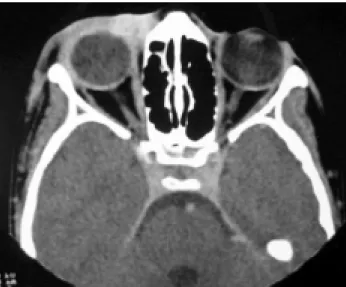
Ophthalmological investigation and computerized tomography of the orbit revealed preseptal cellulitis and a well-defined mass in the lower medial portion of the right orbit (Figure 2). Diagnostic hypotheses raised were infected tumor or burst dermal cyst. Treatment was begun with intravenous oxacillin and chloramphenicol and 1% topical chloramphenicol eye drops. After 13 days there was clinical improvement in the infection and the patient was discharged from hospital with a prescription for oral cephalexin.
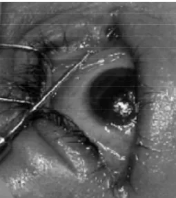
On the following day, however, the child presented fever peaks and worsening of the palpebral edema. On examination a larva was observed in the medial portion of the lacrimal sac. Surgical extraction of the larva, identified as Dermatobia hominis, was curative (Figure 3). A lesion
of the upper lacrimal canal caused by the larva was observed (Figure 4). Canalicular reconstruction was performed by means of intubation with a Crowford probe.
Discussion
Cutaneous myiasis often presents with an injury similar to a furuncle, with a small orifice causing purulence and pain.12 In the case described there was no injury to the eyelid observed which would suggest that the larva had entered and it is therefore probable that invasion had been via the conjunctiva. It is, however, important to point out that there may be ocular involvement even when the entry point is not proximal to the ocular region, as described by Engelbrecht in a description of external ophthalmomyiasis in which the primary site was in the cheek.11
Figure 1 - Preseptal cellulitis Figure 2 - Computerized tomography of the orbit revealed a well-defined mass in the lower medial portion of the right orbit
Jornal de Pediatria - Vol. 81, No.1, 2005 87
Figure 4 - Lesion of the upper lacrimal canal caused by the larva
Periorbital tissues that have been compromised by surgery, tumors, infections or ischaemia may predispose towards myiasis since the flies feed on exudates, blood, secretions and decomposing tissue.9
When the causes of lacrimal drainage system injuries are analyzed, canalicular lacerations are most frequent, in general associated with traumas (dog bites, collisions, falls, etc.).13 Parasitic infestation is a rare cause of tear duct disorders. Other parasitic infestations, equally rare, such as by Candida albicans or Aspergillus sp, only cause chronic dacryocystitis.14 No references to canalicular wall destruction, as in the present case, simulating traumatic laceration, were found in the literature investigated.
Surgical extraction is recommended as the treatment of choice. Other techniques are frequently used in developing countries, such as covering the wound with oily substances, like vaseline. These are not to be recommended however since the larva remains in situ, dying and causing granuloma of the foreign body, which may inflame or progress to calcification.12 Recently, the use of ivermectin was described for myiasis treatment.15-17
Although uncommon, ophthalmomyiasis should be considered as a possible differential diagnosis when cellulitis is refractive to conventional treatment, particularly in endemic areas. Emborsky & Faden published a similar case of ophthalmomyiasis that was diagnosed after unsuccessful clinical treatment for periorbital
References
1. Glasgow BJ, Maggiano JM. Cuterebra ophthalmomyiasis. Am J Ophthalmol. 1995;119:512-4.
2. Wilhelmus K. Myiasis palpebrarum. Am J Ophthalmol. 1986;101: 496-8.
3. Sivaramasubramanyam P, Sadanand AV. Ophthalmomyiasis. Br J Ophthalmol. 1968;52:64-5.
4. Bosniak SL, Schiller JD. Ophthalmomyiasis in an eyelid reconstruction. Am J Ophthalmol. 1990;109:101-2.
5. Torres RJA, Shiokawa N, Torres RJA. Oftalmomiíase interna posterior: relato de dois casos de larva viva no espaço sub-retiniano. Arq Bras Oftalmol. 2001;64:139-41.
6. Esteves JF, Maestri MK. Oftalmomiíase intra-ocular presumida. Arq Bras Oftalmol. 1993;56:62-4.
7. Rodrigues MM, Weiss CB, Muncy DW. Ophthalmomyiasis of the eyelid caused by Cuterebra larva. Am J Ophthalmol. 1974;78:1024-6.
8. Goodman RL, Montalvo MA, Reed JB, Scribbick FW, McHugh CP, Beatty RL, et al. Photo essay: anterior orbital myiasis caused by human botfly (Dermatobia hominis). Arch Ophthalmolol. 2000;118:1002-3.
9. Balasubramanya R, Pushker N, Bajaj MS, Rani A. Massive orbital and ocular invasion in ophthalmomyiasis. Can J Ophthalmol. 2003;38:297-8.
10. Weinand FS, Bauer C. Ophthalmomyiasis externa acquired in Germany: case report and review of the literature. Ophthalmologica. 2001;215:383-6.
11. Engelbrecht NE, Yeatts RP, Slansky F. Palpebral myiasis causing preseptal cellulites. Arch Ophthalmol. 1998;116:684-6. 12. Tsuda S, Nagaji J, Kurose K, Miyasato M, Sasai Y, Yoneda Y.
Furuncular cutaneous myiasis caused by Dermatobia hominis
larvae following travel to Brazil. Int J Dermatol. 1996;35:121-3. 13. Reifler DM. Management of canalicular laceration. Surv
Ophthalmol. 1991;36:113-32.
14. Duke-Elder S. Diseases of the lacrimal passages. In: Duke-Elder S, MacFaul PA, editors. System of Ophthalmology. 1st ed. London: Henry Kimpton; 1974. p. 675-773.
15. Victoria J, Trujillo R, Barreto M. Myiasis: a successful treatment with topical ivermectin. Int J Dermatol. 1999;38:142-4. 16. Matayoshi S, Fernandes JB, Lovaton ME, Aoki L, Nicoletti AB.
Ivermectina no tratamento de miíase orbitária - relato de caso. Arq Bras Oftalmol. 2003;66:519-21.
17. MacDonald PJ, Chan C, Dickson J, Jean-Louis F, Heath A. Ophthalmomyiasis and nasal myiasis in New Zealand: a case series. N Z Med J. 1999;112:445-7.
18. Emborsky ME, Faden H. Ophthalmomyiasis in a child. Pediatr Infect Dis J. 2002;21:82-3.
Ophthalmomyiasis as a cause of canalicular lesion Saraiva FP et alii
Corresponding author: Fábio Petersen Saraiva
Rua Dr. Ovídio Pires de Campos, 171/408, Cerqueira César CEP 05403-010 São Paulo, SP
Brazil
Phone: +55 (11) 3891.0925 E-mail: fabiopetersen@bol.com.br
cellulitis.18 Delays in seeking medical attention can lead to dramatic cases of total loss of the ocular bulb as described in two descriptions from India.3,9