172
Rev Odonto Cienc 2011;26(2):172-175Received: December 8, 2010 Accepted: February 14, 2011
Conflict of Interest Statement: The authors state that there are no financial and personal conflicts of interest that could have inappropriately influenced their work.
Copyright: © 2011 Herrera et al.; licensee EDIPUCRS. This is an Open Access article distributed under the terms of the Creative Commons Attribution-Noncommercial-No Derivative Works 3.0 Unported License.
Case Report
Large apical periodontitis healing following
root canal dressing with calcium hydroxide:
a case report
Reparo de lesão periapical extensa após utilização de curativo
de demora à base de hidróxido de cálcio: relato de caso
Henry Herrera a Helen Herrera a
Francisco Wanderley G de Paula e Silva a Mário R Leonardo a
Léa A B Silva a
a Departamento de Clínica Infantil, Odontologia
Preventiva e Social, Faculdade de Odontologia de Ribeirão Preto, Universidade de São Paulo, Ribeirão Preto, SP, Brasil
Correspondence:
Léa Assed Bezerra da Silva Avenida do Café, s/n Ribeirão Preto, RP – Brasil 14040-904
E-mail: [email protected]
Abstract
Purpose: The objective of this paper is to report the clinical case of a patient who presented a chronic apical periodontitis, arising from internal inflammatory resorption followed by pulp necrosis, and a long-term success of a root canal therapy using calcium hydroxide as root canal dressing.
Case description: A 20-year-old male patient presented for routine dental treatment. By radiographic examination we noted an extensive radioluscent area, laterally to the permanent maxillary right lateral incisor, with possibility of communication with the lateral periodontium, suggestive of a chronic apical periodontitis. Due to external root resorption detection, we used a calcium hydroxide root canal dressing, changed every 15 days, for a period of 2 months. Root canal filling was performed using gutta-percha cones by lateral condensation technique Radiographic follow up held after 19 years of treatment indicated a periodontium in conditions of normality, with the presence of lamina dura.
Conclusion: Calcium hydroxide is a suitable material to be used as root canal dressing in teeth with apical periodontitis. Long-term evaluation demonstrated the satisfactory clinical outcome following root canal treatment.
Key words: Apical periodontitis; root canal dressing; calcium hydroxide; dental trauma
Resumo
Objetivo: O objetivo deste trabalho é relatar o caso clínico de um paciente que apresentava lesão periapical crônica decorrente de necrose de pulpar e o sucesso a longo prazo de uma terapia endodôntica utilizando o hidróxido de cálcio como curativo de demora.
Descrição do caso: O paciente, do gênero masculino, com 20 anos de idade, compareceu à Clínica Odontológica para tratamento de rotina. Por meio de exames radiográficos observamos uma área radioluscente extensa, lateralmente ao incisivo lateral superior direito, com possibilidade de comunicação com o periodonto lateral, sugestivo de lesão periapical. Devido à presença de reabsorção radicular externa, foi utilizado um curativo de demora à base de hidróxido de cálcio (Calen®), trocado a cada 15 dias, por um período de 2 meses. A obturação do canal radicular foi realizada com cones de guta-percha pela técnica de condensação lateral ativa. O acompanhamento radiográfico realizado após 19 anos do tratamento endodôntico indicou um periodonto em condições de normalidade, com integridade da lamina dura.
Conclusão: O hidróxido de cálcio é um material adequado para ser usado como curativo de demora em dentes com lesão periapical, uma vez que a avaliação a longo prazo demonstrou resultados clínicos satisfatórios após o tratamento endodôntico.
Rev Odonto Cienc 2011;26(2):172-175
173
Herrera et al.Introduction
Long-term apical periodontitis infection occurs due to the
predominance of anaerobic gram-negative microorganisms,
both in main root canal and also disseminated throughout the
apical delta (1,2). These microorganisms present different
virulence factors and generate toxic products and
by-products to apical tissues, mostly because of the presence of
endotoxin in their cellular wall (3). Endotoxin carries a series
of important biological effects that lead to an inlammatory
reaction and periapical bone resorption (4).
Clinical studies have demonstrated that teeth with chronic
apical periodontitis present microbial niches in cementum
craters and periapical tissue, areas considered inaccessible
to root canal shaping (5,6). Endodontic bioilm can only
be removed by mechanical instrumentation if it is located
in the main root canal whereas apical bioilm necessitate a
therapeutic conduct to be eliminated (3,7).
Biomechanical root canal cleaning and shaping can be
performed employing bactericide washing solutions and
manual or rotary instrumentation, aimed to combating
infection by removing bacteria present in the main root canal
(1,3). During root canal treatment substances must be used
to combat not only infection in main root canal or root canal
walls, but also to reach those located deeply and diffusely
by tooth structure, areas inaccessible to the biomechanical
preparation or the body’s defense system (8). The objective
of this paper is to report the clinical case of a patient who
presented a chronic apical periodontitis, arising from internal
inlammatory resorption followed by pulp necrosis, and the
long-term success of a root canal therapy using calcium
hydroxide as root canal dressing.
Case Description
A 20-year-old male patient presented for routine dental
treatment. Anamnesis indicated a dental trauma history by
accidental fall in the right lateral incisor when the patient
was 15-year-old. The patient reported recurrent episodes of
edema in the buccal mucosa and referred usage of antibiotic
medication. The patient sought dental treatment only
after 5 years from the occurrence of the trauma. Clinical
examination indicated the presence of a silver amalgam
restoration in the palatal face of the tooth. Periodontal
tissues presented normal aspect and painful sensitivity was
reported to percussion and palpation. Pulp vitality tests were
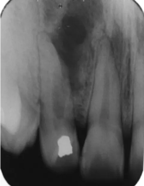
negative. By radiographic examination we noted an extensive
radioluscent area, laterally to the tooth, with possibility of
communication with the lateral periodontium, suggestive
of a chronic apical periodontitis. The main root canal was
calciied at its apical portion (Fig. 1). Root canal treatment
was established. The entire procedure was carried out with
rubber dam isolation of the operatory ield and aseptic
maintenance. After staging cavity access, neutralization of
septic/toxic content was performed using #K iles compatible
with diameter of the main root canal in a crown-apex
direction without pressure. During cleaning and shaping,
the root canal lateral communication was conirmed. The
root canal was prepared using manual instrumentation under
irrigation, inundation and aspiration with a 2.5% sodium
hypochlorite solution. The inal irrigation was performed
with an EDTA solution, under constant agitation with a thin
#K ile to remove the smear layer. As external root resorption
was detected, therefore we used a calcium hydroxide root
canal dressing (Calen
®), changed every 15 days, for a period
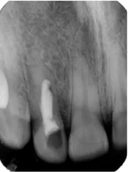
of 2 months. Root canal illing was performed after external
resorption was ceased, by means of Sealapex
®sealer using
gutta-percha cones by lateral condensation technique (Fig. 2).
Fig. 1. Initial radiograph: extensive radioluscent area, laterally to the tooth, suggestive of a chronic apical periodontitis. The main root canal was calcified at its apical portion.
174
Rev Odonto Cienc 2011;26(2):172-175 Apical periodontitis and calcium hydroxideTooth was restored with composite resin. Radiographic
follow up was carried out annually within the irst 2 years,
when complete bone repair was achieved (Fig. 3). The last
radiographic control was held after 19 years, when we
observed the periodontium in conditions of normality, with
the presence of lamina dura (Fig. 4).
Discussion
Several substances have been proposed to be used as root
canal dressing, including the phenolic derivatives, aldehydes,
corticosteroids in combination or not with antibiotics, and
calcium hydroxide (9,10). However, current studies have
proved unsatisfactory biological and antimicrobial of most
of them (3).
Topical drugs used as root canal dressing, which have
antimicrobial properties, anti-inlammatory and mineralizing
induction properties present a beneicial effect on the living
tissue of the periapical region. Calcium hydroxide is the
medication of choice, used in dentistry since the beginning
of the 20
thcentury, which presents ability to improve apical
healing, has a recognized action in soaking up the luid
when edema occurs, and induces mineralization (11). Also,
it penetrates through the dentin tubules and increases the pH
in the areas of cementum resorption (12), therefore, impairing
external root resorption (13). The ability of a calcium
hydroxide based formulation in removing the periapical
exsudate may be explained by its hygroscopic characteristic.
These properties are the main responsible for the long term
clinical and radiographic success observed in this study.
Fig. 3. Radiographic follow up 2 years following root canal filling indicating improved bone repair.
Fig. 4. Radiographic control after 19 years of root canal filling: periodontium in conditions of normality, with the presence of lamina dura.
The alkalinity of calcium hydroxide induces a zone
of supericial tissue necrosis and this necrotic tissue
separates the material from the vital tissues (14). The
ability to induce the formation of mineralized tissue
occurs by stimulating enzymes, such as alkaline phos-
phatase, and inhibiting acid phosphatase and osteoclasto-
genesis (15).
Recent studies have demonstrated that the infection of
root canals system induces the migration of immune and
inlammatory cells to the periapical bone area determining
the drop of the pH in the area and the presence of cytokines
which initiate and perpetuate bone and cementum resorption.
As shown in this case report, calcium hydroxide is a suitable
material to be used as root canal dressing in teeth with
apical periodontitis. Long-term evaluation demonstrated
the satisfactory clinical outcome following root canal
treatment.
Acknowledgments
The authors wish to thank Fundação de Amparo à
Pesquisa do Estado de São Paulo (FAPESP) for inancial
Rev Odonto Cienc 2011;26(2):172-175
175
Herrera et al.References
1. Assed S, Leonardo MR, Silva LAB, Ito IY. Prevalência de microrganismos em canais radiculares de dentes com necrose pulpar e reação periapical crônica – imunofluorescência indireta: efeito do preparo biomecânico e do curativo de demora pela cultura. Rev Bras Odontol 1996;53:24-8.Ruviére DB, Leonardo MR, da Silva LA, Ito IY, Nelson-Filho P. Assessment of the microbiota 2.
in root canals of human primary teeth by checkerboard DNA-DNA hybridization. J Dent Child 2007;74:118-23.
Leonardo MR. Endodoncia: Tratamiento de conductos radiculares. Principios técnicos y 3.
biológicos. São Paulo: Artes Médicas-Latino América; 2005.
Silva LAB, Nelson-Filho P, Leonardo MR, Rossi MA, Pansani CA. Effect of calcium hydroxide 4.
on bacterial endotoxin in vivo. J Endod 2002;28:94-8.
Leonardo MR, Rossi MA, Silva LAB, Ito IY, Bonifácio KC. EM evaluation of bacterial 5.
biofilm and microorganisms on the apical external root surface of human teeth. J Endod 2002;28:815-8.
Rocha CT, Rossi MA, Leonardo MR, Rocha LB, Nelson-Filho P, Silva LA. Biofilm on the apical 6.
region of roots in primary teeth with vital and necrotic pulps with or without radiographically evident apical pathosis. Int Endod J 2008;41:664-9.
Faria G, Nelson-Filho P, Freitas AC, Assed S, Ito IY. Antibacterial effect of root canal 7.
preparation and calcium hydroxide paste (Calen) intracanal dressing in primary teeth with apical periodontitis. J Appl Oral Sci 2005;13:351-5.
Paula-Silva FW, da Silva LA, Kapila YL. Matrix metalloproteinase expression in teeth with 8.
apical periodontitis is differentially modulated by the modality of root canal treatment. J Endod 2010;36:231-7.
Cohenca N, Heilborn C, Johnson JD, Flores DS, Ito IY, da Silva LA. Apical negative 9.
pressure irrigation versus conventional irrigation plus triantibiotic intracanal dressing on root canal disinfection in dog teeth. Oral Surg Oral Med Oral Pathol Oral Radiol Endod 2010;109:e42-6.
Siqueira JF Jr, Lopes HP. Mechanisms of antimicrobial activity of calcium hydroxide: a 10.
critical review. Int Endod J 1999;32:361-9.
Paula-Silva FW, Ghosh A, Arzate H, Kapila S, da Silva LA, Kapila YL. Calcium hydroxide 11.
promotes cementogenesis and induces cementoblastic differentiation of mesenchymal periodontal ligament cells in a CEMP1- and ERK-dependent manner. Calcif Tissue Int 2010;87:144-57.
Tronstad L, Andreasen JO, Hasselgren G, Kristerson L, Riis I. pH changes in dental tissues 12.
after root canal filling with calcium hydroxide. J Endod 1981;7:17-21.
Paula-Silva FW, Santamaria M Jr, Leonardo MR, Consolaro A, da Silva LA. Cone-beam 13.
computerized tomographic, radiographic, and histologic evaluation of periapical repair in dogs’ post-endodontic treatment. Oral Surg Oral Med Oral Pathol Oral Radiol Endod 2009;108:796-805.
Schroder U. Effects of calcium hydroxide-containing pulp-capping agents on pulp cell 14.
migration, proliferation and differentiation. J Dent Res 1985;64:541-8.
Silva RA, Leonardo MR, da Silva LA, de Castro LM, Rosa AL, de Oliveira PT. Effects of the 15.