E
ditorials89 0021-7557/10/86-06/445
Jornal de Pediatria
Copyright © 2010 by Sociedade Brasileira de Pediatria
445
C
aring for children brings many joys to the day of a pediatrician: sound nutritional guidance to foster growth, immunization to prevent infectious disease, timely use of antibiotics for an acute illness. But the encounter with an infant with jaundice and pale stools breaks a smooth day and heightens the fear of danger: does the infant have biliary atresia? Often, theseclinical signs develop in otherwise asymptomatic infants and during a time when parents are enjoying the new family member. Yet, they may represent the hallmarks of a disease of devastating consequences to the infant if untreated. And even when treated, the diagnosis of biliary atresia
brings challenging times for the infant, for the family, and for the pediatrician.
As the most common cause of pathologic jaundice in
infants, biliary atresia is an inlammatory cholangiopathy
that destroys the epithelial lining of bile ducts, disrupts bile
low, and promotes a ibrosing obstruction of extrahepatic bile ducts. The etiology of biliary atresia is yet to be deined,
but the pathogenic mechanisms of the disease are tightly linked to a robust response of the immune system that targets the bile ducts.1,2 Among the elements of the system,
CD8+ and natural killer (NK) lymphocytes have been shown to be key regulators of the biliary atresia phenotype in a mouse model of disease.3,4
The clinical and laboratory hallmarks of the disease are jaundice secondary to direct (conjugated) hyperbilirubinemia, pale stools, variable hepatosplenomegaly, high
gamma-glutamyl transpeptidase, and a liver histopathology
suggestive of biliary obstruction. The inal diagnosis is obtained at the time of exploratory cholangiography, which demonstrates obstruction of the extrahepatic bile duct. The only hope to restore bile low is the resection of biliary
remnants and the creation of an intestinal conduit that is
anastomosed to the hilum in a roux-en-Y
fashion as originally designed by Kasai & Suzuki.5 Clinical course and treatment
response obey the basic principles of illnesses: earlier diagnosis is associated with better response and outcome. The reproducibility of this paradigm across patient populations suggests that the biological basis of clinical
phenotype and response to treatment are less inluenced
by geographical boundaries. If this statement is correct, is it worth studying biliary atresia in individual countries? The article by Carvalho et al.6 in this issue of Jornal de
Pediatria shows that the answer is yes. The article reports the current state of diagnosis and treatment of infants with
biliary atresia in Brazil and identiies goals for the future
that are tailored to the national clinical practice.
Carvalho et al. reviewed the clinical presentation, treatment, and outcome of 513 children with biliary atresia. The data were obtained from large clinical centers in different geographical regions of Brazil. In general, the clinical presentation with direct hyperbilirubinemia, high gamma-glutamyl transpeptidase, and a histopathology with ductal proliferation and plugs was typical in the cohort. The mean age at diagnosis and portoenterostomy was 82.6±32.8
See related article on page 473
Biliary atresia in Brazil:
where we are and where we are going
Jorge a. Bezerra*
* MD. The Pediatric Liver Care Center, Division of Pediatric Gastroenterology, Hepatology and Nutrition, Cincinnati Children’s Hospital Medical Center, Department of Pediatrics, College of Medicine, University of Cincinnati, Cincinnati, OH, USA.
No conflicts of interest declared concerning the publication of this editorial. Financial support: this work was supported by NIH grant DK83781.
446 Jornal de Pediatria - Vol. 86, No. 6, 2010
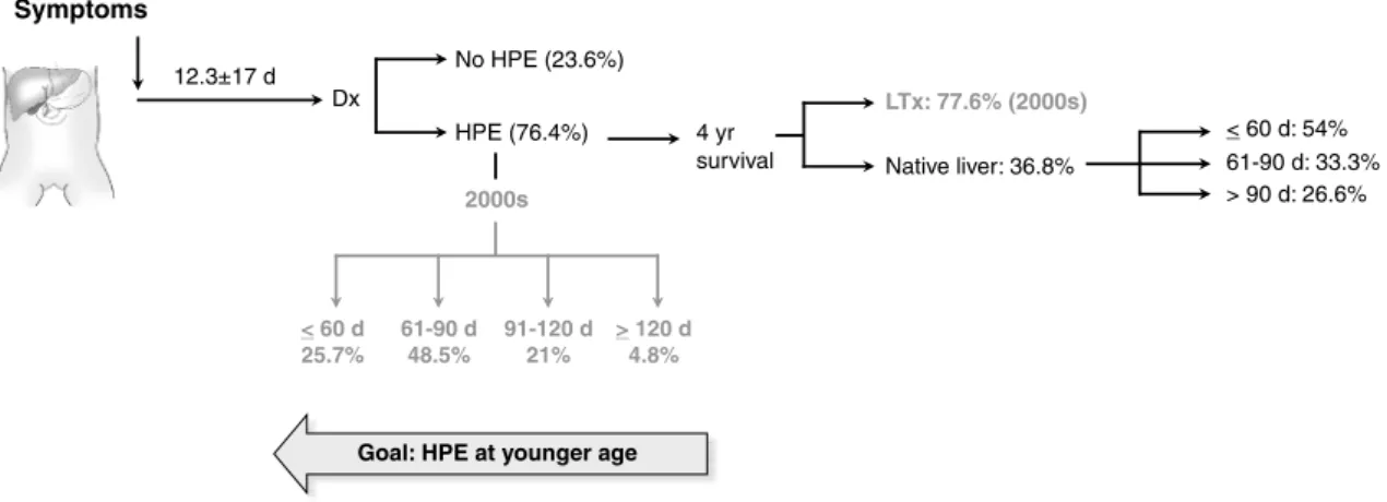
Figure 1 - Natural history of onset of symptoms, surgical intervention, and outcome with the native liver in children
with biliary atresia. The igure depicts the average age at onset of symptoms, the percentage of infants
undergoing hepatoportoenterostomy (HPE), and survival with the native liver by 4 years of age (overall and according to age groups at diagnosis). In gray is the percentage of infants undergoing HPE according to age groups and the survival (HPE + liver transplantation) in the last decade (2000s)
d = days; Dx = diagnosis; HPE = hepatoportoenterostomy; LTx = liver transplantation; yr = year.
Biliary atresia in Brazil - Bezerra JA
days, with surgery performed in 76.4% of the infants. In those infants treated with portoenterostomy, the 4-year survival with the native liver was 36.8%, with higher survival at 54% if the portoenterostomy was performed in infants
≤ 60 days of age. The combination of portoenterostomy
and liver transplantation increased the overall survival to 73.4%. However, only 46.6% of all patients underwent transplantation – a low rate compared to other countries,
in which access to transplantation increases to ≥ 60% of
children.7,8
How do the indings help understand the continuum of
care (diagnosis, treatment options, outcome) for infants with biliary atresia in Brazil? First, it is true that the mean age at
portoenterostomy at 82.6±32.8 days exceeds the practice
in other nations, which is around 60 days.7,9-11 However,
an analysis of the data for the most recent decade (2000 to 2010) in Brazil shows a shift of diagnosis to younger ages, with a greater percentage of infants diagnosed between 61-90 days (48.5 vs. 35% in the 80s) and a lower percentage of diagnosis beyond 120 days (4.8 vs. 20% in the 80s). In agreement with this concept, although a 4-year survival with the native liver for those infants treated with portoenterostomy was reported at 36.8%, survival improved
to a robust 54% if the infants were ≤ 60 days at the time
of surgery. Thus, there is a real trend to earlier diagnosis during the current decade, which is already translated into a greater number of children surviving with the native liver beyond 4 years.
The study also identifies two areas for future improvement. First, there is almost 2 weeks separating the age at onset of symptoms (12.3±17 days) and the age at portoenterostomy (82.6±32.8 days). While the reasons
for such delay in diagnosis and surgical intervention were not obvious, they may have been related to community awareness of the disease, recognition by primary care physicians, and access to specialized care. Pediatric hepatologists have partnered with the Brazilian Society of Pediatrics and the Brazilian Health Ministry to increase community awareness by the incorporation of a stool color card in the Childhealth Booklet distributed by the Ministry to the parents of every neonate. The goal is to aid parents realize that acholic stools (pale, clay-colored stools) is abnormal and seek medical help as soon as the abnormal color is noted – hopefully at early stages of the disease.
These are the infants that are presumed to beneit the most
from current treatment modalities.
The second area for improvement relates to the use of liver transplantation to improve survival when the child develops advanced liver disease. In the centers participating in the study, survival of children treated with portoenterostomy and later with liver transplantation increased in the 2000s to 77.6%. Despite this success, only 46.6% of the patients underwent liver transplantation. The simple answer is to increase the access of children with progressive liver disease to liver transplant centers. While
simple, this solution is largely dependent on an expansion
in the number of accredited transplant centers, support for transplant-related cost, and appropriate follow-up care. To become a reality, these factors must become priorities for
the ield of pediatrics and for the society as a whole.
Jornal de Pediatria - Vol. 86, No. 6, 2010 447
references
1. Santos JL, Carvalho E, Bezerra JA. Advances in biliary atresia: from patient care to research. Braz J Med Biol Res. 2010;43:522-7. 2. Sokol RJ, Shepherd RW, Superina R, Bezerra JA, Robuck P,
Hoofnagle JH. Screening and outcomes in biliary atresia: summary of a National Institutes of Health workshop. Hepatology. 2007;46:566-81.
3. Shivakumar P, Sabla G, Mohanty S, McNeal M, Ward R, Stringer K, et al. Effector role of neonatal hepatic CD8+ lymphocytes in
epithelial injury and autoimmunity in experimental biliary atresia.
Gastroenterology. 2007;133:268-77.
4. Shivakumar P, Sabla GE, Whitington P, Chougnet CA, Bezerra JA.
Neonatal NK cells target the mouse duct epithelium via Nkg2d
and drive tissue-speciic injury in experimental biliary atresia. J Clin Invest. 2009;119:2281-90.
5. Kasai M, Suzuki S. A new operation for ”non-correctable” biliary atresia, hepatic portoenterostomy [in japanese]. Shujutsu. 1959;13:733-9.
6. Carvalho E, dos Santos JL, da Silveira TR, Kieling CO, Silva LR,
Porta G, et al. Biliary atresia: the Brazilian experience. J Pediatr
(Rio J). 2010;86:473-9.
7. Schreiber RA, Barker CC, Roberts EA, Martin SR, Alvarez F, Smith L, et al. Biliary atresia: the Canadian experience.J Pediatr. 2007;151:659-65, 665 e1.
8. Wildhaber BE, Majno P, Mayr J, Zachariou Z, Hohlfeld J, Schwoebel M, et al. Biliary atresia: Swiss national study, 1994-2004. J Pediatr Gastroenterol Nutr. 2008;46:299-307.
9. Hsiao CH, Chang MH, Chen HL, Lee HC, Wu TC, Lin CC, et al.
Universal screening for biliary atresia using an infant stool color card in Taiwan. Hepatology. 2008;47:1233-40.
10. Serinet MO, Broue P, Jacquemin E, Lachaux A, Sarles J, Gottrand F, et
al. Management of patients with biliary atresia in France: results of a decentralized policy 1986-2002. Hepatology. 2006;44:75-84. 11. Shneider BL, Brown MB, Haber B, Whitington PF, Schwarz K, Squires
R, et al. A multicenter study of the outcome of biliary atresia in the United States, 1997 to 2000. J Pediatr. 2006;148:467-74.
Correspondence: Jorge A. Bezerra
Division of Pediatric Gastroenterology, Hepatology, and Nutrition Cincinnati Children’s Hospital Medical Center
3333 Burnet Ave
45229-3031 - Cincinnati, OH - USA Tel.: +1 (513) 636.3008
Fax: +1 (513) 636.5581
E-mail: Jorge.bezerra@cchmc.org
are going.” It will be important for pediatric hepatologists to broaden their investigative network to include greater representation of the geographical and cultural differences
in Brazil, for they are likely to inluence the natural history
of disease and the quality of care. It will also be important for them to continue to collect clinical data prospectively, store tissues to study the pathogenesis of the disease, and to pursue clinical trials. To be successful, their effort must be matched by an investment by the society, perhaps through funds from federal research agencies, to create a sound research infrastructure. Only then will we diagnose
early and ind new therapies to block disease progression
and save children with their own livers.