Abstract
Objective: To evaluate epidemiological, clinical and prognostic characteristics of children with biliary atresia. Methods: Data regarding portoenterostomy, liver transplantation (LTx), age at last follow-up and survival were collected from the records of patients followed up in six Brazilian centers (1982-2008) and compared regarding decades of surgery.
Results: Of 513 patients, 76.4% underwent portoenterostomy [age: 60-94.7 (82.6±32.8) days] and 46.6% underwent LTx. In 69% of cases, LTx followed portoenterostomy, whereas in 31% of cases LTx was performed as the primary surgery. Patients from the Northeast region underwent portoenterostomy later than infants from Southern (p = 0.008) and Southeastern (p = 0.0012) Brazil, although even in the latter two regions age at portoenterostomy was higher than desirable. Over the decades, LTx was increasingly performed. Overall survival was 67.6%. Survival increased over the decades (1980s vs. 1990s, p = 0.002; 1980s vs. 2000s, p < 0.001; 1990s vs. 2000s, p < 0.001). The 4-year post-portoenterostomy survival, with or without LTx, was 73.4%, inversely correlated with age at portoenterostomy (80, 77.7, 60.5% for ≤ 60, 61-90, > 90 days, respectively). Higher survival rates were observed among transplanted patients (88.3%). The 4-year native liver survival was 36.8%, inversely correlated with age at portoenterostomy (54, 33.3, 26.6% for ≤ 60, 61-90, > 90 days, respectively).
Conclusions: This multicenter study showed that late referral for biliary atresia is still a problem in Brazil, affecting patient survival. Strategies to enhance earlier referral are currently being developed aiming to decrease the need for liver transplantation in the irst years of life.
J Pediatr (Rio J). 2010;86(6):473-479: Biliary atresia, portoenterostomy, hepatic, surgery, diagnosis, differential, prognosis.
O
RiginAlA
RtiCleCopyright © 2010 by Sociedade Brasileira de Pediatria
473
introduction
Biliary atresia (BA), characterized by obliteration of the extrahepatic bile ducts, remains the main indication for liver transplantation (LTx) in children.1 BA clinical picture
starts in the irst weeks of life and displays a universal
distribution with variable incidence in different regions
of the world.2-5 The etiology of BA remains elusive and
several mechanisms have been proposed to explain its progressive cholangiopathy.6,7 Early diagnosis of BA and
surgical relief of the mechanical impediment to bile low
by portoenterostomy, preferably before 60 days of life, are
Biliary atresia: the Brazilian experience
elisa de Carvalho,1 Jorge luiz dos Santos,2 themis Reverbel da Silveira,2 Carlos Oscar Kieling,3 luciana Rodrigues Silva,4 gilda Porta,5 irene Kazue Miura,5
Adriana Maria Alves De tommaso,6 Maria Ângela Bellomo Brandão,6
Alexandre Rodrigues Ferreira,7 José Roberto de Deus Macêdo,8 José tenório de Almeida neto,8 grupo de estudos em Hepatologia Pediátrica do Brasil
1. PhD. Hospital de Base do Distrito Federal (HBDF), Brasília, DF, Brazil.
2. PhD. Hospital de Clínicas de Porto Alegre (HCPA), Universidade Federal do Rio Grande do Sul (UFRGS), Porto Alegre, RS, Brazil. 3. MD. HCPA, UFRGS, Porto Alegre, RS, Brazil.
4. PhD. Universidade Federal da Bahia (UFBA), Salvador, BA, Brazil.
5. PhD. Hospital Sírio-Libanês, São Paulo, SP, Brazil. Hospital A. C. Camargo, São Paulo, SP, Brazil. 6. PhD. Universidade Estadual de Campinas (UNICAMP), Campinas, SP, Brazil.
7. PhD. Universidade Federal de Minas Gerais (UFMG), Belo Horizonte, MG, Brazil. 8. MD. HBDF, Brasília, DF, Brazil.
No conflicts of interest declared concerning the publication of this article.
Suggested citation: de Carvalho E, dos Santos JL, da Silveira TR, Kieling CO, Silva LR, Porta G, et al. Biliary atresia: the Brazilian experience. J Pediatr (Rio J). 2010;86(6):473-479.
Manuscript submitted Apr 07 2010, accepted for publication Sep 28 2010.
crucial, since delay leads to the need for LTx or death in the
irst 3 years of life.8-10 Prognosis of patients with BA has
improved in the last decades, reaching 90% of cases,10 due
to timely performance of portoenterostomy and LTx followed by adequate immunosuppression. More than 50% of patients who undergo portoenterostomy become jaundice-freeand, among them, several patients reach adolescence without LTx. For those who present complications such as portal hypertension and cirrhosis, LTx provides a good quality of life.11 The experience of several countries regarding BA
has been described,5,12-17 but there are only a few isolated
reports on the experience with BA in Brazil.18,19 This study
aimed to evaluate clinical, epidemiological and prognostic characteristics of Brazilian children with BA.
Patients and methods
Retrospective review of the medical records of patients with BA followed up between July 1982 and December 2008
in six Brazilian reference centers: Hospital de Clínicas de
Porto Alegre at Universidade Federal do Rio Grande do Sul
(UFRGS), South region; Hospital Sírio Libanês and Hospital
do Câncer, São Paulo, Southeast region; Universidade Federal
da Bahia (UFBA), Northeast region; Hospital de Base do Distrito Federal (HBDF), Midwest region; Universidade de
Campinas (UNICAMP), Southeast region; and Universidade Federal de Minas Gerais (UFMG), Southeast region.
Data collected from the records included: sex; place of origin (Brazilian region) and category of town (capital city or countryside); birth weight; date of birth; onset of jaundice; hospital admission; presence of associated extrahepatic congenital anomalies; results from laboratory tests, ultrasound, biliary scintigraphy, biopsies from liver
and porta hepatis; and surgical indings. Regarding therapy,
data included the performance, or not, of surgical procedures (portoenterostomy and LTx) and date of procedures and postoperative outcomes, including date of last follow-up. Diagnosis of BA was based on clinical, biochemical, histological, imaging and surgical data, as previously reported elsewhere.6,11
Data were initially analyzed as a single set, being subsequently divided into three categories according to the decade in which portoenterostomy was performed: 1980s (1982-1989); 1990s (1990-1999); and 2000s (2000-2008).
Age at portoenterostomy was classiied according to four age groups: ≤ 60 days; 61-90 days; 91-120 days; and > 120 days. In order to analyze the overall and native liver survival, 91-120 days and > 120 days age groups were
evaluated as a single group. Information was collected by investigators from each participating center and sent to
the Data Registry Center, in Brasília, Brazil, where data
were entered on a single platform for statistical analysis. Cases initially followed in a center and then transferred to another hospital to perform LTx were considered as a
single case from only one of these centers. Since this was a retrospective study, a complete data collection including all patients was not feasible.
Categorical variables were described in igures and
tables of frequency distributions, and continuous variables were expressed as mean ± standard deviation (SD) or median and interquartile (IQ) range. The chi-square test and Student’s t test were used for comparisons. Survival curves were built using the Kaplan-Meier method and the Cox model and compared by calculating the hazard ratio. Overall survival was based on date of birth and date of death or last follow-up. Native liver survival was based on date of birth, LTx, and death or last follow-up. P values < 0.05
were considered to be statistically signiicant. Microsoft Excel
2007 (Microsoft Corp, Redmond, WA, USA) and SPSS 15.0 (SPSS Inc, Chicago, IL, USA) were used for data processing and statistical analysis.
This study was approved by the Research Ethics Committees of the participating institutions.
Results
Data on patients with BA (n = 513, 283 female and 230 male) included in this study were sent by individual
centers to the Data Registry Center, in Brasília, Brazil.
The number of patients according to individual centers
was: 187 (36.5%), UFRGS; 151 (29.4%), Hospital Sírio Libanês and Hospital do Câncer; 63 (12.3%), UFBA; 53 (10.3%), HBDF; 37 (7.2%), UNICAMP; and 22 (4.3%),
UFMG. Distribution of patients according to Brazilian regions was: 185 (36.1%), South; 169 (32.9%), Southeast; 96 (18.7%), Northeast; 50 (9.8%), Midwest; and 13 (2.5%),
North. Forty-ive (8.8%), 133 (25.9%) and 335 (65.3%)
patients were admitted in the 1980s, 1990s and after 2000, respectively.
Mean birth weight of patients was 3,138.6 (±499.3) grams and the onset of jaundice occurred at 12.3 (±17.0) days of life. Extrahepatic congenital anomalies were found in 61 (11.8%) patients, including splenic (n = 10), gastrointestinal (n = 25), cardiovascular (n = 25), urinary (n = 6), and teratoma (n = 1) anomalies. Six patients had more than one anomaly. Findings suggestive of BA splenic malformation syndrome occurred in 17 patients, including polysplenia (n = 5), abdominal situs inversus (n = 11), and dextrocardia (n = 1).
Blood chemistry measurements were: total bilirubin, 11.9 (±6.2) mg/dL; serum direct bilirubin, 9.1 (±5.8) mg/dL; gamma-glutamyltransferase (GGT), 15.2 times the normal value (x N) (±16.4); aspartate aminotransferase (AST), 6.1 x N (±4.7); and alanine aminotransferase (ALT), 4.7 x N (±4.8). Regarding liver histopathology, most patients had bile plugs (93.8%), ductular/ductal proliferation (93.8%),
a complete data collection including all patients was not feasible, and results of biliary scintigraphy and abdominal ultrasound, among other tests, could not be analyzed.
Portoenterostomy
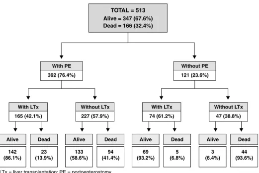
Portoenterostomy was performed in 392 (76.4%) patients; among these, for 12 patients age at portoenterostomy could not be determined (Figure 1). These patients underwent LTx and their post-transplant follow-up data were available for analysis. Age at portoenterostomy for the remaining 380 patients was 82.6±32.8 days [median = 78.5 (60.0-94.7) days].
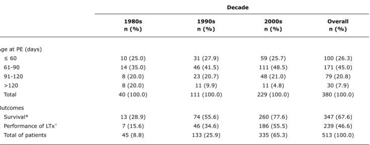
Only 100 (26.3%) patients were operated on until 60 days of life. Most (45.0%) patients underwent portoenterostomy between 61 and 90 days, 79 (20.8%) patients were operated on between 91 and 120 days, and 30 (7.9%) patients after 120 days (Table 1). An increasing number of patients underwent portoenterostomy in the subsequent decades under study. When comparing the three decades with regard to age at portoenterostomy, there were increasing rates of patients undergoing the procedure between 61 and 90 days of life (p = 0.047) and decreasing rates after 120 days (p = 0.020).
When comparing the rates of patients undergoing or not portoenterostomy according to their distribution across Brazilian regions (Table 2), most non-operated infants were from the North (69.2%) and Northeast (45.8%), while only 11.9% of patients were from Southern Brazil.Regarding
category of town, capital city or countryside, no signiicantly
different rates were observed (24.8 vs. 23.0%).
Regarding age at portoenterostomy, according to Brazilian regions (Table 2), infants from the Northeast and North were operated on later [92.3 (±36.1) and 102.2 (±36.4) days, respectively] than infants from the Midwest [84.9 (±29.9) days], South [80.8 (±36.6) days] and Southeast [79.5
(±25.4) days]. Age at portoenterostomy was signiicantly
different when South and Northeast regions (p = 0.008) and Southeast and Northeast regions (p = 0.012) were
compared. However, there were no differences when other
regions were compared or when patients from capital cities [82.2 (±35.9) days] and from countryside [82.8 (±31.3) days] were compared (p = 0.639).
Liver transplant
Of all patients, 239 (46.6%) underwent LTx. A comparison over the three decades revealed increasing rates of performance of LTx in Brazil (1980s vs. 1990s, p = 0.016; 1980s vs. 2000s, p < 0.001; 1990s vs. 2000s, p < 0.001), as described in Table 1. In 69.0% of cases, LTx followed portoenterostomy, whereas in 31.0% of cases LTx was performed as the primary surgery (Figure 1). Among patients previously subjected to portoenterostomy, age at LTx ranged from 0.8 to 2.6 (2.6±3.1) years. The remaining children were transplanted earlier, at around
0.6-1.5 (1.2±0.8) years, revealing a signiicant difference
between groups (p < 0.001).
Survival rates
The overall survival was 67.6%, and the highest survival rates occurred in transplanted patients (without previous
Place of origin
Portoenterostomy South Southeast Midwest northeast north Capital Countryside
Yes, n (%) 163 (88.1) 133 (78.7) 40 (80.0) 52 (54.2) 4 (30.8) 121 (75.2) 271 (77.0)
Total, n (%) 185 (100.0) 169 (100.0) 50 (100.0) 96 (100.0) 13 (100.0) 161 (100.0) 352 (100.0)
Age (days)
Mean 80.8† 79.5* 84.9 92.3*† 102.2 82.2 82.8
±SD ±36.6 ±25.4 ±29.9 ±36.1 ±36.4 ±35.9 ±31.3
Median 74.0 77.0 80.5 90.0 103.0 79.0 77.5
Min-max 59.0-93.0 60.0-90.0 60.7-102.0 70.7-120.0 66.7-137.0 60.0-92.0 60.0-96.0
table 2 - Rates and age of patients undergoing portoenterostomy according to place of origin (Brazilian region and capital city or countryside)
Max = maximum; min = minimum; SD = standard deviation. * p = 0.012.
† p = 0.008.
portoenterostomy, 93.2%; with previous portoenterostomy, 86.1%). The highest mortality rates occurred in non-operated patients (93.6%), among which only 3 children (6.4%), all younger than 2 years, remain alive (Figure 1). The overall survival of transplanted patients (88.3%) was higher than that of non-transplanted patients (49.6%, p < 0.001).Table 1 shows increasing overall survival rates over the three decades under study (1980s vs. 1990s,
p = 0.002; 1980s vs. 2000s, p < 0.001; 1990s vs. 2000s, p < 0.001).
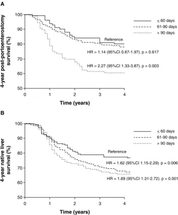
The 4-year post-portoenterostomy survival, including patients that eventually underwent LTx, was 73.4%. Figure 2A shows that 4-year survival was inversely correlated with age at surgery, with rates of 80.0, 77.7 and 60.5% in
children operated at ages ≤ 60 days, 61-90 days, and > 90
days, respectively. Within the group of children operated at
Decade
1980s 1990s 2000s Overall
n (%) n (%) n (%) n (%)
Age at PE (days)
≤ 60 10 (25.0) 31 (27.9) 59 (25.7) 100 (26.3)
61-90 14 (35.0) 46 (41.5) 111 (48.5) 171 (45.0)
91-120 8 (20.0) 23 (20.7) 48 (21.0) 79 (20.8)
>120 8 (20.0) 11 (9.9) 11 (4.8) 30 (7.9)
Total 40 (100.0) 111 (100.0) 229 (100.0) 380 (100.0)
Outcomes
Survival* 13 (28.9) 74 (55.6) 260 (77.6) 347 (67.6)
Performance of LTx† 7 (15.6) 46 (34.6) 186 (55.5) 239 (46.6)
Total of patients 45 (8.8) 133 (25.9) 335 (65.3) 513 (100.0)
table 1 - Comparison over decades of performance of portoenterostomy, age at procedure, survival rates and performance of liver transplantation
LTx = liver transplantation; PE = portoenterostomy.
* Between decades: 1980s vs. 1990s, p = 0.002; 1980s vs. 2000s, p < 0.001; 1990s vs. 2000s, p < 0.001.
Figure 2 - Four-year survival and 4-year native liver survival according to age at portoenterostomy. A) 4-year survival curve. Log rank (Mantel-Cox) = 13.99, p = 0.001; B) 4-year native liver survival curve. Log rank (Mantel-Cox) = 13.38, p = 0.001
95%CI = 95% confidence interval; HR = hazard ratio.
age > 90 days, there was a signiicant difference when the
4-year post-portoenterostomy survival rate was compared
to that of other age groups (≤ 60 days and 61-90 days)
(p = 0.003).
Native liver survival ranged from 0.8 to 3.2 (3.24±4.48) years. Among patients who died, it ranged from 0.8 to 1.7 (2.0±3.0) years; in transplanted patients, from 0.7 to 2.0 (2.1±2.7) years; and in patients who remain alive with their native liver, it ranged from 1.3 to 10.7 (6.4±6.2) years.
Figure 2shows that 4-year native liver survival reached 36.8% of cases and was inversely correlated with age at portoenterostomy: 54.0% in children operated on until 60 days of life, 33.3% in those operated on between 61 and 90 days, and 26.6% in patients operated on after 90 days,
with signiicant differences between age groups (≤ 60 vs. 61-90 days, p = 0.006; ≤ 60 vs. > 90 days, p = 0.001).
Discussion
This study comprises a large series of children with BA, including patients from all Brazilian regions, and evaluates data regarding portoenterostomy, LTx, and survival. In this
series there was a slight predominance of female infants (1.2:1.0), in agreement with the literature,20 and a low rate
of associated congenital anomalies (12.0%), indicating, as observed in other studies,21 the prevalence of the perinatal
form of BA.
This investigation showed that, currently in Brazil, most patients are not operated as needed or undergo late portoenterostomy, after 60 days of life, a situation that can negatively affect their native liver survival. The observed age at portoenterostomy, which was higher than desirable, reveals the occurrence of late referral, a problem that has been previously described in our country.18,19 Thus, in Brazil,
late referral of patients with BA for portoenterostomy remains a problem to be solved nationwide, regardless of region and category of town, whether capital city or countryside. Early patient referral for portoenterostomy in due time remains a challenge worldwide,22-26 but it has been attained
in some countries,27 with a trend toward decreasing age
of referral over the years. In developed countries, age at portoenterostomy is around 60 days.13,14,28
The 4-year survival rate among Brazilian patients who underwent portoenterostomy (73.4%) was similar to that observed in other centers, such as in Canada (79.0%)5 and
France (75.3%).13 The 4-year native liver survival rate of
Brazilian patients (36.8%) was similar to that of patients from Canada (36.0%)5 and Switzerland (37.4%),15 but
lower than that observed in the United Kingdom (51.0%)14
and Japan (63% in 5 years).28
In this study, 4-year survival rate and 4-year native liver survival rate were both inversely correlated with age at portoenterostomy, reinforcing the detrimental effect of age on the postoperative prognosis.8,26,29 Age at portoenterostomy
is known to affect native liver survival. According to Serinet
et al.,30 its impact remains until adolescence and, if all
patients with BA underwent portoenterostomy before 46 days of life, 5.7% of all LTx performed in France in patients younger than 16 years could be avoided. The best surgical results are reached when the procedure is performed
within the irst 30 days of life.5 It is still debatable whether
portoenterostomy should be performed in patients older than 90 days of life. In this study, the 4-year native liver survival in patients operated later, after 90 days, was 26.6%, similar to that observed in Canada (23.0% in 4 years)5 and
France (25.0% in 5 years).31 Furthermore, in the United
Kingdom, the 5-year native liver survival rate of patients operated on after 100 days reached 45.0%.32 Such data
suggest that portoenterostomy should be attempted even in children around 90 days of life, provided they present neither decompensated liver disease nor complications of portal hypertension.
References
1. Balistreri WF, Grand R, Hoofnagle JH, Suchy FJ, Ryckman FC, Perlmutter DH, et al. Biliary atresia: current concepts and research directions. Summary of a symposium. Hepatology. 1996;23:1682-92.
2. Nio M, Ohi R, Miyano T, Saeki M, Shiraki K, Tanaka K; Japanese Biliary Atresia Registry. Five- and 10-year survival rates after surgery for biliary atresia: a report from the Japanese Biliary Atresia Registry. J Pediatr Surg. 2003;38:997-1000.
3. Yoon PW, Bresee JS, Olney RS, James LM, Khoury MJ. Epidemiology of biliary atresia: a population-based study. Pediatrics. 1997;99:376-82.
4. McKiernan PJ, Baker AJ, Kelly DA. The frequency and outcome of biliary atresia in the UK and Ireland. Lancet. 2000;355:25-9. 5. Schreiber RA, Barker CC, Roberts EA, Martin SR, Alvarez F,
Smith L, et al. Biliary atresia: the Canadian experience. J Pediatr. 2007;151:659-65.
6. de Carvalho E, Ivantes CA, Bezerra JA. Extrahepatic biliary atresia: current concepts and future directions. J Pediatr (Rio J). 2007;83:105-20.
7 Santos JL, Carvalho E, Bezerra JA. Advances in biliary atresia: from patient care to research. Braz J Med Biol Res. 2010;43:522-7. 8. Mieli-Vergani G, Howard ER, Portman B, Mowat AP. Late referral
for biliary atresia--missed opportunities for effective surgery.
Lancet. 1989;1:421-3.
9. Nio M, Ohi R. Biliary atresia. Semin Pediatr Surg. 2000;9:177-86.
10. Chardot C, Carton M, Spire-Bendelac N, Le Pommelet C, Golmard JL, Auvert B. Prognosis of biliary atresia in the era of liver transplantation: French national study from 1986 to 1996.
Hepatology. 1999;30:606-11.
can provide a 20-year native liver survival for 21.0% of patients,33 while native liver survival of non-operated children
with BA decreases dramatically in the irst years of life.13
In the long run, however, most patients with BA ultimately need a LTx.34 In this study, 46.6% of patients underwent
LTx, a low rate in comparison with other countries, such as Switzerland (64.6%)15 and Canada (60.0%).5 The low
LTx rates observed herein may relect socioeconomic and cultural dificulties of the population in some Brazilian
regions, in which access to centers where LTx is performed
is not always feasible. On the other hand, LTx was the irst
surgical therapy in 31.0% of patients, a rate higher than that observed in other centers,2,4,13-15,26,33 which may relect
the late referral for diagnosis of BA. Patients who underwent portoenterostomy were transplanted later (2.6±3.1 years) than children not undergoing the procedure (1.2±0.8 years) (p < 0.001), suggesting that it is worth performing
portoenterostomy as the irst-choice surgical treatment.
Although portoenterostomy can not alter the number of LTx in patients with BA throughout life, it can postpone its performance.35 In developed countries, sequential
performance of portoenterostomy and LTx leads to overall survival rates around 90.0%,36 higher than that observed
in this study (67.6%). However, increasing survival rates
have been observed over the last three decades, reaching a rate of 77.6% in the last decade, a number similar to that obtained in Canada5 and Japan.2 These higher
survival rates coincided with increasing LTx performance due to the collaborative efforts of some centers, included
in the Brazilian public Uniied Health System, where LTx is
performed. Figure 1 shows that performance of LTx reduced mortality rates among Brazilian children with BA, with a post-transplant survival reaching 88.3%, a rate similar to that observed in Canada,5 USA,12 and United Kingdom.35
Another factor that may negatively impact overall survival rates, in addition to availability of LTx, is late referral of patients with BA. In Brazil, late referral of patients with BA
might relect dificult access to reference centers or lack of
diagnostic suspicion by parents and non-specialized pediatric services, since, at onset, patients are in good condition and present appropriate weight for age. Jaundice, which may
be less evident, mainly in dark-skinned patients, can be overlooked, thus delaying diagnosis. Experiences from other
countries have proved that late referral can be changed by improving medical practices and health policies. The United Kingdom adopted a policy of centralization in 1999, limiting treatment of patients with BA to three reference centers,14
while France, in 1997, introduced a collaborative effort among their various national centers.13 Other countries have
adopted measures to raise awareness of their population with warning signs and screening systems for BA, such as “Yellow Alert” campaigns37 and the use of a colorimetric
scale to identify acholic stools.38,39 Among these strategies,
the national stool color screening system, which integrated
the infant stool color card into the Child-health Booklet
given to every neonate, has proven to be effective and easily applicable.40
In summary, this multicenter study on patients with BA showed that, in Brazil, overall survival of affected patients is below the desired level already attained by other groups, but post-transplant results are similar to those attained in
developed countries. However, the number of LTx is still
below demand. A timely performance of portoenterostomy increases postoperative survival rates, decreasing the need
for LTx in the irst years of life. Late referral of patients
with BA remains a nationwide problem in Brazil. Currently, Brazilian pediatric hepatologists involved in the treatment of patients with BA are trying to develop collaborative strategies in order to improve the situation of affected children. These professionals, in concert with the Brazilian Society of Pediatrics, have included the stool color card
into the Child-health Booklet distributed by the Brazilian Ministry of Health to parents of every neonate and launched
11. Kelly DA, Davenport M. Current management of biliary atresia.
Arch Dis Child. 2007;92:1132-5.
12. Shneider BL, Brown MB, Haber B, Whitington PF, Schwarz K, Squires R, et al. A multicenter study of the outcome of biliary atresia in the United States, 1997 to 2000. J Pediatr. 2006;148:467-74. 13. Serinet MO, Broué P, Jacquemin E, Lachaux A, Sarles J, Gottrand F, et
al. Management of patients with biliary atresia in France: results of a decentralized policy 1986-2002. Hepatology. 2006;44:75-84. 14. Davenport M, De Ville de Goyet J, Stringer MD, Mieli-Vergani G,
Kelly DA, McClean P, et al. Seamless management of biliary atresia in England and Wales (1999-2002). Lancet. 2004; 363:1354-7. 15. Wildhaber BE, Majno P, Mayr J, Zachariou Z, Hohlfeld J, Schwoebel
M, et al. Biliary atresia: Swiss national study, 1994-2004. J Pediatr Gastroenterol Nutr. 2008;46:299-307.
16. Lai HS, Chen WJ, Chen CC, Hung WT, Chang MH. Long-term prognosis and factors affecting biliary atresia from experience over a 25 year period. Chang Gung Med J. 2006;29:234-9. 17. Tiao MM, Tsai SS, Kuo HW, Chen CL, Yang CY. Epidemiological
features of biliary atresia in Taiwan, a national study 1996-2003.
J Gastroenterol Hepatol. 2008;23:62-6.
18. dos Santos JL, da Silveira TR, Almeida H, Carvalho PA, Cerski CT. Colestase neonatal – atraso no encaminhamento de crianças para diagnóstico diferencial. J Pediatr (Rio J). 1997;73:32-6. 19. Kieling CO, Santos JL, Vieira SM, Ferreira CT, Linhares AR, Lorentz
AL, et al. Biliary atresia: we still operate too late. J Pediatr (Rio J). 2008;84:436-41.
20. Narkewicz MR. Biliary atresia: an update on our understanding of the disorder. Curr Opin Pediatr. 2001;13:435-40.
21. Sokol RJ, Mack C, Narkewicz MR, Karrer FM. Pathogenesis and outcome of biliary atresia: current concepts. J Pediatr Gastroenterol Nutr. 2003;37:4-21.
22. Sookpotarom P, Vejchapipat P, Chittmittrapap S, Sookpotarom P, Vejchapipat P, Chittmittrapap S, et al. Short-term results of Kasai operation for biliary atresia: experience from one institution. Asian J Surg. 2006;29:188-92.
23. Mshelbwala PM, Sabiu L, Lukong CS, Ameh EA. Management of biliary atresia in Nigeria: the ongoing challenge. Ann Trop Paediatr. 2007;27:69-73.
24. Lee WS, Chai PF, Lim KS, Lim LH, Looi LM, Ramanujam TM.
Outcome of biliary atresia in Malaysia: a single-centre study. J Paediatr Child Health. 2009;45:279-85.
25. Sanghai SR, Shah I, Bhatnagar S, Murthy A. Incidence and prognostic factors associated with biliary atresia in western India.
Ann Hepatol. 2009;8:120-2.
26. Karrer FM, Lilly JR, Stewart BA, Hall RJ. Biliary atresia registry, 1976 to 1989. J Pediatr Surg. 1990;25:1076-80.
27. Sokol RJ, Shepherd RW, Superina R, Bezerra JA, Robuck P, Hoofnagle JH. Screening and outcomes in biliary atresia:
summary of a National Institutes of Health workshop. Hepatology. 2007:46:566-81.
28. Shinkai M, Ohhama Y, Take H, Kitagawa N, Kudo H, Mochizuki K, et al. Long-term outcome of children with biliary atresia
who were not transplanted after the Kasai operation: >20-year
experience at a children’s hospital. J Pediatr Gastroenterol Nutr. 2009;48:443-50.
29. Subramaniam R, Doig CM, Bowen J, Bruce J. Initial response to portoenterostomy determines long-term outcome in patients with biliary atresia. J Pediatr Surg. 2000;35:593-7.
30. Serinet MO, Wildhaber BE, Broué P, Lachaux A, Sarles J, Jacquemin E, et al. Impact of age at Kasai operation on its results in late childhood and adolescence: a rational basis for biliary atresia screening. Pediatrics. 2009;123:1280-6.
31. Chardot C, Carton M, Spire-Bendelac N, Le Pommelet C, Golmard J, Reding R, et al. Is the Kasai operation still indicated in children older than 3 months diagnosed with biliary atresia? J Pediatr. 2001;38:224-8.
32. Davenport M, Puricelli V, Farrant P, Hadzic N, Mieli-Vergani G, Portmann B, et al. The outcome of the older (> or =100 days)
infant with biliary atresia. J Pediatr Surg. 2004;39:575-81. 33. López Santamaría M, Gámez M, Murcia J, Díez-Pardo J,
Vázquez J, Migliazza L, et al. Kasai operation in the age of liver
transplantation. Healing or merely palliative technique? Cir Pediatr. 2000;13:102-5.
34. Lykavieris P, Chardot C, Sokhn M, Gauthier F, Valayer J, Bernard O. Outcome in adulthood of biliary atresia: a study of 63 patients who survived over 20 years with their native liver. Hepatology. 2005;41:366-71.
35. McKiernan PJ, Baker AJ, Lloyd C, Mieli-Vergani G, Kelly DA. British paediatric surveillance unit study of biliary atresia: outcome at 13 years. J Pediatr Gastroenterol Nutr. 2009;48:78-81
36. Chardot C, Serinet MO. Prognosis of biliary atresia: what can be further improved? J Pediatr. 2006;148:432-5.
37. Mowat AP, Davidson LL, Dick MC. Earlier identiication of biliary
atresia and hepatobiliary disease: selective screening in the third
week of life. Arch Dis Child. 1995;72:90-2.
38. Matsui A, Dodoriki M. Screening for biliary atresia. Lancet. 1995;345:1181.
39. Chen SM, Chang MH, Du JC, Lin CC, Chen AC, Lee HC, et al.
Screening for biliary atresia by infant stool color card in Taiwan.
Pediatrics. 2006;117:1147-54.
40. Hsiao CH, Chang MH, Chen HL, Lee HC, Wu TC, Lin CC, et al.
Universal screening for biliary atresia using an infant stool color card in Taiwan. Hepatology. 2008;47:1233-40.
Correspondence: Elisa de Carvalho
SQSW 300, bloco N, ap 603 – Sudoeste CEP 70673-048 – Brasília, DF – Brazil
Tel.: +55 (61) 9984.4058, +55 (61) 3257.1501 Fax: +55 (61) 3257.1501