Insulin Action in Peripheral Glucose Uptake
-The Molecular Perspective
Acção Periférica da Insulina na Captação de
Glucose – a Perspectiva Molecular
Ricardo A. Afonso, PhD1,2
1CEDOC, Faculdade de Ciências Médicas, Universidade Nova de Lisboa 2Departamento de Ciências Biomédicas e Medicina, Universidade do Algarve
Correspondência: Ricardo A. Afonso › Área de Ensino e Investigação em Ciências Funcionais e Alvos Terapêuticos (Departamento de Fisiologia) › Faculdade de Ciências Médicas › Universidade Nova de Lisboa › Campo Mártires da Pátria, 130, 1169-056 LISBOA › ricardo.afonso@fcm.unl.pt
RESUMO
Nos últimos anos, a insulinorresistência tem sido objecto de inúmeros estudos e um dos prin-cipais alvos da pesquisa e intervenção farmacológicas. Como consequência, importantes pas-sos têm sido dados a um ritmo acelerado, com vista à compreensão dos mecanismos asso-ciados à acção da insulina, bem como às suas alterações. Deste modo, a tomada de conhe-cimento dos mais recentes avanços nesta área e de como eles se encaixam na panorâmica global da acção da insulina parece ser útil, tanto do ponto de vista da pesquisa como do ponto de vista clínico.
O presente é o primeiro de dois mini-artigos de revisão acerca da acção da insulina no apor-te periférico de glucose. Esta primeira revisão apor-tem como objectivo dar uma visão geral dos eventos intracelulares conducentes à captação de glucose insulino-dependente, enquanto que na segunda revisão será efectuada uma abordagem da acção da insulina numa perspec-tiva fisiológica, ie, integrativa, dando particular ênfase às diferenças na acção da insulina de acordo com o estado prandial.
Assim, na presente publicação será dada uma visão sumária e geral das principais vias de transdução de sinal da insulina, envolvidas no aporte de glucose por tecidos periféricos (extra-hepáticos). Apesar de neste artigo não se fazer uma abordagem farmacológica, espe-ra-se que constitua uma boa base para compreender os mecanismos associados à fisiopato-logia e farmacofisiopato-logia das alterações na acção da insulina.
PALAVRAS-CHAVE
Insulina; Acção da insulina; Receptor de insulina; Transdução de sinal da insulina; Aporte de glucose.
ABSTRACT
glu-INTRODUCTION
Insulin is probably the most important anabolic hormone in the human organ-ism1. At the cellular level, its action is char-acterized by several effects, which suggests the involvement of multiple signaling path-ways initiated by the binding of insulin to the receptor.
The present review aims to provide a brief overview of the major insulin signaling path-ways involved in glucose uptake via GLUT4 translocation, in particular in adipose tissue and skeletal muscle, last of which is responsi-ble for about 75 % of the insulin-dependent glucose uptake2. Transposition from the cel-lular to the physiological level (ie, whole-body) will be essayed in a second review, resulting in a broad outline of insulin action on glucose metabolism and on glucose uptake in particular.
INSULIN RECEPTOR
The insulin receptor is ubiquitous in ver-tebrate tissues, although it may be expressed in different concentrations in different tis-sues3. A general schematic representation of the insulin receptor is provided in figure 1.
Structurally, insulin receptor is an het-erotetrameric glicoprotein, composed of two
α-subunits and by two β-subunits, with N-terminal complex carbohydrates capped by terminal sialic acid residues4,5. Insulin recep-tor structure is stabilized by 3 dissulphide
bonds that link the two α-subunits to each other and to the β-subunits, presenting a (αβ)2 organization6,7. The α-subunits are
entirely located in the outside the cell, whereas β-subunits contain one extracellu-lar portion, one transmembrane region and an intracellular region, last of which includes a juxtamembrane domain, a regu-latory domain (activation domain) and a C-terminal domain6,8, with different func-tional roles.
Presently, there are two types of insulin receptor described: types A and B. The differ-ence between these two isoforms is the pres-ence of a 12 aminoacid sequpres-ence between
cose uptake, whereas in the second review insulin action will be approached in a whole-body per-spective, giving particular emphasis to differences in insulin action according to the prandial state. Thus, in the present review, we will provide a brief overview of the major insulin signaling pathways involved in peripheral (extra-hepatic) glucose uptake. Although this article does not aim pharma-cological therapeutic, we hope that it may launch some minimum comprehensive basis to better understand the mechanism behind the pathophysiology and pharmacology of insulin action.
KEYWORDS
Insulin; Insulin action; Insulin receptor; Insulin signalling pathway; Glucose uptake.
FIGURE 1
positions 716 and 717 of the α-subunits of type A insulin receptor9Type B insulin recep-tor is highly specific for insulin and promi-nent in the major target-tissues for insulin action, such as liver, skeletal muscle and adipose tissue10. Type A insulin receptor pro-motes binding of IGF-2 (insulin-like growth factor 2) instead of insulin and is present in many fetal tissues, central nervous system and haematopoietic cells10. Patients with accummulation of type A receptor in skele-tal muscle seem to be more prompt to the development of insulin resistance11.
INSULIN BINDING AND ACTIVA-TION OF THE RECEPTOR
Insulin binds to one of the α-subunits of the insulin receptor, bringing the two α -sub-units closer upon disruption of the
α2-dimer6,7. Although there are two major binding sites (in the two α-subunits – figure 1), only one insulin molecule binds to the insulin receptor with high affinity, present-ing a negative cooperativity for insulin con-centrations lower than 0.1 μmol/dm3 12.
Insulin binding to the α-subunit induces tyrosine kinase activity in the regulatory domain of the intracellular portion of the
β-subunit, promoting phosphorylation of tyrosine residues of this domain and concomitant activation of the insulin receptor -autophosphorylation.
Autophosphorylation of the insulin receptor is the key step in the initiation of the intracellular signalling and it may occur at seven different tyrosine residues, located in the three regions of the β-subunits with tyrosine kinase activity (juxtamembrane, regulatory or activation and C-terminal)3. However, the process seems to be initiated by phosphorylation of the tyrosine1162 residue of the regulatory domain6(figure 1).
Insulin binding induces conformational changes in the regulatory (or activation) domain that allow binding to ATP, favoring the initial phosphorylation of the
tyro-sine1162 residue (regulatory domain) and, subsequently, the remaining tyrosine residues of the regulatory domain of the insulin receptor6. Phosphorylation of tyro-sine residues in the insulin receptor allow the recruitment, docking and activation of the efector proteins involved in the signal-ing cascade that present SH2 (Src-2 homolo-gy) domains2,13,14. Many of these efector pro-teins are small adaptive molecules, such as p85, which is the regulatory subunit of the enzyme phosphatidylinositol-3-kinase (PI3K) and of CrkII, a small protein G acti-vation molecule2.
After insulin binding and activation of the insulin receptor, the complex insulin-insulin receptor is internalized and incorpo-rated into endossomes, still in an active form, which facilitates the binding of the cytoplasmatic substrates15.
Interestingly, in the absence of insulin,
α-subunits seem to exert a negative effect upon the regulatory domains, thus blocking the signal transduction cascade2,3,6,16,17.
This unusual form of activation seems to allow small molecules to interact with the insulin receptor in distinct sites from the activation domains of insulin18.
INSULIN SIGNALING PATHWAYS INVOLVED IN GLUCOSE UPTAKE
Both insulin receptor and the majority of the proteins involved in insulin signalling are activated by tyrosine residues phospho-rylation13.
and IGF-1 receptors19.
Aditionally, APS, Cbl and IRS proteins, in particular, have been associated with the process of glucose uptake through stimula-tion of glucose trasporters-4 (GLUT4) translocation2.
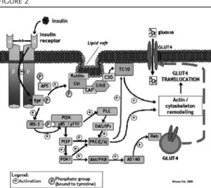
Figure 2 summarizes the major insulin signaling pathways that involve these sub-strates, leading to GLUT4 translocation and glucose uptake.
The most relevant mediators of insulin action in glucose uptake by skeletal muscle and adipocytes are the IRS proteins, in par-ticular IRS-1 and IRS-2. In mammals, four major proteins of the IRS family were described: IRS-1, expressed in skeletal mus-cle and adipose tissue; IRS-2, present in the brain, ovary, liver and adipose tissue; IRS-3, expressed in adipose tissue, presumably in rodents only; and IRS-4, present in the thy-mus and kidney13. IRS proteins present an amine terminal, with binding domains for the insulin receptor and a carboxyl termi-nal, with tyrosine phosphorylation sites20.
Following tyrosine phosphorylation, IRS
protein activates PI3K, which plays a central role in GLUT4 translocation. IRS activates PI3K by binding to p85 regulatory subunit, which presents two SH2 domains that bind to phosphorylated residues in IRS proteins2. Besides p85 subunit, PI3K presents a p110 catalitic subunit, responsible for phospho-inositides phosphorylation at position 3, producing phospholipidic compounds of the phosphatidylinositol-3-phosphate (PI3P) family, namely phosphatidylinositol-3,4,5-triphosphate (PtdInsP3)2.
PI3P (and PtdInsP3 in particular)
acti-vate phosphoinositide-dependent kinase 1 (PDK1), which in turn activates protein kinase B (Akt/PKB) and the atypical protein kinase C (PKCζand PKCλ)2. It has also been described that PtdInsP3can bind directly to
PKC (PKCζ and PKCλ)13 and to Akt/PKB, activating them2, therefore not requiring PDK1 as an intermediate.
Active Akt/PKB then promotes phospho-rylation of Akt Substract of 160 kDa protein (AS160)21, which is constitutively associated to GLUT4 vesicles22and in particular to Rab proteins, small G proteins involved in the processes of transport and fusion of GLUT4 vesicles to plasma membrane23. Thus, AS160 phosphorylation by Akt/PKB pro-motes activation of the Rab proteins22,24, leading to a higher rate of GLUT4 transloca-tion23- this topic will be further explored in the next section. On the other hand, PI3K can activate phospholipase C (PLC), result-ing in the production of the second messen-gers DAG and inositol triphosphate (IP3), which activate PKCζ, thus stimulating glu-cose uptake22(figure 2).
An additional insulin signalling path-way contributing to GLUT4 translocation and somehow independent of IRS phospho-rylation and PI3K activation is described1, and also presented in figure 2.
Such pathway involves phosphorylation of both APS (adaptive protein with SH2 and PH domains, last of which is present in Akt/PKB, allowing this enzyme to bind
FIGURE 2
PtdInsP3) and Cbl protooncogene (Casitas
b-lineage lymphoma, c-Cbl)2,25directly by the IR1,2,25. APS is involved in Cbl recruitment for the insulin receptor26. In the majority of insulin-sensitive cells Cbl is associated with the adaptive protein CAP (Cbl-associated protein)2. Following phosphorylation, the Cbl-CAP complex is transported into lipid rafts in the plasma membrane, where it binds to flotilin and recruits CrkII protein2. CrkII then forms a complex with the guanyl nucleotide exchange protein C3G27, which activates TC1022,28. TC10 is a GTP-binding protein present in the lipid rafts that con-tributes to GLUT4 translocation and their docking at the plasma membrane26,28, possi-bly though regulation of actin microfila-ments dynamics22,29,30.
Although the TC10 pathway can be seen as an separate pathway from the PI3K-dependent one, some studies have suggest-ed that TC10 activates PI3P31 and others have described that atypical PKC (PKCζand PKCλ) are also able to promote TC10 activa-tion26,32. Thus, atypical PKC may represent a point of convergence for the PI3K and TC10 signaling pathways19, both of which con-tributing synergistically to GLUT4 transloca-tion (figure 2).
GLUT4 TRANSLOCATION
GLUT4, present mostly in skeletal mus-cle and adipocyte, are located within vesi-cles that move in a cyclic manner between the intracellular storing sites and plasma membrane. Insulin promotes the presence of GLUT4 at the plasma membrane in two distinct, but synergistic ways: by increasing the rate of GLUT4 exocytosis and by reduc-ing their internalization rate2,33.
As in the case of the insulin secretory granules in β-pancreatic cells, GLUT4 vesi-cles also seem to be translocated towards the plasma membrane by means of a sys-tem involving mycrotubules network and actin polymerization2,22. Actin remodeling is
required not only for translocation of the GLUT4 vesicles, but also to their fusion with the plasma membrane2.
As stated in the previous section and presented in figure 2, the remodeling or reorganization of the actin filaments in response to insulin binding to the receptor appears to be modulated by both the TC10 and IRS/PI3K pathways, through activation of the Rab proteins22.
Rab proteins have been shown to be nec-essary effectors in vesicle trafficking, docking and fusion. In particular, Rabs 2A, 8A, 10, and 14 are expressed in insulin-sensitive tis-sues and appear to be substrates of the AS160 GAP domain (IRS/PI3K pathway) and are associated with insulin-responsive GLUT4-containing vesicles34-36. AS160 thus may represent a convergence between insulin signaling and vesicle trafficking22. AS160 is a negative regulator of basal GLUT4 exocytosis, ie, in basal conditions, AS160 associates with GLUT4 vesicles, main-taining Rab proteins in their inactive form (Rab-GDP)33,34. Insulin-stimulated phospho-rylation of S160 (PI3K pathway) inhibits AS160 negative effect on Rab proteins, caus-ing a shift towards Rabs activation (Rab-GTP complex formation) and allowing for Rab-dependent GLUT4 translocation to occur33,34,37.
receptors (SNARE), namely SNAP-23, syn-taxin 4, Synip, Munc18c and vesicle-associ-ated membrane protein-2 (VAMP2) and and the plasma membrane proteins synapto-some-associated 25-kDa protein and syn-taxin-1A22,39-41.
INHIBITION OF THE INSULIN SIG-NALING CASCADE
Besides tyrosine phosphorylation (figure 2), both insulin receptor and IRS proteins have the potential to be phosphorylated at serine or threonine residues , which blocks or impairs the insulin signaling pathway42-44. Such inhibitory effect of serine/threonine phosphorylation is achieved by reducing the number of phosphorylated tyrosine residues2,43,45, by dissociating IRS proteins from their receptor, hindering tyrosine residues phosphorylation46, by releasing IRS from the intracellular complexes that main-tain them in close proximity to the recep-tor47, by promoting IRS degradation48, or by inducing IRS interaction with other proteins rather than with the tyrosine kinase catalyt-ic site of PI3K2,49.
These inhibitory (serine/threonine) phosphorylations constitue a physiological feedback mechanism in insulin signaling50 and allow the establishment of cross-talk mechanisms with different pathophysiolog-ical pathways that promote insulin resist-ance2,43,50. Indeed, most of the stress and/or inflammation pathways studied so far stim-ulate serine/threonine phosphorylation of either IRS or insulin receptor (or both) as a way to induce insulin resistance51.
Several kinases are known to be involved in the process of serine/threonine phosphorylation-dependent regulation, namely PI3K, Akt/PKB, glycogen synthase kinase-3 (GSK3) and mammalian target of rapamycin (mTOR)2, as well as PKC43 and the inhibitor of nuclear factor κ (IκB) kinase2; these last two (PKC and IκB) have been suggested to be involved in the
obesi-ty-induced insulin resistance2,52,53.
Insulin action is also attenuated by pro-tein tyrosine phosphatases (PTPases) that promote tyrosine dephosphorylation of the insulin receptor and its substrates2,54, a mech-anism that seems to be augmented in many insulin resistant conditions2,55, particularly in those associated with inflammation56. Indeed, in studies using transgenic knockout of PTP1B models was observed an increase in the number of phosphorylated tyrosine residues, in both the receptor and IRS pro-teins, as well as an amelioration of insulin sensitivity in muscle2and liver57,58, improving or avoiding the diabetic condition59.
CONCLUSION
Insulin plays a central role in carbohy-drate metabolism. Although insulin presents different effects in different target-organs, one can consider that its major role in extra-hepatic tissues, such as skeletal muscle and adipose tissue, is to promote glucose uptake. The knowledge of the molecular aspects of insulin action is important to understand the mechanism underlying pathophysiology and pharmacology of insulin resistance. In the present mini-article, we provided a brief review of the main signaling pathways that ensure insulin-stimulated glucose uptake.
target molecule for the study and/or modu-lation of different insulin signaling path-ways involved in glucose uptake, since GLUT4 compliance should always be ensured in order to allow insulin-dependent glucose uptake.
The molecular aspects summarized herein constitute the basis for a second review, in which insulin action will be approached from a whole-body physiologi-cal perspective, more directed to the clinic.
ACKNOWLEDGEMENTS
A word of acknowledgement to Professors M. Paula Macedo and Helena Cardoso, for the help revising the present manuscript and for recognizing its scientific merit.
REFERENCES
1. Saltiel AR, Pessin JE (2002) Insulin signaling pathways in time and space. Trends Cell Biol
12,65-71.
2. Saltiel AR, Kahn CR (2001) Insulin signalling and the regulation of glucose and lipid metab-olism. Nature414,799-806.
3. White MF, Kahn CR (1994) The insulin signal-ing system. J Biol Chem269,1-4.
4. Edge ASB, Kahn RC, Spiro RG (1990) Insulin Receptor Carbohydrate Units Contain Poly-N-Acetyllactosamine Chains. Endocrinology
127,1887-1895.
5. Bjornholm M, Zierath JR (2005) Insulin signal transduction in human skeletal muscle: identi-fying the defects in Type II diabetes. Biochem. Soc. Trans.33,354-357.
6. White MF (1997) The insulin signalling system and the IRS proteins. Diabetologia 40 Suppl 2,S2-17.
7. Ottensmeyer FP, Beniac DR, Luo RZ, et al.
(2000) Mechanism of transmembrane signal-ing: insulin binding and the insulin receptor.
Biochemistry39,12103-12.
8. Yip RG, Goodman HM (1999) Growth hormone and dexamethasone stimulate lipolysis and
acti-vate adenylyl cyclase in rat adipocytes by selec-tively shifting Gi alpha2 to lower density mem-brane fractions. Endocrinology140,1219-27. 9. Lawrence MC, McKern NM, Ward CW (2007)
Insulin receptor structure and its implications for the IGF-1 receptor. Current Opinion in Structural Biology Catalysis and regulation / Proteins17,699-705.
10. Mosthaf L, Grako K, Dull TJ, et al. (1990) Functionally distinct insulin receptors generat-ed by tissue-specific alternative splicing. Embo J9,2409-13.
11. Savkur RS, Philips AV, Cooper TA (2001) Aberrant regulation of insulin receptor alterna-tive splicing is associated with insulin resistance in myotonic dystrophy. Nat Genet29,40-7. 12. De Meyts P (1994) The structural basis of
insulin and insulin-like growth factor-I receptor binding and negative co-operativity, and its relevance to mitogenic versus metabolic sig-nalling. Diabetologia37 Suppl 2,S135-48. 13. White TW, Srinivas M, Ripps H, et al.(2002)
Virtual cloning, functional expression, and gat-ing analysis of human connexin31.9. Am J Physiol Cell Physiol283,C960-70.
14. Whitehead JP, Clark SF, Urso B, et al. (2000) Signalling through the insulin receptor. Curr Opin Cell Biol12,222-8.
15. Carpentier JL, Paccaud JP, Backer J, et al.(1993) Two steps of insulin receptor internalization depend on different domains of the beta-sub-unit. J Cell Biol122,1243-52.
16. Shoelson SE, White MF, Kahn CR (1988) Tryptic activation of the insulin receptor. Proteolytic truncation of the alpha-subunit releases the beta-subunit from inhibitory con-trol. J Biol Chem263,4852-60.
17. Villalba M, Wente SR, Russell DS, et al.(1989) Another version of the human insulin receptor kinase domain: expression, purification, and characterization. Proc Natl Acad Sci U S A
86,7848-52.
18. Zhang B, Salituro G, Szalkowski D, et al. (1999) Discovery of a small molecule insulin mimetic with antidiabetic activity in mice. Science
284,974-7.
19. Chang L, Chiang SH, Saltiel AR (2004) Insulin signaling and the regulation of glucose trans-port. Mol Med10,65-71.
20. Liu YF, Herschkovitz A, Boura-Halfon S, et al.
phosphotyrosine binding domain inhibits insulin receptor substrate 1 function and pro-motes insulin resistance. Mol Cell Biol24 ,9668-81.
21. Kane S, Sano H, Liu SC, et al.(2002) A method to identify serine kinase substrates. Akt phos-phorylates a novel adipocyte protein with a Rab GTPase-activating protein (GAP) domain.J Biol Chem277,22115-8.
22. Brozinick JT, Jr., Berkemeier BA, Elmendorf JS (2007) "Actin"g on GLUT4: membrane & cytoskeletal components of insulin action. Curr Diabetes Rev3,111-22.
23. Jordens I, Marsman M, Kuijl C, et al. (2005) Rab proteins, connecting transport and vesicle fusion. Traffic6,1070-7.
24. Sano H, Kane S, Sano E, et al.(2003) Insulin-stimulated phosphorylation of a Rab GTPase-activating protein regulates GLUT4 transloca-tion. J Biol Chem278,14599-602.
25. Ribon V, Saltiel AR (1997) Insulin stimulates tyrosine phosphorylation of the proto-onco-gene product of c-Cbl in 3T3-L1 adipocytes.
Biochem J324 (Pt 3),839-45.
26. Saito M, Lessard SJ, Rivas DA, et al. (2008) Activation of atypical protein kinase C[zeta] toward TC10 is regulated by high-fat diet and aerobic exercise in skeletal muscle. Metabolism
57,1173-1180.
27. Chiang SH, Baumann CA, Kanzaki M, et al.
(2001) Insulin-stimulated GLUT4 translocation requires the CAP-dependent activation of TC10. Nature410,944-8.
28. Watson RT, Shigematsu S, Chiang SH, et al.
(2001) Lipid raft microdomain compartmental-ization of TC10 is required for insulin signaling and GLUT4 translocation. J Cell Biol154,829-40. 29. Kanzaki M, Watson RT, Hou JC, et al. (2002) Small GTP-binding protein TC10 differentially regulates two distinct populations of filamen-tous actin in 3T3L1 adipocytes. Mol Biol Cell
13,2334-46.
30. Jiang ZY, Chawla A, Bose A, et al. (2002) A phosphatidylinositol 3-kinase-independent insulin signaling pathway to N-WASP/Arp2/3/F-actin required for GLUT4 glucose transporter recycling. J Biol Chem277,509-15.
31. Maffucci T, Brancaccio A, Piccolo E, et al.
(2003) Insulin induces phosphatidylinositol-3-phosphate formation through TC10 activation.
Embo J22,4178-89.
32. Kanzaki M, Mora S, Hwang JB, et al. (2004) Atypical protein kinase C (PKC{zeta}/{lambda}) is a convergent downstream target of the insulin-stimulated phosphatidylinositol 3-kinase and TC10 signaling pathways. J. Cell Biol. 164,279-290.
33. Eguez L, Lee A, Chavez JA, et al. (2005) Full intracellular retention of GLUT4 requires AS160 Rab GTPase activating protein. Cell Metab2,263-72.
34. Larance M, Ramm G, Stockli J, et al. (2005) Characterization of the role of the Rab GTPase-activating protein AS160 in insulin-regulated GLUT4 trafficking. J Biol Chem280,37803-13. 35. Miinea CP, Sano H, Kane S, et al.(2005) AS160,
the Akt substrate regulating GLUT4 transloca-tion, has a functional Rab GTPase-activating protein domain. Biochem J391,87-93.
36. Elmendorf JS, Pessin JE (1999) Insulin signaling regulating the trafficking and plasma mem-brane fusion of GLUT4-containing intracellular vesicles. Exp Cell Res253,55-62.
37. Zeigerer A, McBrayer MK, McGraw TE (2004) Insulin stimulation of GLUT4 exocytosis, but not its inhibition of endocytosis, is dependent on RabGAP AS160. Mol Biol Cell15,4406-15. 38. Inoue M, Chang L, Hwang J, et al.(2003) The
exocyst complex is required for targeting of Glut4 to the plasma membrane by insulin.
Nature422,629-33.
39. Rothman JE (1994) Mechanisms of intracellular protein transport. Nature372,55-63.
40. Sudhof TC (1995) The synaptic vesicle cycle: a cascade of protein-protein interactions. Nature
375,645-53.
41. Kawanishi M, Tamori Y, Okazawa H, et al.
(2000) Role of SNAP23 in insulin-induced translocation of GLUT4 in 3T3-L1 adipocytes. Mediation of complex formation between syn-taxin4 and VAMP2. J Biol Chem 275,8240-7. 42. Tanti JF, Gremeaux T, Van Obberghen E, et al.
(1994) Insulin receptor substrate 1 is phosphory-lated by the serine kinase activity of phos-phatidylinositol 3-kinase. Biochem J 304 (Pt 1),17-21.
43. Waraich RS, Weigert C, Kalbacher H, et al.
of IRS proteins, insulin action, and insulin resistance. Am J Physiol Endocrinol Metab
296,E581-591.
45. Tanti JF, Gremeaux T, van Obberghen E, et al.
(1994) Serine/threonine phosphorylation of insulin receptor substrate 1 modulates insulin receptor signaling. J Biol Chem269,6051-7. 46. Aguirre V, Werner ED, Giraud J, et al. (2002)
Phosphorylation of Ser307 in insulin receptor substrate-1 blocks interactions with the insulin receptor and inhibits insulin action. J Biol Chem
277,1531-7.
47. Tzatsos A, Kandror KV (2006) Nutrients sup-press phosphatidylinositol 3-kinase/Akt signal-ing via raptor-dependent mTOR-mediated insulin receptor substrate 1 phosphorylation.
Mol Cell Biol26,63-76.
48. Greene MW, Sakaue H, Wang L, et al.(2003) Modulation of insulin-stimulated degradation of human insulin receptor substrate-1 by Serine 312 phosphorylation. J Biol Chem 278, 8199-211.
49. Craparo A, Freund R, Gustafson TA (1997) 14-3-3 (epsilon) interacts with the insulin-like growth factor I receptor and insulin receptor substrate I in a phosphoserine-dependent manner. J Biol Chem272,11663-9.
50. Boura-Halfon S, Zick Y (2009) Serine kinases of insulin receptor substrate proteins. Vitam Horm
80,313-49.
51. Tanti JF, Jager J (2009) Cellular mechanisms of insulin resistance: role of stress-regulated ser-ine kinases and insulin receptor substrates (IRS) serine phosphorylation. Curr Opin Pharmacol. 52. Yuan M, Konstantopoulos N, Lee J, et al.
(2001) Reversal of obesity- and diet-induced insulin resistance with salicylates or targeted disruption of Ikkbeta. Science293,1673-7. 53. Kim JK, Kim YJ, Fillmore JJ, et al. (2001)
Prevention of fat-induced insulin resistance by salicylate. J Clin Invest108,437-46.
54. Venable CL, Frevert EU, Kim YB, et al. (2000) Overexpression of protein-tyrosine phos-phatase-1B in adipocytes inhibits insulin-stimu-lated phosphoinositide 3-kinase activity with-out altering glucose transport or Akt/Protein kinase B activation. J Biol Chem275,18318-26. 55. Delibegovic M, Zimmer D, Kauffman C, et al.
(2009) Liver-specific deletion of protein-tyrosine phosphatase 1B (PTP1B) improves metabolic syndrome and attenuates diet-induced
endo-plasmic reticulum stress. Diabetes58,590-9. 56. Zabolotny JM, Kim YB, Welsh LA, et al.(2008)
Protein-tyrosine phosphatase 1B expression is induced by inflammation in vivo. J Biol Chem
283,14230-41.
57. Haj FG, Zabolotny JM, Kim YB, et al. (2005) Liver-specific protein-tyrosine phosphatase 1B (PTP1B) re-expression alters glucose homeosta-sis of PTP1B-/-mice. J Biol Chem280,15038-46. 58. Delibegovic M, Bence KK, Mody N, et al. (2007) Improved Glucose Homeostasis in Mice with Muscle-Specific Deletion of Protein-Tyrosine Phosphatase 1B, 10.1128/MCB.00959-07. Mol. Cell. Biol.27, 7727-7734.
59. Xue B, Kim YB, Lee A, et al. (2007) Protein-tyrosine phosphatase 1B deficiency reduces insulin resistance and the diabetic phenotype in mice with polygenic insulin resistance. J Biol Chem282,23829-40.
60. Afonso RA (2009) Sensibilidade à Insulina Pós-prandial: Mecanismos Fisiológicos de Activação e Fisiopatologia na Obesidade.