Glucagon-like peptide 1 improves insulin resistance
in vitro
through anti-in
fl
ammation of macrophages
C. Guo, T. Huang, A. Chen, X. Chen, L. Wang, F. Shen and X. Gu
Department of Endocrinology, The First Affiliated Hospital of Wenzhou Medical University, Wenzhou, Zhejiang, China
Abstract
Glucagon-like peptide 1 (GLP-1), a kind of gut hormone, is used in the treatment of type 2 diabetes (T2D). Emerging evidence indicates that GLP-1 has anti-inflammatory activity. Chronic inflammation in the adipose tissue of obese individuals is a cause of insulin resistance and T2D. We hypothesized that GLP-1 analogue therapy in patients with T2D could suppress the inflammatory response of macrophages, and therefore inhibit insulin resistance. Our results showed that GLP-1 agonist (exendin-4) not only attenuated macrophage infiltration, but also inhibited the macrophage secretion of inflammatory cytokines including TNF-b, IL-6, and IL-1b. Furthermore, we observed that lipopolysaccharide (LPS)-induced macrophage conditioned media could impair insulin-stimulated glucose uptake. This effect was compensated by treatment with the conditioned media from macrophages treated with the combination of LPS and exendin-4. It was also observed that exendin-4 directly inhibited the activation of NF-kB in macrophages. In conclusion, our results indicated that GLP-1 improved inflammatory macrophage-derived insulin resistance by inhibiting NF-kB pathway and secretion of inflammatory cytokines in macrophages. Furthermore, our observations suggested that the anti-inflammatory effect of GLP-1 on macrophages can contribute to GLP-1 analogue therapy of T2D.
Key words: Diabetes; Glucagon-like peptide; Macrophage infiltration; Adipose inflammation; Insulin resistance
Introduction
Obesity and type 2 diabetes (T2D) are considered chronic pro-inflammatory diseases (1). Adipose tissue macrophages (ATMs) produce inflammatory cytokines and play an impor-tant role in such chronic inflammatory responses (2,3). Macrophages that are mostly polarized have either an M1 or an M2 phenotype (4). In general, the activation status of M1 or M2 phenotype are influenced by local microenvironments (5,6). Th1 cytokines (such as IFN-g) induce activation of M1 macrophages, which produce inflammatory mediators. Th2 cytokines (such as IL-4 and IL-13) induce the alternative activation of M2 macrophages, which regulate anti-infl am-matory responses. Accumulated evidence has proven that macrophage polarization plays a critical role in the develop-ment of T2D. It is considered that the imbalance in the M1/M2 macrophage ratio leads to chronic inflammation in adipose tissue of T2D patients (1). The majority of ATMs in lean individuals exhibit an anti-inflammatory M2 polarity. Accumulation of M1 macrophages in obesity individuals are associated with insulin resistance (1,7,8).
Obesity-related insulin resistance plays a critical role in the cause and development of diabetic pathophysiology (9). The infiltration of macrophages into adipose tissue is the initial event of inflammation in the adipose tissues (10). M2 macrophages produce anti-inflammatory cytokines,
such as IL-10. It is reported that M2 macrophages can increase insulin-dependent glucose uptake in adipocytes of lean people (11). M1 macrophages, which represent a pro-inflammatory state, can secrete inflammatory cyto-kines such as tumor necrosis factor (TNF)-a, interleukin-6 (IL-6) and IL-1b that interfere with insulin signaling, and cause adipocyte dysfunction and insulin resistance (4,10). NF-kB pathway plays a key role in inflammation by its ability to induce transcription of pro-inflammatory genes (12). The NF-kB family is formed by several members, including NF-kB1 (p50/p105), NF-kB2 (p52/p100), p65 (RelA), RelB, and c-Rel (13). The activity of NF-kB is regulated by inhibitors ofkB (IkB) proteins and IkB kinase (IKK). NF-kB is located in the cytoplasm in an inactive form, which associates with IkB. The most important form of IkB is IkBa, IkBb, and IkBE. Phosphorylation of IkB that is mediated by IKK is an important step in NF-kB, which causes dissociation of NF-kB and IkB and its translocation into nuclei (14). As one of the most important regulators of pro-inflammatory gene expression, NF-kB regulates the synthesis of many inflammatory cytokines including TNF-a, IL-1b, IL-6 and IL-8 (14,15).
Glucagon-like peptide-1 (GLP-1), a kind of incretin hormone, is secreted by intestinal L cells in response to
Correspondence: X. Gu:<[email protected]>
nutrients. Then, GLP-1 stimulates the release of insulin from pancreaticb-cells (16). It has been reported that the secretion of GLP-1 decreases in patients with T2D (17). Now, the GLP-1 analogues have been used in combina-tion with insulin for patients with T2D. It has also been reported that a GLP-1 agonist plays an anti-inflammatory function in cultured human macrophages (18). Moreover, GLP can inhibit adipose tissue macrophage infiltration and inflammation in an obese mouse model of diabetes (19). However, whether GLP-1 affects insulin resistance by suppressing macrophage inflammation is not clear.
In the present study, we evaluated the effects of exendin-4, a kind of GLP-1 analogue, in the infiltration of macrophages, and in the secretion of inflammatory cyto-kines including TNF-a, IL-6, and IL-1b by inactivation of NF-kB pathway. The inhibitory effect of GLP-1 on macro-phage inflammation further prevents the impairment of insulin sensitivity induced by lipopolysaccharide (LPS)-stimulated inflammation. Our results that GLP-1 improved insulin resistance by anti-inflammation of macrophages provide a new biological mechanism for the clinical thera-peutics of T2D.
Material and Methods
Cell culture and cell transfection
Mouse peritoneal macrophages were prepared, iso-lated and cultured as described previously (20). The mouse macrophage cell line (RAW264) and mouse preadipocyte (3T3-L1s) were obtained from Shanghai Institute of Chi-nese Academy of Sciences (China). RAW264 cells were maintained in DMEM (Gibco, USA), supplemented with 10% fetal bovine serum at 37°C in a humidity incubator within 5% CO2. 3T3-L1 cells were cultured in DMEM,
supplemented with 10% calf serum at 37°C in a humidity incubator within 5% CO2. RAW264 cells were grown in
6-well plates with a 75% confluence at 24 h before trans-fection. Cell transfection was carried out using lipofecta-mine2000 (Invitrogen, USA) according to the manufacturer’s description. RAW264 cells and mouse peritoneal macro-phages were transfected with siRNA of GLP-1 receptor (GLP-1R) and scramble siRNA. The sequences of siRNA were described in previous studies (21,22): GLP-1R forward, 50-AUA AUG AGC CAG UAG UUC AUG UUGG-30 and
reverse, 50-CCA ACA UGA ACU ACU GGC UCA UUAU-30;
negative control (scramble) forward, 50-UUC UCC GAA CGU
GUC ACG UTT-30; reverse, 50-ACG UGA CAC GUU CGG
AGA ATT-30. These sequences were synthesized by
Gene-Pharma Co. (China).
Protein extraction and western blot
Protein extraction and western blot were performed as previously described (23). Briefly, total proteins were extracted from RAW264 cells and mouse peritoneal macrophages using a RIPA lysis buffer (Beyotime Biotech Inc., China) supplemented with Complete EDTA-free
protease inhibitor cocktail tablets (Roche, USA) according to the manufacturer’s instructions. Total protein concen-trations were assayed with the BCA protein assay kit (Applygen, China). The protein concentration was diluted to 2mg/mL in every sample. Approximately 40mg of protein in every sample were fractionated by gel electrophoresis, and transferred to a polyvinylidene fluoride membrane (Millipore, USA), blocked with 5% skim milk for 1 h, and then incubated with primary antibodies including anti-IkBa, anti-p-IkBa, anti-NF-kB, anti-GLP-1R, and anti-b -actin (Santa Cruz, USA) at 4°C overnight. The membrane was next incubated with horseradish peroxidase-conju-gated secondary antibodies for 1 h after three washes with TBST. Signals were tested by enhanced chemilumines-cence detection reagent (Thermo Scientific, USA) and protein expressions were calculated by normalization to b-actin. All detection reactions were repeated three times. Western blots showed in thefigures are representative of three independent experiments.
RNA extraction and qPCR
Total RNA was isolated from RAW264 cells using Trizol (Invitrogen) under the manufacturer’s instructions and then cDNA was generated from RNA using a First Strand cDNA Synthesis Kit (Fermentas, USA). Quantita-tive real-time PCR (qRT-PCR) was performed using 2TransStart Green qPCR SuperMix (TransGen Biotech Co., China) on an ABI 7300 instrument as previously described (24). mRNA levels were normalized to the level ofb-actin. PCRs were performed in duplicates, and error bars in the charts represent the corresponding standard deviations. The primers used to detect mouse GLP-1R (22) were forward: 50-TTG GGG TGA ACT TCC TCA
TC-30, reverse: 50-CTT GGC AAG TCT GCA TTT GA-30;
b-actin (25) forward: 50-GCC AAC CGT GAA AAG ATG
ACC-30, and reverse: 50-CCC TCG TAG ATG GGC ACA
GT-30.
Transwell assays
Transwell migration assay of macrophage was per-formed as previously described (25). Briefly, 2105
Macrophage-conditioned media (CM) preparation and inflammatory factors detection
Seventy to eighty percent confluence of RAW264 cells or mouse peritoneal macrophages in 6-well plates were starved with serum-free medium, or serum-free DMEM containing LPS (200 ng/mL) with or without exendin-4 (2.5 nM; Sigma, USA) overnight. Then the cells were washed for three times using PBS, and incubated in serum-free medium for 24 h. The cells cultured in con-ditioned media were collected, centrifuged at 500 g for 5 min at 4oC,filtered through a 0.22-mm syringefilter, and stored at 4°C before being used for the experiments. RAW264 cells or mouse peritoneal macrophages starved with serum-free medium were defined as macrophage-conditioned media (CM). CM from macrophages treated with serum-free DMEM containing LPS with or without exendin-4 (2.5 nM; Sigma) were defined as CM-LPS-Ex4 or CM-LPS, respectively.
The levels of TNF-a, IL-6, and IL-1bin conditioned media were investigated using enzyme-linked immunosorbent assay (ELISA). The concentration of these factors was mea-sured using a Human Quantikine ELISA kit (R&D Systems, USA), according to the manufacturer’s instructions.
Insulin-stimulated glucose uptake
3T3-L1 adipocytes were used for determining insulin-stimulated glucose uptake as previously described (26). Briefly, the 3T3-L1 preadipocytes were differentiated into adipocytes as described in a previous report (25). After differentiation, the medium was switched to low-glucose DMEM containing 0.3% bovine serum albumin (BSA) alone (control group) or with CM, CM-LPS, or CM-LPS-Ex4 and incubated at 37°C for 16 h. Then, the medium was switched to a KRBH buffer containing 10 nM of insulin with or without vehicles (DMSO), and with methanol and water extracts, and further incubated at 37°C for 30 min. After incubation, 0.1 lCi 2-deoxy-D-[3H] glucose was added into the KRBH buffer for 10 min. At the end of the
incubation, the buffer was removed and the cells were washed three times with ice-cold PBS. The radioactivity of 3H was counted using a Wallac Liquid Scintillation Counter (USA) to determine glucose uptake. Non-specific glucose uptake was measured in cells treated with vehi-cles and with methanol and water extracts without insulin.
Statistical analyses
Data are reported as means±SD. Student’st-test was performed to assess differences between two groups. One-way ANOVA was conducted to assess differences among multiple groups. All statistical calculations were carried out using SPSS 19.0 software (USA) and Po0.05
was considered statistically significant.
Results
GLP-1R knockdown with siRNA in macrophages
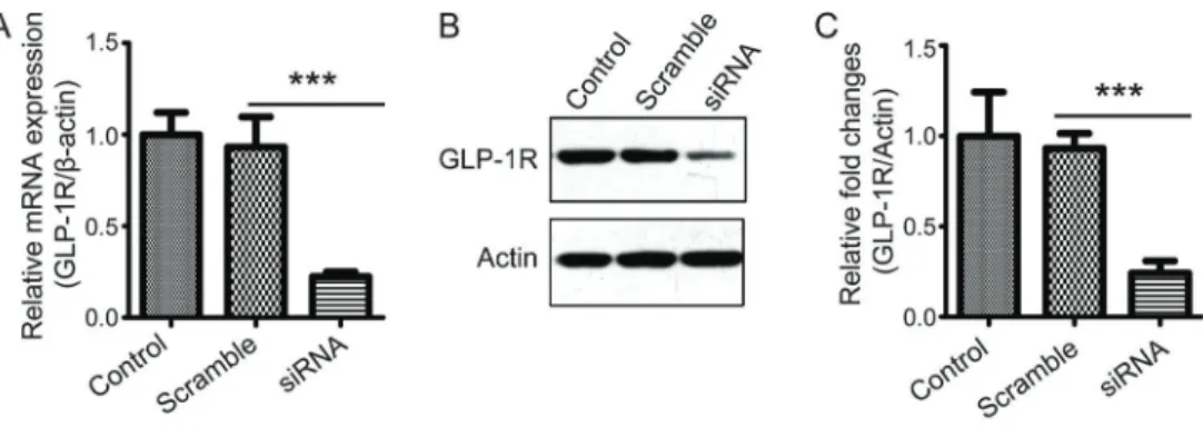
To evaluate the function of GLP-1, siRNA was used to knockdown GLP-1R in macrophages. Then, the knock-down effect of GLP-1R siRNA was detected by qPCR and western blot. The results showed that the expression levels of GLP-1R were significantly decreased (Figure 1A–C). Therefore, siRNA for GLP-1R can be used to analyze the function of GLP in macrophages.
Exendin-4 inhibited the migration of macrophages
The infiltration of macrophages initiates low-grade inflammation in the adipose tissues, which is an important factor in the development of diabetes (27). To determine whether GLP affects macrophage infiltration, we treated RAW264 cells or mouse peritoneal macrophages with LPS, and tested whether LPS treatment-induced macro-phages migration was inhibited by exendin-4 that is a long-acting potent agonist of GLP-1R (28). Our results showed that LPS led to an increased transwell migra-tion of RAW264 cells and mouse peritoneal macrophages, and this effect was reversed by exendin-4 treatment
(Figure 2A–D). Moreover, knocking down of GLP-1R using siRNA could rescue the inhibitory effect of exendin-4 on macrophage migration (Figure 2A–D). These results sug-gest that GLP can suppress LPS-induced macrophage infiltration.
Exendin-4 inhibited the secretion of inflammatory factors
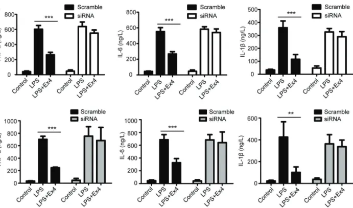
Inflammatory cytokines such as TNF-a, IL-6, and IL-1b can cause adipocyte dysfunction, which is involved in the development of diabetes. To study the effect of GLP-1 on the macrophage-secreted inflammatory cytokines, we detected the levels of TNF-a, IL-6, and IL-1bin RAW264 cells and mouse peritoneal macrophages after treatment with exendin-4. As showed in Figure 3A and B, LPS treatment induced the upregulation of TNF-a, IL-6, and IL-1b in RAW264 and mouse peritoneal macrophages, which could be rescued by exendin-4 treatment. In addi-tion, knocking down GLP-1R in RAW264 cells and mouse peritoneal macrophages using siRNA could reverse the inhibitory effect of exendin-4 on secretion of TNF-a, IL-6, and IL-1b(Figure 3A and B). Taken together, all of these results suggest that GLP could reduce LPS-induced inflammatory cytokines secretion of macrophages.
Exendin-4 increased insulin-stimulated glucose uptake by targeting inflammatory macrophages
It was reported that adipose tissue inflammation in the prediabetic state is related to increased insulin resistance (29,30). To explore the effect of exendin-4 on macrophage-secreted inflammatory cytokines mediated insulin resistance, we tested the insulin-stimulated glucose uptake using 3T3-L1 adipocytes which were incubated with LPS or exendin-4-treated macrophage CM from RAW264 cells or mouse peritoneal macrophages. Our results showed that macro-phage CM and LPS-treated macromacro-phage CM reduced insulin-stimulated glucose uptake (Figure 4A and B). More-over, CM from exendin-4 and LPS-treated macrophages could rescue the inhibitory effect of CM-LPS on insulin-stimulated glucose uptake, which could be abrogated by GLP-1R knockdown in macrophages (Figure 4A and B). Taken together, these results suggest that GLP inhibited macrophage-secreted inflammatory factors and induced insulin resistancein vitro.
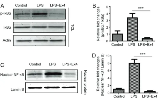
Exendin-4 inhibited activation of NF-kB in macrophages
of macrophages. It was reported that activation of NF-kB is involved in the secretion of inflammatory cytokines. To investigate the mechanism of GLP-suppressed infl amma-tion, we examine the expression and phosphorylation level of IkBa, and the nuclear translocation of NF-kB in RAW246 cells and mouse peritoneal macrophages. Our results showed that LPS did not affect the expression levels of
IkBa, but increased the phosphorylation of IkBa and the nuclear translocation of NF-kB (Figures 5 and 6), which suggests that LPS could activate the transcription factor NF-kB. Furthermore, we found that exendin-4 treatment could reverse the activation of NF-kB induced by LPS (Figures 5 and 6). Thus, we infer that GLP likely inhibits the secretion of inflammatory cytokines by inactivation of NF-kB pathway. Figure 3.Effect of exendin-4 (Ex4) on the secretion of inflammatory macrophage cytokines. RAW264 cells (A) or mouse peritoneal macrophages (B) were treated with lipopolysaccharide (LPS) or a combination of LPS and Ex4 overnight, and then incubated in serum-free medium for 24 h. The secretion of TNF-a, IL-6, and IL-1bwas evaluated using enzyme-linked immunosorbent assay (ELISA). Data are reported as means±SD. **Po0.01; ***Po0.001 (ANOVA).
Discussion
T2D represents a significant threat to global health and human life (31). Insulin resistance is an important con-tributor of T2D. Chronic inflammation is a major cause of insulin resistance (27,32). The infiltration and accumula-tion of macrophages drive inflammation in the adipose tissues, and induced the secretion of inflammatory cyto-kines such as TNFa, which in turn lead to adipocyte dys-function (33). Thus, inhibition of macrophage infiltration and adipose inflammation is an important target for clinical treatment of insulin resistance and T2D.
GLP-1 is a gut hormone, which can increase pancrea-tic secretion of insulin and be used in the clinical therapy of T2D (16). In the current study, we found that exendin-4 showed an inhibitory effect on migration of RAW264 macrophages. This result suggests that GLP could directly target macrophages and suppress its infiltration. Consis-tently, previous studies have reported that GLP-1 inhibits macrophage infiltration in adipose tissue (19), liver and vessel wall (34).
Chronic inflammation plays a critical role in the develop-ment of insulin resistance. Proinflammatory cytokines are mainly secreted by inflammatory macrophages in adipose tissue (16). Our results showed that GLP-1 treatment reduced the release of the inflammatory cyto-kines TNF-a, IL-6, and IL-1b in LPS-stimulated macro-phages. These results are consistent with previous observations that GLP-1 inhibits the secretion of IL-1b
and TNF-a in vitro(18) as well as IL-1b and IL-6in vivo
(35). Our observations taken together with previous studies suggest that GLP-1 has anti-inflammatory properties in macrophages. It has been indicated that proinflammatory cytokines can directly affect insulin signaling pathway and impair insulin sensitivity (36). Bouzakri and Zierath (37) reported that TNF-aleads to insulin resistance by directly targeting muscle insulin signaling. Accordingly, our results showed that LPS-treated macrophage CM decreased the insulin-stimulated glucose uptake in 3T3-L1 adipocytes, and this effect was reversed by CM from macrophages treated with exendin-4. These results suggest that GLP-1 increases insulin sensitivity by inhibiting the production of inflammatory cytokines in macrophages.
including TNF-a, IL-1b, IL-6, and IL-8 (14). Taken together, our results and previous observations suggest that GLP-1 inhibits inflammatory response of macrophages and the production of TNF-a, IL-6, and IL-1bby cAMP/PKA/NF-kB signaling pathway.
Although the improving effect of GLP-1 on insulin resistance by targeting inflammatory macrophages was demonstrated in this study, only RAW246 macrophage cell line, mouse peritoneal macrophages and 3T3-L1 adipocytes were used in vitro. Therefore, there are some limitations in our study. In future researches, whether GLP-1 inhibits inflammatory response of macrophages and therefore
improves insulin resistance in vivo needs to be further investigated.
In conclusion, ourfindings suggest that GLP-1 analogue had inhibitory effects on macrophage-mediated adipose tissue inflammation and could be used for therapy in patients with T2D.
Acknowledgements
This work was supported by the National Science Foundation of China (No. 81000356) and Zhejiang Pro-vincial Natural Science Foundation (LY16H070006).
References
1. Kraakman MJ, Murphy AJ, Jandeleit-Dahm K, Kammoun HL. Macrophage polarization in obesity and type 2 diabetes: weighing down our understanding of macrophage function? Front Immunol2014; 5: 470, doi: 10.3389/fimmu.2014.00470. 2. Weisberg SP, McCann D, Desai M, Rosenbaum M, Leibel RL, Ferrante AW Jr. Obesity is associated with macrophage accumulation in adipose tissue. J Clin Invest 2003; 112: 1796–1808, doi: 10.1172/JCI200319246.
3. Virtue S, Vidal-Puig A. Adipose tissue expandability, lipo-toxicity and the Metabolic Syndrome - an allostatic per-spective. Biochim Biophys Acta 2010; 1801: 338–349,
doi: 10.1016/j.bbalip.2009.12.006.
4. Brown BN, Ratner BD, Goodman SB, Amar S, Badylak SF. Macrophage polarization: an opportunity for improved
outcomes in biomaterials and regenerative medicine. Bio-materials2012; 33: 3792–3802, doi: 10.1016/j.biomaterials.
2012.02.034.
5. Mantovani A, Sica A, Sozzani S, Allavena P, Vecchi A, Locati M. The chemokine system in diverse forms of macrophage activation and polarization. Trends Immunol 2004; 25: 677–686, doi: 10.1016/j.it.2004.09.015.
6. Gordon S, Taylor PR. Monocyte and macrophage hetero-geneity.Nat Rev Immunol2005; 5: 953–964, doi: 10.1038/
nri1733.
7. Fujisaka S, Usui I, Bukhari A, Ikutani M, Oya T, Kanatani Y, et al. Regulatory mechanisms for adipose tissue M1 and M2 macrophages in diet-induced obese mice. Diabetes2009; 58: 2574–2582, doi: 10.2337/db08-1475.
8. Fujisaka S, Usui I, Kanatani Y, Ikutani M, Takasaki I, Tsuneyama K, et al. Telmisartan improves insulin resistance and modulates adipose tissue macrophage polarization in high-fat-fed mice. Endocrinology 2011; 152: 1789–1799,
doi: 10.1210/en.2010-1312.
9. Manna P, Kalita J. Beneficial role of vitamin K supplementa-tion on insulin sensitivity, glucose metabolism, and the reduced risk of type 2 diabetes: A review.Nutrition2016; 32: 732–739, doi: 10.1016/j.nut.2016.01.011.
10. Lumeng CN, Bodzin JL, Saltiel AR. Obesity induces a phenotypic switch in adipose tissue macrophage polarization. J Clin Invest2007; 117: 175–184, doi: 10.1172/JCI29881.
11. Sun K, Kusminski CM, Scherer PE. Adipose tissue remodeling and obesity.J Clin Invest2011; 121: 2094–2101, doi: 10.1172/
JCI45887.
12. Baldwin AS Jr. The NF-kappa B and I kappa B proteins: new discoveries and insights.Annu Rev Immunol1996; 14: 649–683, doi: 10.1146/annurev.immunol.14.1.649.
13. Hunter JE, Butterworth JA, Zhao B, Sellier H, Campbell KJ, Thomas HD, et al. The NF-kappaB subunit c-Rel regu-lates Bach2 tumour suppressor expression in B-cell lym-phoma. Oncogene 2016; 35: 3476–3484, doi: 10.1038/
onc.2015.399.
14. Tak PP, Firestein GS. NF-kappaB: a key role in inflammatory diseases. J Clin Invest 2001; 107: 7–11, doi: 10.1172/ JCI11830.
15. Aupperle KR, Bennett BL, Boyle DL, Tak PP, Manning AM, Firestein GS. NF-kappa B regulation by I kappa B kinase in primaryfibroblast-like synoviocytes.J Immunol1999; 163: 427–433.
16. Hogan AE, Gaoatswe G, Lynch L, Corrigan MA, Woods C, O’Connell J, et al. Glucagon-like peptide 1 analogue therapy directly modulates innate immune-mediated inflammation in individuals with type 2 diabetes mellitus.Diabetologia2014; 57: 781–784, doi: 10.1007/s00125-013-3145-0.
17. Vilsboll T, Krarup T, Sonne J, Madsbad S, Volund A, Juul AG, et al. Incretin secretion in relation to meal size and body weight in healthy subjects and people with type 1 and type 2 diabetes mellitus.J Clin Endocrinol Metab2003; 88: 2706–2713, doi: 10.1210/jc.2002-021873.
18. Buldak L, Machnik G, Buldak RJ, Labuzek K, Boldys A, Belowski D, et al. Exenatide (a GLP-1 agonist) expresses anti-inflammatory properties in cultured human monocytes/ macrophages in a protein kinase A and B/Akt manner. Pharmacol Rep2016; 68: 329–337, doi: 10.1016/j.pharep. 2015.10.008.
19. Lee YS, Park MS, Choung JS, Kim SS, Oh HH, Choi CS, et al. Glucagon-like peptide-1 inhibits adipose tissue macrophage infiltration and inflammation in an obese mouse model of diabetes. Diabetologia 2012; 55: 2456–2468,
doi: 10.1007/s00125-012-2592-3.
20. Arakawa M, Mita T, Azuma K, Ebato C, Goto H, Nomiyama T, et al. Inhibition of monocyte adhesion to endothelial cells and attenuation of atherosclerotic lesion by a glucagon-like peptide-1 receptor agonist, exendin-4.Diabetes2010; 59: 1030–1037, doi: 10.2337/db09-1694[pii].
21. Yang Y, Tong Y, Gong M, Lu Y, Wang C, Zhou M, et al. Activation of PPARbeta/delta protects pancreatic beta cells from palmitate-induced apoptosis by upregulating the expression of GLP-1 receptor.Cell Signal 2014; 26: 268–
278, doi: 10.1016/j.cellsig.2013.11.019.
22. Gupta NA, Mells J, Dunham RM, Grakoui A, Handy J, Saxena NK, et al. Glucagon-like peptide-1 receptor is present on human hepatocytes and has a direct role in decreasing hepatic steatosis in vitro by modulating elements of the insulin signaling pathway. Hepatology 2010; 51: 1584–1592, doi: 10.1002/hep.23569.
23. Wang H, Wang L, Song L, Zhang YW, Ye J, Xu RX, et al. TNNI3K is a novel mediator of myofilament function and phosphorylates cardiac troponin I.Braz J Med Biol Res 2013; 46: 128–137, doi: 10.1590/1414-431X20122515. 24. Wang L, Wang H, Ye J, Xu RX, Song L, Shi N, et al.
Adenovirus-mediated overexpression of cardiac troponin I-interacting kinase promotes cardiomyocyte hypertro-phy. Clin Exp Pharmacol Physiol 2011; 38: 278–284,
doi: 10.1111/j.1440-1681.2011.05499.x.
25. Wang H, Chen Y, Lu XA, Liu G, Fu Y, Luo Y. Endostatin prevents dietary–induced obesity by inhibiting
adipogene-sis and angiogeneadipogene-sis. Diabetes 2015; 64: 2442–2456,
doi: 10.2337/db14-0528.
26. Kwon DY, Kim YS, Ryu SY, Choi YH, Cha MR, Yang HJ, et al. Platyconic acid, a saponin from Platycodi radix, improves glucose homeostasis by enhancing insulin sensi-tivity in vitro and in vivo. Eur J Nutr2012; 51: 529–540,
doi: 10.1007/s00394-011-0236-x.
27. Wang P, Mariman E, Renes J, Keijer J. The secretory function of adipocytes in the physiology of white adipose tissue.J Cell Physiol2008; 216: 3–13, doi: 10.1002/jcp.21386.
28. Edwards CM, Stanley SA, Davis R, Brynes AE, Frost GS, Seal LJ, et al. Exendin-4 reduces fasting and postprandial glucose and decreases energy intake in healthy volunteers. Am J Physiol Endocrinol Metab 2001; 281: E155–E161,
doi: 0193-1849/01.
29. Temelkova-Kurktschiev T, Siegert G, Bergmann S, Henkel E, Koehler C, Jaross W, et al. Subclinical inflammation is strongly related to insulin resistance but not to impaired insulin secretion in a high risk population for diabetes.Metabolism2002; 51: 743-749, doi: S002604950242029X.
30. Festa A, Hanley AJ, Tracy RP, D’Agostino R Jr, Haffner SM. Inflammation in the prediabetic state is related to increased insulin resistance rather than decreased insulin secretion. Circulation2003; 108: 1822–1830, doi: 10.1161/01.CIR.000
0091339.70120.53.
31. Tuomilehto J, Lindstrom J, Eriksson JG, Valle TT, Hamalainen H, Ilanne-Parikka P, et al. Prevention of type 2 diabetes mellitus by changes in lifestyle among subjects with impaired glucose tolerance.N Engl J Med2001; 344: 1343–1350, doi: 10.1056/ NEJM200105033441801.
32. Scherer PE. Adipose tissue: from lipid storage compart-ment to endocrine organ.Diabetes2006; 55: 1537–1545,
doi: 10.2337/db06-0263.
33. Chen XH, Zhao YP, Xue M, Ji CB, Gao CL, Zhu JG, et al. TNF-alpha induces mitochondrial dysfunction in 3T3-L1 adipo-cytes. Mol Cell Endocrinol 2010; 328: 63–69, doi: 10.1016/
j.mce.2010.07.005.
34. Wang Y, Parlevliet ET, Geerling JJ, van der Tuin SJ, Zhang H, Bieghs V, et al. Exendin-4 decreases liver inflammation and atherosclerosis development simultaneously by redu-cing macrophage infiltration. Br J Pharmacol 2014; 171: 723–734, doi: 10.1111/bph.12490.
post-myocardial infarction remodelling via specific actions on inflammation and the extracellular matrix. Basic Res Cardiol2015; 110: 20, doi: 10.1007/s00395-015-0476-7. 36. Lang J. Molecular mechanisms and regulation of insulin
exocytosis as a paradigm of endocrine secretion. Eur J Biochem 1999; 259: 3–17, doi: 10.1046/j.1432-1327.
1999.00043.
37. Bouzakri K, Zierath JR. MAP4K4 gene silencing in human skeletal muscle prevents tumor necrosis factor-alpha-induced insulin resistance. J Biol Chem 2007; 282: 7783–7789, doi: 10.1074/jbc.M608602200.
38. Brubaker PL, Drucker DJ. Minireview: Glucagon-like peptides regulate cell proliferation and apoptosis in the
pancreas, gut, and central nervous system.Endocrinology 2004; 145: 2653–2659, doi: 10.1210/en.2004-0015[pii].
39. Aronoff DM, Canetti C, Serezani CH, Luo M, Peters-Golden M. Cutting edge: macrophage inhibition by cyclic AMP (cAMP): differential roles of protein kinase A and exchange protein directly activated by cAMP-1.J Immunol2005; 174: 595–599, doi: 174-2/595 [pii].
40. Mogi C, Tobo M, Tomura H, Murata N, He XD, Sato K, et al. Involvement of proton-sensing TDAG8 in extracel-lular acidification-induced inhibition of proinflammatory cyto-kine production in peritoneal macrophages.J Immunol2009; 182: 3243–3251, doi: 10.4049/jimmunol.0803466182/5/