UNIVERSIDADE ESTADUAL PAULISTA “JULIO DE MESQUITA FILHO” FACULDADE DE MEDICINA – CAMPUS DE BOTUCATU
ALTERAÇÕES GENÉTICAS RELACIONADAS À OBESIDADE: DANOS NO DNA, PERFIL DE EXPRESSÃO E POLIMORFISMOS GÊNICOS
DANIELLE CRISTINA DE ALMEIDA DIONÍZIO
DRA. DAISY MARIA FÁVERO SALVADORI
BOTUCATU – SP
2013
UNIVERSIDADE ESTADUAL PAULISTA “JULIO DE MESQUITA FILHO” FACULDADE DE MEDICINA – CAMPUS DE BOTUCATU
ALTERAÇÕES GENÉTICAS RELACIONADAS À OBESIDADE: DANOS NO DNA, PERFIL DE EXPRESSÃO E POLIMORFISMOS GÊNICOS
DANIELLE CRISTINA DE ALMEIDA DIONÍZIO
DRA. DAISY MARIA FÁVERO SALVADORI
BOTUCATU – SP
2013
Dedico este trabalho aos meus pais Antonio e Cristina que sempre me incentivaram e não mediram esforços para me darem a melhor educação.
Ao meu marido, por me apoiar, me escutar, vibrar com as minhas vitórias e me dar a mão nas derrotas.
Agradeço em primeiro lugar à Deus, pois sem Sua permissão nada aconteceria. À minha irmã Karen, ao meu cunhado Rafael e à minha sobrinha Rafinha, sei que mesmo de longe, torcem muito por mim!!
À minha sogra Sô, que sempre me ajudou e incentivou muito.
À todos os meus amigos, em especial Pri, Dan, Karen, Rodolfo, Ana, Ví, Marcos, Véio e Tita, e parentes vó, primos, primas, tios e tias que mesmo sem entender muito bem sempre me apoiaram e fizeram com que os momentos de folga fossem relaxantes e revigorantes.
Ao pessoal do laboratório, todos sem exceção (incluo aqui, Tony, Camilinha e Amandinha) foram muito importantes para a conclusão dessa etapa, mas agradeço especialmente àuma pessoinha, Elaininha, por sua amizade incondicional!!
À Daisy, pela sua orientação, ajuda, compreensão e por sempre compartilhar seu conhecimento e suas experiências.
À professora Maria Rita, por toda ajuda nas conversas e discussões de resultados.
À Patrícia, pela paciência e ajuda com as amostras, que não foram poucas! Aos Dr. Irineu, Dr. Celso e Dr. Evandro pela ajuda com as coletas, com certeza sem essa parceria, esse trabalho não seria possível!
Índice
1. Revisão da Literatura ...01
2. Objetivos ………...08
3. Artigo 1: GHRL, GHSR, LEP, LEPR, 5-HT2C and IL-6 gene polymorphisms and DNA damage in morbid obese women ………...09
3.1 Introduction ………...11
3.2 Material and Methods ………...13
3.3 Results ………...16
3.4 Discussion ……….21
3.5 References ……….25
4. Artigo 2:Different gene expression profiling between morbid obese and eutrophic women ………29
4.1 Introduction ……...31
4.2 Material and Methods ...33
4.3 Results ...36
4.4 Discussion ...41
4.5 References ...47
5. Considerações Finais ...52
6. Conclusões ...56
7. Referências ...57
Resumo
Abstract
Apesar do acúmulo de conhecimentos sobre a obesidade e comorbidades associadas, a prevalência, em diversos países, atingiu níveis recordes (Chaput et al., 2012). Nos últimos anos, a obesidade e o sobrepeso chegaram a níveis epidêmicos, com cerca de 1 bilhão de pessoas com sobrepeso e 300 milhões consideradas obesas. Desse total, pelo menos 2,6 milhões vão a óbito todos os anos em decorrência das complicações oriundas do excesso de peso (WHO, 2005).
Sabe-se que a obesidade tem afetado não somente habitantes de países desenvolvidos e industrializados, mas, também, de países subdesenvolvidos e em desenvolvimento. Censo realizado no Brasil entre 2002 e 2003, com 95,5 milhões de brasileiros com idade acima de 20 anos, mostrou que 38,8 milhões (40,6%) apresentavam sobrepeso e 10,5 milhões (11%) eram obesos (IBGE, 2004). O número crescente de indivíduos obesos tem levado ao aumento do número de cirurgias bariátricas realizadas mundialmente, uma vez que parece ser esta a alternativa realmente efetiva para a perda significativa de peso e, consequente, para o controle da obesidade mórbida (Bushwald, 2004). Este tratamento cirúrgico tem como objetivo a diminuição do consumo energético, com consequente perda de peso, melhoria da qualidade de vida e das comorbidades associadas (Campos et al., 2008). No entanto, o resultado da cirurgia bariátrica, no que se refere à perda de peso, depende de diversos fatores nem sempre conhecidos, sendo que o indivíduo pode recuperar o peso, geralmente entre o 3º e o 5º ano após a cirurgia (Capella & Capella, 1996; Fobi, 2004; Kaplan, 2005; Brolin et al., 1994).
distintos: o tecido adiposo branco e o tecido adiposo marrom. Nos mamíferos, o tipo predominante é o branco, constituído, principalmente, por adipócitos, pré-adipócitos (células ainda não contendo lipídeos), células endoteliais, fibroblastos e leucócitos, especialmente macrófagos (Tilg & Moschen, 2006). O excesso de energia resultado do desequilíbrio entre o consumo de alimento e o gasto de energia é armazenado nas células do tecido adiposo, que aumentam em tamanho e quantidade, levando a problemas clínicos não só pelo aumento de peso e de gordura corporal, mas, também, pelo aumento da produção de ácidos graxos livres e de peptídeos secretados pelas células do tecido(Bray, 2004).
Realmente, o tecido adiposo, além da estocagem de gordura, é reconhecido como importante órgão endócrino, com papel crucial na regulação de diversos processos patológicos (Tilg & Moschen, 2006; Federico et al., 2010). Nesse sentido, inúmeros estudos vêm sendo realizados com o objetivo de identificar e caracterizar a relação entre a obesidade e algumas doenças, especialmente aquelas relacionadas ao sistema imunológico. Ao que parece, essa relação seria orquestrada por complexa rede de mediadores derivados das células do sistema imune e dos adipócitos (Wellen & Hotamisligil, 2005). Dentre as substâncias secretadas pelo tecido adiposo estão as adipocitocinas (adiponectina, leptina, visfatina e resistina), que têm importante papel na obesidade com relação à resistência a insulina e desordens inflamatórias (Calle & Kaaks, 2004; Kusminski et al., 2005; Weisberg, 2006). Muitos outros produtos também secretados pelo tecido adiposo já foram identificados, incluindo o fator de necrose tumoral - α (TNF-α) -, as interleucinas 1 e 6 (IL-1 e IL-6) e mediadores envolvidos no processo de coagulação (Wellen & Hotamisligil, 2005; Calle & Kaaks, 2004).
grande número de macrófagos presentes no tecido adiposo branco parece ser a principal fonte de TNF-α; por outro lado, os adipócitos contribuem com um terço da produção de IL-6 circulante em obesos (Weisberg et al., 2003; Xu et al., 2003; Fantuzzi, 2005).
Além do fato dessas citocinas serem potentes estimuladores da produção de espécies reativas de oxigênio (ERO) e de nitrogênio (ERN), os adipócitos possuem capacidade secretora de angiotensina II, a qual estimula a atividade da NADPH (nicotinamida adenina dinucleotídeo fosfatase), que é a principal via para a produção de ERO nos adipócitos (Fernandez-Sanchez et al., 2011). Em 2006, de La Maza et al. demonstraram que amostras de músculo esquelético de pacientes com sobrepeso apresentavam níveis aumentados de 8-OHdG, 4HNE e TNF-α (biomarcadores de dano oxidativo e inflamação), quando comparados a indivíduos com peso normal e estável. De forma semelhante, Al-Aubaidy & Jelinek (2011) descreveram que maiores níveis de 8-OHdG podiam ser observados em pacientes diabéticos e pré-diabéticos, e que estes estavam positivamente relacionados ao Índice de Massa Corporal (IMC), sugerindo que a obesidade poderia contribuir para o aumento de danos oxidativos no DNA. Embora muitos estudos já tenham relacionado diversas doenças à indução de estresse oxidativo e danos no DNA, são raras as avaliações sobre a frequência ou níveis de lesões genotóxicas em indivíduos com obesidade mórbida (Thavanati & Ortega, 2005).
em um artigo de revisão, descreveram que a administração da grelina é capaz de estimular o apetite e aumentar a circulação de GH, ACTH (hormônio adrenocorticotrófico), cortisol, prolactina e glicose tanto em indivíduos saudáveis como em obesos, diabéticos, pacientes com câncer, com disfunção na glândula pituitária e com distúrbios alimentares.
O receptor da grelina, formado por meio de splicing alternativo do mRNA do gene do receptor do hormônio secretagogo do crescimento (GHS-R), também tem papel fundamental na biologia da obesidade. Duas variantes são geradas nesse splicing: o GHS-R tipo 1a, que atua como receptor da grelina, e o tipo 1b, que é uma forma farmacologicamente inativa do receptor. Em seres humanos, o mRNA do GHS-R1a já foi identificado em células da glândula pituitária, da tireóide, do pâncreas, baço, miocárdio e das glândulas adrenais (Depoortere, 2009).
lado, a alimentação após o jejum, a insulina, os glicocorticóides e as citocinas pró-inflamatórias podem, também, estimular a expressão do gene e a produção do hormônio (Trayhurn, 2007). Os primeiros estudos sobre a função fisiológica da leptina foram realizados em camundongo e mostraram que animais com mutação no gene da leptina eram incapazes de produzir o hormônio de forma funcional e tornavam-se obesos, atingindo peso corporal quatro vezes maior que seu peso normal (Friedman & Haalas, 1998). Por outro lado, os benefícios terapêuticos do tratamento de indivíduos obesos com leptina exógena são ainda controversos. Em 1998, Friedman & Hallaas, observaram que a administração de leptina por quatro semanas consecutivas reduzia significativamente o peso de pacientes obesos e de eutróficos, porém a perda somente era observada nos indivíduos que não apresentavam hiperleptinemia.
O apetite e outros processos fisiológicos como o sono, o humor, a temperatura corporal e a secreção de outros hormônios são também regulados pela serotonina (5-HT)(Barsh & Shwartz, 2002; Rosmond et al., 2002). Enquanto em níveis normais desse neurotransmissor a saciedade é facilmente atingida, com maior controle sobre a ingestão de açúcares, baixos níveis estão relacionados ao aumento do desejo de ingerir doces e carboidratos, (Lam & Heisler, 2007; Neves & Paschoal, 2007; Lam et al., 2008). Assim sendo, o sistema serotoninérgico tem sido alvo terapêutico para o controle de peso. Diversos subtipos de receptores de serotonina foram identificados, dentre os quais o 5-HT1B e o 5-HT2C, que têm sido reconhecidos por induzir a saciedade (Lam et al., 2008; Ward et al., 2008). O receptor 5-HT2C parece ser o mais importante na relação entre a ingestão alimentar e o balanço energético, uma vez que foi observado que camundongos desprovidos do gene que codifica para esse receptor tornam-se obesos e epiléticos (Ward et al., 2008; Bickerdike, 2003).
A serotonina envolvida na modulação do comportamento alimentar tem sua atividade avaliada pelo nível de seu principal metabólito, o ácido 5-hidroixindolacético (HIAA). Foi observado que em pacientes com anorexia nervosa a concentração de 5-HT apresenta-se diminuída, e que essa redução poderia estar associada à menor ingestão de aminoácidos essenciais como o triptofano (precursor da serotonina), bem como à sensibilidade do receptor serotoninérgico 5-HT2C (Hermsdorff et al., 2006; Luras, 2009).
que predispõem à obesidade teriam “vantagem seletiva” em populações nas quais seus ancestrais experimentaram períodos de fome (Neel, 1962). Portanto, os indivíduos que herdaram tais genes apresentariam superexpressão de elementos relacionados à fome, não apenas ganhando peso, mas se tornando extremamente obesos. Mais tarde, essa teoria foi comprovada por estudo realizado nos Estados Unidos, que demonstrou a desproporção entre afro-americanos e hispano-americanos obesos em relação a caucasianos (Crossrow & Falkner, 2004).
Nas últimas décadas foram inúmeros os estudos que buscaram identificar genes associados a doenças, dentre as quais à obesidade mórbida. Uma das grandes vantagens advindas do uso de técnicas de biologia molecular foi a possibilidade de se caracterizar variantes gênicas associadas à predisposição a determinada enfermidade. Em relação à obesidade, há pesquisas que mostram que polimorfismos de genes que codificam hormônios relacionados à fome e à saciedade podem ter papel fundamental para essa disfunção metabólica (Duarte et al., 2007). A maioria dos genes que predispõem à obesidade codifica componentes moleculares de sistemas fisiológicos que regulam o balanço energético (Duarte et al., 2007; Yurtcu et al., 2009). Genes como os dos receptores da leptina (LEPR) e da melanocortina-4 (MC4R) e as proteínas desacopladoras de prótons 2 e 3 (UCP2 e UCP3) têm sido avaliados como potencialmente relacionados à fisiopatologia da obesidade e suas complicações (Jacobson et al., 2006). Além desses, o gene da grelina (GHRL), dos receptores da grelina (GHSR) e o da serotonina (5-HT2C) também parecem ter relevância tanto na gênese como nas complicações e consequências da obesidade (Leibel et al., 1995; Doucet et al., 2000; Rosmond et al., 2002 Garcia et al., 2008).
comportamentais mostraram que o receptor 5-HT2C está envolvido na regulação do apetite em seres humanos (Walsh et al., 1994; Sargent et al., 1997), e que o polimorfismo -759C/T da região promotora do gene tem forte associação com a obesidade (Yuan et al., 2000) e ganho de peso durante o início do tratamento com medicamentos antipsicóticos (Reynolds et al., 2002). Da mesma forma, Ramírez-López et al. (2012), observaram que adolescentes mexicanos com os polimorfismos 597G>A, -572G>C e -174G>C do gene da IL-6 tinham risco aumentados para o desenvolvimento de obesidade e hiperglicemia.
Diante das informações apresentadas sobre a gênese e desenvolvimento da obesidade, este estudo propôs:
determinar a prevalência dos polimorfismos dos genes da grelina (GHRL) e da leptina (LEP), assim como de seus receptores (GHSR e LEPR), do receptor da serotonina (5-HT2C) e da interleucina 6 (IL-6) em uma população de mulheres com obesidade mórbida (IMC < 40,0);
avaliar os níveis de danos no DNA de linfócitos das mulheres obesas;
avaliar a relação entre os polimorfismos gênicos e os níveis de danos no DNA;
avaliar o perfil de expressão gênica em células de sangue periférico de mulheres obesas e eutróficas;
GHRL
,
GHSR
,
LEP
,
LEPR
,
5-HT2C
and
IL-6
gene
polymorphisms and DNA damage in morbid obese women
Almeida, DC1; Luperini, BCO1; Oliveira, MRM2, Salvadori, DMF1
Abstract
Obesity is characterized by increased adipose tissue mass with low-grade systemic inflammation, resulting from a chronic energy imbalance between energy intake and expenditure. Food intake regulation is a behavioral-physiological interaction between hypothalamic receptors and released hormones. Herein, we aimed to evaluate if ghrelin (GHRL – rs26802), ghrelin receptor (GHSR – rs572169), leptin (LEP – rs7799039), leptin receptor (LEPR – rs 1137101), serotonin receptor (5-HT2C- rs 3813929) and interleukine-6 (IL-6 – rs1800796) gene polymorphisms, and also oxidative DNA damage are related to obesity. The relationship between the gene variants and genotoxicty (detected by the comet assay) was also investigated. A total of 300 morbid obese and 300 healthy weight (controls) women was recruited for this study. The results demonstrated no statistically significant difference in the frequencies of GHRL, GHSR, LEP, LEPR, 5-HT2C and IL-6 gene polymorphisms (p>0.05) between obese and control groups. In addition, data showed higher (p<0.05) amount of genetic damage (DNA strand breaks and oxidative lesions) in morbid obese women than in controls. Nevertheless, no relationship was detected between the genotypes and the amount of DNA damage. In conclusion, independent on the genotype, obesity was associated with increased DNA damage in lymphocytes, what may be caused by the inflammatory state related to this metabolic dysfunction.
1. Introduction
The etiology of obesity is associated to a complex interaction of genetic, diet, metabolism and physical activity, leading to a chronic imbalance between energy intake and energy expenditure (Tatarani & Ravussin, 2002). The genetic approach considers obesity as an inherited disease caused by mutations and/or gene polymorphisms. On the other hand, food intake regulation is mainly a behavioral-physiological interaction between individual and environment, with hypothalamic receptors responding to peripherally released (an)orexigenic hormones, such as peptide YY (PYY), glucagon-like peptide 1 (GLP-1), ghrelin and leptin, the latter two being key mediators for body weight regulation (den Hoed et al., 2008; Nakazato et al., 2001; Halaas et al., 1995; Zhang et al., 1994).
Ghrelin was discovered at the end of 1999 as an endogenous ligand for the growth hormone secretagogue receptor (GHS-R) (Kojima et al., 1999). It is produced predominantly in the stomach, and its concentration in human plasma rises during fasting and falls post-prandially, making this hormone known as “hunger hormone” (Cummings et al., 2001; Cummings, 2006). In 2001, Ukkola et al. analyzing Swedish morbid obese and lean subjects have observed that the -501A>C polymorphism of the ghrelin genewas more frequent in obese. Latter,Gueorguiev et al. (2009) genotyped 10 common GHSR SNPs in 1275 obese subjects and in 1059 subjects from the general population, and have observed that the SNP rs572169 was associated to obesity. These same authors have also detected associations between the ghrelin variant g.A265T (rs4684677) and obesity, between ghrelin variant gA-604G (rs27647) and insulin levels at 2-h post-oral glucose tolerant test (OGTT), and between the eating behavior “overeating” and the GHSR SNP rs2232169 in obese subjects.Leptin, a 167-amino acid protein coded by the LEPgene and produced in the white adipose tissue, is an important hormone for the regulation of adiposity and body weight. It acts by inhibiting food intake and stimulating energy expenditure (Paracchini et al., 2005). Similarly, leptin receptor, coded by the LEPRgene, is also important in the pathophysiology of human obesity (Yiannakouris et al., 2001). Functional mutations in the LEPR gene, resulting in a truncated receptor, have been associated to obesity and diabetes in rodents and humans (Lee et al., 1996; Takaya et al., 1996; Clément et al., 1998).
itself, can reduce food intake, decrease body weight and enhance energy expenditure (Lam & Heisler, 2007). It has been demonstrated that intracerebroventricular (ICV) injections of p-chlorophenylalanine, which inhibits tryptophan hydroxylase and thereby reduces brain serotonin, induces hyperphagia and weight gain in rats (Breisch et al., 1976). Among the serotonergic most investigated genes are tryptophan hydroxylase 1 and 2 (TPH 1 and TPH 2; responsible by serotonin synthesis), serotonin receptors 2A and 2C (5-HT2A and 5-HT2C; post-synaptic receptors) and serotonin transporter ( 5-HTT; serotonin re-uptake) (Walter & Bader, 2003). Many pharmacological and behavioral studies have showed that 5-HT2C is involved in appetite regulation in humans (Reynolds et al., 2002; Reynolds et al., 2003). Furthermore, literature has also reported that the SNP -759C/T in the promoter region of this gene has strong association with obesity (Walsh et al., 1994) and weight gain in the beginning of antipsychotic treatment (Sargent et al., 1997; Yuan et al., 2000).
Other peptides, such as anti- and pro-inflamatory adipokines and adiponectin,have been also linked to adiposity (Guerre Millo, 2008). Interleukin-6 (IL-6), produced by fibroblasts, macrophages, endothelial cells and adipocytes, for instance, can inhibit the lipoprotein lipase activity and stimulates glucose and fatty-acid oxidation and glucagon and cortisol liberation (Mohamed-Ali et al., 1998). Obesity itself is associated with a chronic low-grade inflammatory state, able to produce free radicals which can interact with DNA, causing genotoxicity. In fact, experimental and clinical observations have demonstrated oxidative stress as an important mechanism in obesity-associated metabolic syndrome (Furukawa et al., 2004; Grattagliano et al., 1998).
2. Material and Methods Subjects
The Ethics Committee for Human Research of the Botucatu Medical School – UNESP approved this study protocol (Document n° 3361-2009). Signed informed consent was obtained from all subjects recruited.
Two groups of women were included in this study: 1) morbid obese women (BMI > 40 Kg/m2; n = 300), age range 20-50 years old; 2) health weight women (BMI ≤ 24.9 Kg/m2;control group;n = 300), matched for age and smoking habit. In the obese group were included those women who were registered for bariatric surgery at Center of Gastroenterology and Bariatric Surgery (Hospital dos Fornecedores de Cana), Piracicaba – SP, Brazil. The exclude criteria were alcohol consumption > 40g alcohol/day, presence of genetic syndromes associated to obesity, Cushing Syndrome, hypothyroidism, kidney or liver diseases, neoplasias, HIV, use of corticoids and estrogen replacement. Women for the control group should not be in use of any medicine, should not have exercised at least 24 hours before blood collection and should not have diabetes, hypercholesterolemia, hypertension or obesity, neither family history for these diseases.
Blood sampling
Peripheral blood sample (10mL) was collected through venipuncture in EDTA tubes. Part of this sample (4 mL) was immediately used for lymphocytes isolation and 6 mL were stored in a freezer (-80°C) for later DNA extraction.
Alkaline comet assay (DNA damage)
mg/ml bovine serum albumin and 0.5 mM EDTA at pH 8). Slides were incubated at 37ºC for 30 min in a moist and dry chamber, with 100 µl of fpg and endo III (1:1000) enzymes which recognize oxidised purine and pyrimidine, respectively. Subsequently, slides were left for 15 min at 4° C and then the coverslips were removed (Braz et al., 2011). All the slides were placed into a horizontal electrophoresis tank filled with freshly prepared alkaline buffer (1mM EDTA and 300 mM NaOH at pH >13). After a 40-min DNA unwinding period, electrophoresis was conducted at 25 V and 300 mA for 30 min. After 15 min neutralization with 0.4 M Tris (pH 7.5), the slides were fixed with absolute ethanol and stored at 4◦C. Slides were stained with 70µL of 2:10000 Sybr Gold solution (Invitrogen, USA) and immediately analyzed in fluorescent microscope, at 400 X magnification. Images from100 nucleoids (two replicateslides)per subject were scored using semi automated image analysis system (Comet Assay IV, Perceptive Instruments, UK).Tail intensity (% DNA in the tail) was used to estimate the extent of DNA damage.
DNA extraction
Genomic DNA was extracted from whole blood cells using Illustra Blood genomic prep mini spin kit (GE, Sweden), according to manufacturer’s protocol. DNA concentration was evaluated in spectrophotometer (NanoVue - GE, Sweden), and each sample was assessed for purity by absorbance at 260 and 280nm (260/280 = 1.9 – 2.1). DNA integrity was assessed by 1.5% agarose gel, using Tris/borate/EDTA (TBE). DNA was stored at -20°C until amplification.
Genotyping
Statistical analysis
3. Results
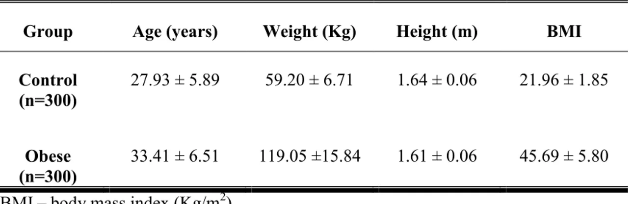
Table 1 shows demographic characteristics (mean age, weight, height and BMI) of the obese and healthy weight groups. Metabolic syndrome was detected in 99/300 (33%) morbid obese women.
Table 1. Demographic characteristics of obese and healthy weight (control) groups
Group Age (years) Weight (Kg) Height (m) BMI
Control
(n=300) 27.93 ± 5.89 59.20 ± 6.71 1.64 ± 0.06 21.96 ± 1.85
Obese
(n=300) 33.41 ± 6.51 119.05 ±15.84 1.61 ± 0.06 45.69 ± 5.80 BMI – body mass index (Kg/m2)
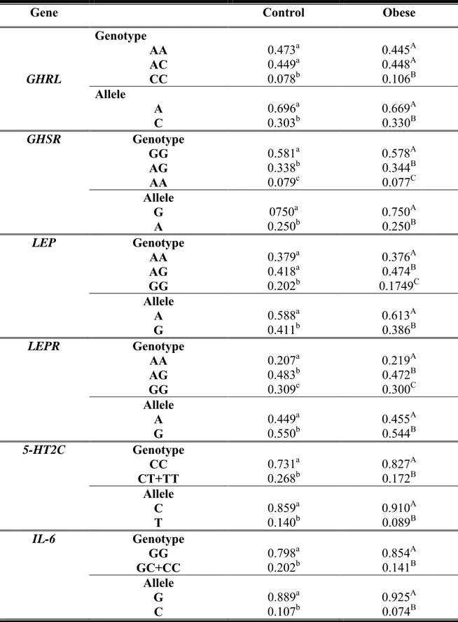
Genotype and allele frequencies of GHRL, GHSR, LEP, LEPR, 5-HT2C and IL-6 genes are presented in Table 2. Both obese and healthy weight populations were in Hardy-Weinberg Equilibrium. No statistically significant difference was detected between the two groups (control vs obese). CC-GHRL, AA-GHSR, GG-LEP, AA-LEPR, TT-5-HT2C and CC-IL-6 were the less frequent genotypes in both obese and control groups. No relationship was found between body weight or BMI and the gene variants (data not shown).
Table 2. Genotype and allelic frequencies in control and obese sampled populations
Gene Control Obese
GHRL
Genotype
AA 0.473a 0.445A
AC 0.449a 0.448A
CC 0.078b 0.106B
Allele
A 0.696a 0.669A
C 0.303b 0.330B
GHSR Genotype
GG 0.581a 0.578A
AG 0.338b 0.344B
AA 0.079c 0.077C
Allele
G 0750a 0.750A
A 0.250b 0.250B
LEP Genotype
AA 0.379a 0.376A
AG 0.418a 0.474B
GG 0.202b 0.1749C
Allele
A 0.588a 0.613A
G 0.411b 0.386B
LEPR Genotype
AA 0.207a 0.219A
AG 0.483b 0.472B
GG 0.309c 0.300C
Allele
A 0.449a 0.455A
G 0.550b 0.544B
5-HT2C Genotype
CC 0.731a 0.827A
CT+TT 0.268b 0.172B
Allele
C 0.859a 0.910A
T 0.140b 0.089B
IL-6 Genotype
GG 0.798a 0.854A
GC+CC 0.202b 0.141B
Allele
G 0.889a 0.925A
C 0.107b 0.074B
Table 3. DNA damage in lymphocytes of morbid obese and healthy weight (control) women
Group Strand
breaks1 Oxidized purines pyrimidines Oxidized
Control (n=70) 16.94 ± 23.51 33.29 ± 33.46 29.79 ± 29.81 Obese (n=70) 28.96 ± 33.12* 46.5 ± 37.06* 44.74 ± 35.5* 1- DNA single and double strand breaks, and alkali-labile sites; * p<0.01 (obese vs control)
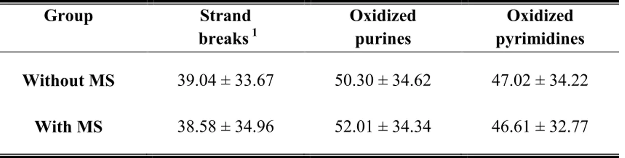
When the obese women were distributed according to the presence or absence of metabolic syndrome, no statistically significant difference in the mean of DNA damage (single and double strand breaks, alkai-labile sites, and oxidized purines and pyrimidines) was detected between the two subgroups (Table 4).
Table 4. DNA damage according to the presence or absence of metabolic syndrome (MS) in the obese group
1- DNA single and double strand breaks, and alkali-labile sites
DNA damage according to the gene polymorphisms are showed in Table 5. For all genes investigated, morbid obese women always had higher level of DNA damage (strand breaks, oxidized purines and pyrimidines) than those from the control group. GHRL-CC, LEP-GG and IL-6-GC + CC genotypes presented the higher amount of strand breaks in both control and obese groups. Differently, this not happened for the GHSR, LEPR and 5-HT2C. Regarding to oxidized DNA damage, only GG-GHSR had similar result (higher amount of oxidized purines and pyrimidines) in both groups.
Group Strand
breaks 1 Oxidized purines pyrimidines Oxidized
Table 5. DNA damage in obese and healthy weight women according to gene polymorphisms
Gene Genotype Group Strand breaks1 Oxidized
purines pyrimidines Oxidized GHRL AA Control 17.68 ± 24.22a 34.7± 33.91a 28.75 ± 28.40a Obese 30.22 ± 32.74#A 47.71 ± 37.44#A 46.79 ± 37.43#A AC Control 16.71 ± 22.37a 31.39 ± 32.05b 27.99 ± 28.45a
Obese 31.66 ± 34.70#A 45.85 ± 36.07#A 43.69 ± 34.00#B CC Control 22.13 ± 23.57b 24.55 ± 24.98c 30.11 ± 28.49a
Obese 44.33 ± 35.83#B 52.70 ± 36.15#B 48.13 ± 34.10#A GHSR AA Control 18.59 ± 18.84a 23.70 ± 21.05a 25.54 ± 25.36a
Obese 33.27 ± 29.68#AB 41.43 ± 32.53#A 44.02 ± 34.17#A AG Control 14.62 ± 21.22b 32.72 ± 33.29b 25.63 ± 27.13a
Obese 35.18 ± 33.87#A 48.03 ± 35.30#B 47.85 ± 34.94#B GG Control 22.35 ± 26.79c 35.12 ± 34.04bc 32.63 ± 30.46b
Obese 32.59 ± 33.25#B 50.10 ± 35.66#BC 48.13 ± 34.27#BC LEP AA Control 16.56 ± 21.79a 28.16 ± 29.39a 29.85 ± 30.79a
Obese 30.75 ± 32.40#A 41.16 ± 34.54#A 43.63 ± 33.41#A AG Control 19.08 ± 24.72b 35.74 ± 33.76b 27.95 ± 27.21b
Obese 34.30 ± 33.74#B 51.02 ± 35.29#B 48.34 ± 34.84#B GG Control 21.10 ± 22.55c 31.94 ± 28.32c 27.80 ± 25.13bc Obese 48.14 ± 33.67#C 55.45 ± 34.63#BC 53.53 ± 33.21#C LEPR AA Control 16.81 ± 21.37a 31.00 ± 30.37a 28.88 ± 28.05ab Obese 43.04 ± 38.34#A 49.60 ± 35.06#A 49.52 ± 33.34#A AG Control 20.07 ± 25.37b 37.01 ± 34.18b 30.66 ± 29.40a
Obese 34.39 ± 32.36#B 52.29 ± 34.40#B 51.07 ± 33.83#AB GG Control 18.19 ± 23.47c 31.21 ± 31.31ac 27.68 ± 28.77b
Gene Genotype Group Strand breaks1 Oxidized
purines pyrimidines Oxidized
5-HT2C
CC Control 17.79 ± 23.36a 39.39 ± 35.19a 31.28 ± 29.54a Obese 33.02 ± 33.69#A 54.38 ± 36.20#A 51.59 ± 35.10#A CT+TT Control 19.66 ± 25.31b 30.92 ± 31.26b 32.09 ± 30.46a
Obese 27.16 ± 32.82#B 41.96 ± 37.00#B 44.93 ± 36.09#B IL-6 GG Control 18.25 ± 23.55a 35.10 ± 33.53a 29.46 ± 28.82a
Obese 28.58 ± 32.07#A 46.54 ± 36.97#A 46.85 ± 34.83#A GC+CC Control 19.75 ± 24.52b 30.89 ± 31.66b 30.51 ± 28.59a
Obese 32.86 ± 33.59#B 53.08 ± 36.53#B 50.78 ± 35.23#B 1- DNA single and double strand breaks and alkali-labile sites; # p<0.05 (obese vs control with the same
4. Discussion
The prevalence of obesity has increased over the past decades in all over the world (Heymsfield et al., 2004). Obesity is a multifactorial disorder that reflects complex interactions of genes, environment and lifestyle. Considering obesity epidemic from a genomic perspective, which takes into account the interactions between genome and environment, studies on molecular epidemiology may have the potential to improve the effectiveness of intervention strategies and obesity prevention (Newell et al., 2007).Based on these premises, we investigated whetherthe GHRL, GHSR, LEP, LEPR, 5-HT2C and IL-6 genotypes were different between obese and healthy weight subjects. Additionally, we also evaluated if some of these gene variants were related or favoring an increase of DNA damage in peripheral lymphocytes. In fact, the relationship between genotoxic events and some diseases has been extensively reported in literature. However, little is known regarding to obesity.
Overall, our data showed higher amount of DNA strand breaks and oxidized purines and pyrimidines in obese than in eutrophic women. However, we did not detect in the obese women any difference between those with and without metabolic syndrome associated. Since obesity is characterized by a low grade inflammatory state, the reactive oxygen species (ROS) generated due to this condition might have been responsible for DNA damage (Sakata et al., 2002; Lopes, 2007; Fernández-Sánchez et al., 2011). Recently, Karbownik-Levinska et al. (2012) performed a study where they evaluated the levels of lipid peroxidation (LPO) and 8-oxodG in obese adults patients. They observed that both LPO and 8-oxodG are positively correlated to BMI, blood pressure, waist/rip circumference and C-reactive protein. Al-Aubaidy & Jelinek (2011) have observed that increased amount of 8-OHdG (a biomarker for oxidative damage) in diabetic and pre diabetic patients were positively related to the BMI. Similarly, high level of 8-OHdG was detected in skeletal muscle of overweight patients. These what can suggest obesity as a contributing status for increasing oxidative stress. (de La Maza et al., 2006).
ghrelin precursor molecules (Ukkola et al., 2001;Baessler et al., 2005). Positive association betweenGHRL variants and mean BMIhas been also reported (Vartiainen et al., 2006). However, no association between fasting plasma total ghrelin concentrations and the SNPs was detected, suggesting that the gene variants analyzed do not play a major role in the overall determination of fasting ghrelin levels in plasma. According to Gueorguiev et al. (2009), GHRL (rs4864677) and GHSR (rs572169) polymorphisms were positively associated to obesity in French but not in a German population. No association among five SNPs of GHRL(including the rs26802) and body fat and serum lipid levels have been also detected in a Canadian population (Ukkola et al., 2001; Martin et al., 2008).
Leptin is another important hormone produced in the adipose tissueand related to body weight control (Roseland et al., 2001).Our resultsshowed that SNPs -2548G>A and 668A>G from leptin and leptin receptor genes, respectively, were not associated to the BMI. However, we detected for both SNPs higher amount of DNA damage in women who presented the GG than in those with AA and AG genotypes. Previous studies have demonstrated that SNPs in the promoter region (-2548G>A) of LEP gene are associated with increased levels of leptin in obese girls (Le Stunff et al., 2000) and in overweight European and Taiwanese aborigines (Wang et al., 2006). Stratigopoulos et al. (2009) have observed that some SNPs in LEPR are related to conservative alterations in the distal part of the membrane leptin receptor, in the extracellular domain, changing its affinity to leptin. Since leptin is a proinflammatory cytokine, its increased availability in obese may stimulates inflammatory process and, consequently, the generation of ROS (Heber & Carpenter, 2001), increasing damage in macromolecules, such as DNA.
in the frequencies of 5-HT2Cpolymorphism between obese and control subjects, our data showed that CC was the most frequent genotype in both groups.
We also evaluated whether some of those gene variants were associated to the amount of DNA damage.First of all, it is important to take in mind that, even when distributed according to genotypes obese women always presented increased amount of genetic lesions. Our data demonstrated that the GHRL-CC (-501A>C SNP; rs26802), LEP-GG (-2548G>A, rs 7799039), LEPR-GG (668A>G, rs 1137101),5-HT2C-CC (759C/T) and IL-6-GG and GC(-572G>C, rs 1800796)genotypes presented the higher level of DNA damage. Literature does not have enough data to confirm and explain possible associations between GHRL and genotoxicity. However, some of its SNPs might be related to a deficient activation of ghrelin precursor molecules (Ukkola et al., 2001) and changes in the protein function. This modification might lead to adiposity and higher inflammatory status and, consequently, to an increased amount of DNA damage. To help the assessment of possible associations among ghrelin activity, obesity and genotoxicity, we also evaluated the ghrelin receptor gene (GHSR). The studied 477G>A (rs572169)GHSR variant is located in the coding region of the gene and, therefore, it can cause changes in the whole amino acid, leading to structural alterations in the ghrelin receptor, affecting its interaction with the ligands (Nakayama et al., 1999). Although our findings did not show any association between the GHSR genotypes and body weight or BMI, obese women with at least one A allele presented increased amount of DNA strand breaks, while those with at least one G allele showed increased level of oxidized DNA purines and pyrimidines. Similarly to the GHRL-CC genotype, these findings might be explained by the enhancement of inflammatory condition and chemical bonding of different generated reactive radicals to DNA.
of ROS generation and, consequently, an oxidative stress environment. Actually, we found higher amount of DNA damage in obese women with the IL-6 GG and GC genotypes, probably because the G allele favored the induction of ROS generation and genetic lesions.
Concluding, our data showed that not only one, but a set genes or gene variants, were related to the increased amount of DNA damage in obese women. Nevertheless, since those gene polymorphisms were also associated to increased damage in eutrophic women, we might suggest that large amount of genetic damage in morbid obese was not exclusively related to the protein isoforms coded by those genes.
5. References
Al-Aubaidy, H. A.; Jelinek, H. F. Oxidative DNA damage and obesity in type 2 diabetes mellitus.Eur. J. Endocrinol., v. 164, p. 899-904, 2011.
Baessler, A. et al. Genetic linkage and association of the Growth Hormone Secretagogue Receptor (Ghrelin Receptor) gene in human obesity. Diabetes, v. 54, p. 259-267, 2005.
Baratta, M. Leptin – from a signal of adiposity to a hormonal mediator in peripheral tissues. Med. Sci. Monit., v. 8, p. 282-292, 2002.
Braz, M. G. et al. Genotoxicity, citotoxicity and gene expression in patients undergoing elective surgery under isoflurane anaesthesia.Mutagenesis, v. 26, n. 3, p. 415-420, 2011.
Breisch, S. T.; Zemlam, F. P.; Hoebel, B. G. Hyperphagia and obesityfollowing serotonin depletion by intraventricular p-chlorophenylalanine. Science, v. 192, p. 382-385, 1976.
Clément, K. et al. A mutation in the human leptin receptor gene causes obesity and pituitary dysfunction.Nature, v. 392, p. 398-401, 1998.
Considini, R. V. et al. Serum immunoreactive leptin concentrations in normal-weight and obese humans.N. Engl. J. Med., v. 334, n. 5, p. 292-295, 1996.
Cummings, D. E. et al. A preprandial rise in plasma ghrelin levels suggests a role in meal initiation in humans. Curr. Med. Lit. Diabetes, v. 50, p. 1714–1719, 2001.
Cummings, D. E. Ghrelin and the short- and long-term regulation of appetite and body weight. Physiol. Behav., v. 89, p. 71–84, 2006.
de La Maza, M. et al. weight increase and overweight are associated with DNA oxidative damage in skeletal muscle. Clin.Nutr., v. 25, p. 968-976, 2006.
den Hoed, M. et al. SNP analyses of postprandial responses I (an)orexigenic hormonesand feelings of hunger reveal long-term physiological adaptations to facilitate homeostasis.Int. J. Obes., v. 32, p. 1790-1798, 2008.
Fernández-Sánchez, A. et al. Inflammation, Oxidative stress and Obesity.Int. J. Mol. Sci., v. 12, n. 3, p. 117-132, 2011.
Feummeler, B. F. et al. Interactions between genotype and depressive symptoms on besity.Behav.Genet., v. 39, p. 296-305, 2009.
Friedman, J. M.; Halaas, J. L. Leptin and the regulation of body weight in mammals.Nature, v. 395, n. 22, p. 763-770, 1998.
Grattagliano, I. et al. Oxidative retinal products and ocular damages in diabetic patients.Free Radic. Biol. Med., v. 25, p. 369-372, 1998.
Gueorguiev, M. et al. Association studies on Ghrelin and Ghrelin Receptor gene polymorphism with obesity. Obesity, v. 17, p. 745-754, 2009.
Guerre Millo, M. Adiponectin: an update.Diabetes Metab., v. 34, p. 12-8, 2008.
Halaas, J. L. et al. Weight-reducing effects of the plasma protein encoded by the obese gene.Science, v. 269, p. 543-546, 1995.
Heber, D.; Carpenter, C. L. Addictive genes and the relationship to obesity and inflammation.Mol. Neurobiol., v. 44, p. 160-165, 2011.
Heymsfield, S. B. et al. Handbook of obesity: Ethiology and Pathophysiology, New York, 2004. p. 33-79.
Karbownik-Levinska, M. et al. Direct contribution of obesity to oxidativedamage to macromolecules.Neuro Endocrinol. Lett., v. 33, p. 453-461, 2012.
Kojima, M. et al. Ghrelin is a growth-hormone-releasing acylated peptide from stomach. Nature, v. 402, p. 656-660, 1999.
Lam, D. D.; Heisler, L. K. sertonin and energy balance: molecular mechanisms and implications for type 2 diabetes. Expert.Ver. Mol. Med., v. 9, n. 5, p. 1-24, 2007. Le Stunff, C. et al. A common promoter variant of the leptin gene is associated with changes in the relationship between serum leptin and fat mass in obese girls. Diabetes, v. 49, p. 2196-2200, 2000.
Lee, G. H. et al. Abnormal splicing of the leptin receptor in diabetic mice. Nature, v. 379, p. 632-635, 1996.
Lopes, H. F. Hipertensão e inflamação: papel da obesidade. Rev. Bras. Hipertens., v. 14, n. 4, p. 239-244, 2007.
Martin, R. G.; Loredo, J. C.; Sun, G. Lack of association of ghrelin precursor gene variants and percentage body fat or serum lipid profiles.Obesity, v. 16, p. 908-912, 2008.
Mohamed-Ali, V.; Pinkney, J. H.; Coppack, S. W. Adipose tissue as na endocrine and paracrine organ. Int. J. Obes. Relat.Metab.Disord., v. 22, p. 1145-1158, 1998.
Mulder, H. et al. The association between HTR2C polymorphisms and obesity in psychiatric patients using antipsychotics: a cross-sectional study. Pharmacogenomics J., v. 7, p. 318-324, 2007.
Nakazato, M. et al. A role for ghrelin in the central regulation of feeding.Nature, v. 409, p. 194-198, 2001.
Newell, A. M. D et al. Addressing the obesity epidemic: A genomics perspective. Prev. Chronic Dis., v. 4, n. 2, p. 1-6, 2007.
Paracchini, V., Pedotti, P.; Taioli, E. Genetics of leptin and obesity> a HuGE review. Am. J. Epidemiol. v. 162, n. 2, p. 101-114, 2005.
Reynolds, G. P.; Zhang, Z. J.; Zhang, X. B. Association of antipsychotic drug-induced weight gain with a 5-HT2C receptor gene polymorphism.Lancet, v. 359, p. 2086-2087, 2002.
Reynolds, G. P.; Zhang, Z. J.; Zhang, X. B. Polymorphism of the promoter region of the serotonin 5-HT2C receptor gene and clozapine-induced weight gain.Am. J. Psychatry, v. 160, p. 677-679, 2003.
Roseland, J. E. et al. Effect of long-term changes in diet and exercise on plasma leptin concentrations. Am. J. Clin. Nutr., v. 73, n. 2, p. 240-245, 2001.
Sakata, I. et al. Ghrelin-producing cells exist as two types of cells, closed- and opened-type cells, in the rat gastrointestinal tract. Peptides, v. 23, p. 531-536, 2008.
Sandrin, V. C.; Tanus-Santos, J. E. O conhecimento em farmacogenômica pode auxiliar no conrole da pressão arterial em pacientes com hipertensão de difícil controle? Rev. Bras. Hipertens., v. 15, n. 1, p. 34-36, 2008.
Sargent, P. A. et al. 5-HT2C receptor activation decreases appetite and body weight in obese subjects. Psychopharmacol., v. 133, p. 309-312, 1997.
Singh, N. P. et al. A simple technique for quantitation of low levels of DNA damage individual cells. Exp. Cell. Res., v. 175, n. 1, p, 84-91, 1988.
Stratigopoulos, G. et al. Functional consequences of the human leptin receptor (LEPR) Q223R transversion.Obesity, v. 17, p. 126-135, 2009.
Takaya, K. et al. Nonsense mutation of leptin receptor in obese spontaneously hypertensive Kolesky rat.Nat. Genet., v. 14, p. 130-131, 1996.
Tataranni, P. A.; Ortega, E. A burning question: does an adipokine-induced activation of the immune system mediate the effect of overnutrition on type 2 diabetes? Diabetes, v. 54, p. 917-927, 2005.
Tataranni, P. A.; Ravussin, E. Energy metabolism and obesity.In Handbook of obesity treatment.New York, 2002. p. 42-72.
Ukkola, O. et al. Mutations in Preproghrelin/ Ghrelin gene associated with obesity in humans. J. Clin. Endocrinol.Metab., v. 86, p. 3996-3999, 2001.
Vartiainen, J.; Kesäniemi, Y. A.; Ukkola, O. Sequencing analysis of ghrelin gene 5’ flanking region: relations between the sequence variants, fasting plasma total ghrelin concentrations, and body mass index. Metab.Clin.Exp., v. 55, p. 1420-1425, 2006. Walsh, A. E. S. et al. m-Chlorophenyl-piperazine decreases food intake in a test meal. Psychopharmacol., v. 116, p. 120-122, 1994.
Walter, D. J.; Bader, M.A unique central tryptophan hydroxylase isoform.Biochem.Pharmacol., v. 66, p. 1673-1680, 2003.
Wang, T. N. et al. G-2548A polymorphism of the leptin gene is correlated with extreme obesity in Taiwanese aborigines. Obesity, v. 14, p. 183-187, 2006.
Yiannakouris, N. et al. The Q223R polymorphism of the leptin receptor gene is significantly associated with obesity and predicts a small percentage of body weight and body composition variability.J. Clin. Endocrinol. Metab., v. 86, p. 4434-4439, 2001. Yuan, X. et al. Identificatio of polymorphic loci in the promoter region of the serotonin 5-HT2C receptor gene and their association with obesity and type 2 diabetes.Diabetologia, v. 43, p. 373-376, 2000.
Yuan, X. et al. identification of polymorphic loci in the promoter region of the serotonin 5-HT2C receptor gene and their association with obesity and Type II diabetes.Diabetologia, v. 43, p. 373-376, 2000.
Different gene expression profiling between morbid obese and
eutrophic women
Almeida, DC1; da Silva, GN2; Salvadori, DMF1
1- Dept. of Pathology, Botucatu Medical School, UNESP – São Paulo State University, Botucatu - Brazil
Abstract
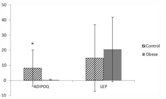
Now-a-days, obesity represents one of the major public health issues. It is known that obesity is accompanied by complex changes in gene expression across various functional categories, making them suitable candidates for a large-scale gene expression analysis. In the present study, using DNA microarrays, we analyzed the gene expression profile in peripheral blood leukocytes of morbid obese and eutrophic women. Additionally, we also evaluated the expression of leptin (LEP) and adiponectin (ADIPOQ) genes in adipocytes by using the real-time qPCR. Our data showed that genes related to food intake, immune system, defense response, DNA repair and too many other different mechanisms were modulated,confirming the multifactorial and multigenic characteristics of obesity. ADIPOQ, but not LEP, was downregulated in adipocytes. Gene networks will be build in order to check relationships among them.
1. Introduction
Obesity can be defined as excessive accumulation of adipose tissue caused by chronic energy imbalance between energy intake and energy expenditure (Tataranni & Ravoussin, 1997). Adipose tissue (AT) has been recognized as an important tissue, not only for energy storage, but also for its endocrine function.This role has emerged in recent years with the increased identification of adipocyte-secreted proteins and their broad effects on whole-body homeostasis (Kim & Moustaid-Moussa, 2001; Milan et al., 2002). In mammals, there are two types of AT: white and brown. Most AT in mammals is white and this is thought to be the site of energy storage. By contrast, brown AT is found mainly in human neonates, and it is important for regulating temperature. In addition to adipocytes, which are the most abundant cells in the white AT, pre-adipocytes (adipocyte that have not yet been loaded with lipids), endothelial cells, fibroblasts, leukocytes and, most importantly, macrophages, can be also found.These macrophages are bone marrow derived and their number in the white AT correlates directly with obesity (Tilg & Moschen, 2006). The expansion of AT in obesity is accompanied by increased secretion of hormones such as cytokines, resistin and adiponectin which affect insulin sensitivity, and angiotensin, that regulates blood pressure (Jones et al., 1997; Kim & Moustaid-Moussa, 2001; Yamauchi et al., 2001; Steppan et al., 2001; Milan et al., 2002;).Adiponectin is an adipocyte-derived hormone,and one of the most abundant circulating proteins (Stumvoll et al., 2002). In contrast to other adipocyte-released hormones,adiponectin seems to protect from insulin resistance and type 2 diabetes (Stumvoll et al., 2002). Leptin, another adipocyte derived hormone, plays a key role in obesity by regulatingfood intake and energy expenditure and, consequently, adipose-tissue mass and body weight (Paracchini et al., 2005).
al., 2008; Greg et al., 2008; Lockstone et al., 2008; Sortiriou & Pusztai, 2009;). Differentially expressed genes, involved in a wide variety of biological processes (immune response, lipid metabolism, energy production, cell adhesion and glucose metabolism), have been detected in abdominal subcutaneous adipose tissue of eutrophic and obese men (Shea et al., 2009).
2. Material and Methods Subjects
The Ethics Committee for Human Research from the Botucatu Medical School – UNESP approved this study protocol (Document nº 3361-2009). Signed informed consent was obtained from all the study subjects.
Two groups were included in the study: 10 morbid obese women (BMI > 40 Kg/m2), with age range from 30 to 50 years; and 10 eutrophic women (BMI ≤ 24.9 Kg/m2), matched by age (control group). Women included in the obese group were registered for bariatric surgery at the Center of Gastroenterology and Bariatric Surgery, - Piracicaba, SP, Brazil. The eutrophic women were registered for abdominoplasty at Misericórdia Hospital, Botucatu – SP, Brazil. From both groups, blood and adipose tissue samples were sampling at the moment of surgery. The exclusion criteria for the obese group were: alcohol consumption (> 40g alcohol/ day), presence of genetic syndromes associated to obesity, Cushing syndrome, hypothyroidism, kidney or liver diseases, neoplasias, HIV infection and use of corticoids or estrogen replacement. For control group, women should not be in use of any medicine, should not have diabetes, hypercholesterolemia, hypertension or obesity, nor family history of one of these diseases, and should not have exercised at least 24h before blood sampling.
Blood Sampling
A peripheral blood sample (10 mL) was collected through venipuncture in PAXgene Blood RNA Tubes (Qiagen/PreAnalitiX, Switzerland), for RNA stabilization. The tubes were maintained at room temperature for 12 h and, then, kept in freezer -20° C, until the procedures.
Adipose tissue sampling and adipocyte isolation
400 g) was performed. The floating adipocytes were transferred to separate tubes (Junior et al., 2005 (20B)).
RNA extraction
From blood samples, RNA was isolated using DNAse treatment and the PAXgene Blood RNA kit, according to manufacturer’s protocol (Qiagen/PreAnalitiX, Switzerland). Total RNA was measured by spectrophotometry (NanoVue – GE, Sweden); purity was assessed by absorbance at 260/280 and between 1.9 and 2.1; integrity was evaluated using the Agilent 2100 Bioanalyzer, under standard conditions. From the isolated adipocytes, total RNA was extracted by using a TRIzol solution, according to the manufacturer’s specifications. During the extraction, RNA was treated with DNAse (RNAse-free DNAse Set, Qiagen) andthen stored at -80°C. RNA was reverse-transcribed (cDNA) using the High Capacity cDNA Reverse Transcription kit (Applied Biosystems – ABI, USA), according to the manufacturer’s protocol.
Microarray
the fragmentation reaction. Hybridizations were performed for 17 hours at 65 ° C using an automated system (SureHyb, GE healthcare). Subsequently, the slides were washed with the wash buffer solutions 1 and 2. The Agilent's Stabilization and Drying solutions were also used to protect the cyanine probes against ozone degradation. The hybridization signals were captured using the GenePix 4000B scanner (Molecular Devices). Data quantification and quality control were performed using the Feature Extraction (FE) software version 10.7 (Agilent Technologies).
Background adjustment was performed by using normexp method and offset=16. Data were log2-transformed and then normalized using a quartile function. Differentially expressed genes were identified using T-test, for comparing control to the obese group. The eBayes of RBioconductor was used to perform these statistical analyses. All clusters of co-regulated genes were submitted to functional analysis using the Biological Networks Gene Ontology tool (BINGO). Score values lower than 0.05 were considered.
Analysis of ADIPOQ and LEP mRNA by quantitative real-time PCR (qRT-PCR)
After reverse transcription, cDNA samples were stored at -20°C until PCR procedures. TaqMan/FAM-MGB probes and primers (Applied Biosystems, USA) for adiponectin (ADIPOQ) (Hs00605917_m1) and leptin (LEP) (Hs00174877_m1) were used for amplification. Reactions were performed at 95°C for 20 seconds, followed by 40 cycles of 95°C for 3 seconds and 60°C for 30 seconds. β-actin gene was used as an endogenous control. Taqman Universal PCR Master Mix was purchased from Applied Biosystems, and quantitative real-time PCR was performed in duplicate using the 7500 FAST PCR system (Applied Biosystems, USA). Relative gene expression data were analyzed using the 2−ΔΔCT method (20C).
Statistical analysis
3. Results
From 40,000 genes analyzed in each array, 2,984 were differentially expressed in the obese group compared to control. From these genes, 1,404 were downregulated, and distributed into 618 different biological processes; other 1,580 were upregulated and belonged to 857 biological processes, according to the BINGO tool. Just to cite a few, these biological processes include: immune/defense response, regulation of metabolic processes, regulation of cytokine production, cellular response to stress and inflammatory response. Table 1 summarizes the biological processes related to the genes differentially expressed (obese X control).
In Table 2 are presented some genes and biological processes that we selected to discuss, because of their probably implication in obesity: stress response and food intake and satiety (HTR3C, GHRLOS, LEPROTL1 and NPY), immune/defense response (MARCO, IL-4R, IL-2RA, IL-5RA and TNFAIP8L2) and DNA repair mechanisms (ERCC2, ERCC4 and ERCC6).
Table 1. Gene Ontology Biological Process classification of differentially expressed genes, as indicated by the microarray data
Biological Process Number of genes
Downregulated genes
Regulation of Metabolic Process 343
Immune System Process 153
Apoptosis 78
Defense Response 101
Response to Stimulus 302
Regulation of Cell Communication 151
Coagulation 40
Inflammatory Response 43
Leukocyte Activation 37
Wound Healing 46
Cell Cycle 77
Regulation of Cytokine Production 34
Regulation of Insulin Secretion 16
Cellular Response to Stress 71
Cell Division 27
Upregulated genes
Response to Stress 268
Immune System Process 186
Response to Stimulus 372
Apoptosis 83
Regulation of Cytokine Production 48
Leukocyte Activation 41
Coagulation 46
Inflammatory Response 76
Cell Differentiation 179
Toll-like receptor 4 Signaling Pathway 12
Regulation of Cell-Cell Adhesion 8
Lipid Oxidation 8
Fatty Acid Oxidation 8
Table 2. Differentially expressed genes in leukocytes of morbid obese women, as indicated by the microarray data
Gene ID
Number Gene Symbol
p-value# Biological Process (GO)*
Upregulated
NM_006770 Macrophage receptor with collagenous structure MARCO 0.013578 Defense response, immune response, regulation of response to stimulus
NR_004431 Ghrelin opposite strand (non-protein coding) GHRLOS 0.003486 Regulation of metabolic processes, food intake
NM_000400 Excision repair cross-complementing rodent repair deficiency, complementation group 2
ERCC2 0.034245 Transcription induction, cell cycle checkpoints
NM_005236 Excision repair cross-complementing rodent repair deficiency, complementation group 4
ERCC4 0.039915 Response to stress, metabolic processes
NM_000124 Excision repair cross-complementing rodent repair deficiency, complementation group 6
ERCC6 0.035088 Cell response to stress, response to reactive oxygen species
NM_000417 Interleukin2 receptor, alpha IL-2RA 0.002635 Immune system, regulation of biological processes, defense response
NM_000418 Interleukin4 receptor IL-4R 0.00955 Immune response, regulation of cellular
response to stress
NM_175725 Interleukin5 receptor, alpha (IL5RA) IL-5RA 0.047961 Immune response, cytokines production
NM_052945 Tumor necrosisfactor receptor superfamily, member13C
TNFSRF13C 0.006153 Immune system, cytokines production, lymphocytes activation
NM_024575 Tumor necrosis factor, alpha-induced protein 8-like 2
TNFAIP8L2 0.012759 Lymphocytes activation, immune response
Downregulated
NM_015344 Leptin receptor overlapping transcript-like 1 LEPROTL1 0.007216 Food intake, immune system
NM_000905 Neuropeptide Y NPY 0.017908 Stress response, food intake, cardiovascular
function, cell proliferation
NM_003853 Interleukin18 receptor accessory protein IL-18RAP 0.00058 Immune system, regulation to stimulus
NM_003855 Interleukin 18 receptor 1 IL-18R1 0.000629 Defense response, immune system
NR_023392 Zinc finger protein 252 ZNF252 0.049975 Regulation of biological processes,
metabolic processes
NM_207333 Zinc finger protein 320 ZNF320 0.049479 RNA metabolic processes, cellular
metabolic processes, regulation of gene expression
4. Discussion
Genetic mechanisms related to obesity or to hyperplasia and hypertrophy of the adipose tissue are not yet completely understood (Gómez-Ambrosi et al., 2003). Therefore, because of the epidemic proportions reached by obesity worldwide, special attention has been directed to identify deregulated genes that can be causes of this metabolic dysfunction (Nadler et al., 2000; Mokdad et al., 2001; Soukas et al., 2001; Jagoe et al., 2002). Herein, we used DNA-microarrays to evaluate gene expression profiling from leukocytes of morbid obese and eutrophic women, and we detected 2,984 differentially expressed genes between these two groups. The downregulated genes (1,404) were involved in 618 processes and the upregulated ones (1,580) were involved in 857 different biological processes. Literature has demonstrated the suitability of peripheral blood mononuclear cells (PBMCs) for obesity-related studies is in part related to their active metabolism (Kussman et al., 2006). Furthermore, gene expression patterns have shown that 86% of the expressed genes in blood cells are also expressed in the adipose tissue(Brattbakk et al., 2013).
pressure have been also described (Kraja et al., 2012). The pro-inflammatory cytokine TNF-α (tumor necrosis factor-α) was the first identified adipocytokine associated to obesity and related to insulin resistance (Hotamisligil et al., 1993). Isolated and differentiated adipocytes are the principal source of elevated TNF-α level in obesity. However, more recently it has been recognized that macrophages from the stromal vascular fraction are the primary source of adipose derived TNF-α (Weisberg et al., 2003). Tumor necrosis factor-α induced protein 8 like-2 (TNFAIP8L2, TIPE2) is the second member of tumor necrosis factor-α induced protein 8 (TNFAIP8) family, which was recently defined as a novel protein involved in negative regulation of both adaptative and innate immune systems, thus manifesting a negative regulatory effect in the maintenance of immune homeostasis (Sun et al., 2008; Li et al., 2009; Zhang et al., 2010). According to some investigators, TIPE2 is highly expressed in inflammatory, but not in normal tissues (Sun et al., 2008). Nevertheless, in murine, this protein is also expressed in lymphoid tissues, including T cells and small group of non-lymphoid tissues, such as endocrine tissues, skeletal muscle and monocyte/macrophage derived cell lines (Zhang et al., 2010). Therefore, the role ofTIPE2in obesity and obesity-related diseases still need to be better investigated.
syndrome due to increased TNF-α production (Carballo et al., 1998). Therefore, the lower ZFP expression observed in our group of morbid obese women confirms the relevance of this gene in obesity, favoring adipogenesis and the inflammatory status.
Among the modulated genes directly related to appetite and food intake control we found GHRLOS(overexpressed),LEPROTL1 (downregulated), andNPY (downregulated).GHRLOSis an antisense gene that seems to be involved in the regulation of the GHRL expression (acting in cis) and in the expression of a large number of genes that are outside its locus and potentially unrelated to the ghrelin (acting intrans). Actually, GHRLOS is a non-coding RNA geneon the opposite strand of GHRL, but its genomic structure, expression pattern and potential function remains to be investigated (Tack et al., 2006). Since ghrelin acts regulating appetite, food intake, gut motility and energy balance (Bednarek et al., 2000; Heijboer at al., 2006; Neary et al., 2006; Tack et al., 2006), we might suppose GHRLOS may play important regulatory and functional roles in the ghrelin axis in obese individuals. However, it is important to notice that we evaluated GHRLOS expression in blood cells and, usually, ghrelin expression occurs in stomach, where it is produced and released. Therefore, GHRLOS overexpression in blood cells might be explained by its role in regulating other genes and not only those related to ghrelin pathway.
Regarding to the neuropeptide Y (NPY), it is a potent hypothalamic orexigenic peptide. Evidence has indicated that the control of NPY expression in the arcuate nucleus (ARC) differs from its regulation in the dorsomedial hypothalamus (DMH). While ARC NPY is under the control of circulating leptin, the controls of DMH NPY are leptin-independent (Bi et al., 2003). A previous study showed that NPY expression is increased in the ARC in response to acute food deprivation, when circulating leptin levels are significantly decreased, whereas DMH NPY expression is significantly increased in rats only after chronic food restriction (Bi et al., 2003). Therefore, our data corroborated literature, once we found NPY downregulation in morbid obese women.
Not directly acting on appetite and food intake control, 5-HTR3C is involved in the transport and re-uptake of serotonin. Serotonin (5-HT) is known to be an important neurotransmitter found in various areas of the central nervous system, being associated to several physiological mechanisms (Veenstra-VanderWeele et al.,2000). This neurotransmitter mediates cellular effects through several proteins that are involved in neurotransmission, synthesis, metabolism and membrane re-uptake (Veenstra-VanderWeele et al.,2000; Cravchik et al., 2000). 5-HT can regulate appetite and food intake, probably by promoting satiety. While 5-HT interacts with multiple subtypes of specific membrane receptors, there is evidence that its effects on appetite and food intake are, in part, mediated, by activation of receptors in the hypothalamus (Kennett & Curzon, 1991; Kennett et al., 1994). It is known that depletion of brain serotonin promotes hyperphagia and obesity, whereas administration of drugs increasing serotonergic transmission, serotonin receptor agonists and serotonin itself all reduce food intake, decrease body weight and enhance energy expenditure (Lam & Heisler, 2007).Since the levels o serotonin mRNA in blood sample did not differ in both groups, we could speculate that the 5-HTR3C overexpression would be related to a compensatory mechanism in attempt to reduce hyperphagia, however this mechanism needs further investigation since few data are found in literature.