Identification and characterization of two critical sequences
in SV40PolyA that activate the green fluorescent protein reporter gene
Honggang Wang
1,2, Wuzhuang Sun
3, Zhu Li
3, Xiufang Wang
1and Zhanjun Lv
1 1Hebei Key Lab of Laboratory Animal, Department of Genetics, Hebei Medical University,
Shijiazhuang, Hebei Province, China.
2
Department of Biochemistry and Molecular Biology, Medical College, Henan University, Kaifeng,
Henan Province, China.
3Department of Respiratory Medicine, The First Hospital of Hebei Medical University, Shijiazhuang,
Hebei Province, China.
Abstract
Alu repeats or Line-1-ORF2 (ORF2) inhibit expression of the green fluorescent protein (GFP) gene when inserted downstream of this gene in the vector pEGFP-C1. In this work, we studied cis-acting elements that eliminated the re-pression of GFP gene exre-pression induced byAlu and ORF2 and sequence characteristics of these elements. We found that sense and antisense PolyA of simian virus 40 (SV40PolyA, 240 bp) eliminated the repression of GFP gene expression when inserted between the GFP gene and theAlu (283 bp) repeats or ORF2 (3825 bp) in pAlu14 (14 tan-dem Alu repeats were inserted downstream of the GFP gene in the vector pEGFP-C1) or pORF2. Antisense SV40PolyA (PolyAas) induced stronger gene expression than its sense orientation (PolyA). Of four 60-bp segments of PolyAas (1F1R, 2F2R, 3F3R and 4F4R) inserted independently into pAlu14, only two (2F2R and 3F3R) eliminated the inhibition of GFP gene expression induced byAlu repeats. Deletion analysis revealed that a 17 nucleotide AT re-peat (17ntAT; 5’-AAAAAAATGCTTTATTT-3’) in 2F2R and the fragment 3F38d9 (5’-ATAAACAAGTTAACAACA ACAATTGCATT-3’) in 3F3R were critical sequences for activating the GFP gene. Sequence and structural analyses showed that 17ntAT and 3F38d9 included imperfect palindromes and may form a variety of unstable stem-loops. We suggest that the presence of imperfect palindromes and unstable stem-loops in DNA enhancer elements plays an important role in GFP gene activation.
Key words: Alu, enhancer, GFP, stem-loop structures, SV40PolyA.
Received: October 26, 2010; Accepted: March 23, 2011.
Introduction
Historically, considerable attention has been given to proteins and their encoding genes. However, with comple-tion of the human and mouse genomes and a better under-standing of eukaryotic gene expression, the noncoding se-quences of genes have attracted increasing attention. Noncoding sequences are widespread in eukaryotic geno-mes and contain important genetic information (Eggleston, 2005; Maeshima and Eltsov, 2007; Satzinger, 2008; Depken and Schiessel, 2009) that includes promoters, enhancers and insulators (Tour and Laemmli, 1988), noncoding RNA that directs DNA methylation (Furey and Haussler, 2003), the regulation of axon formation (Dietzel and Belmont, 2001), and small RNA genes (Eggleston, 2005).
AluandLine-1repeat elements represent about 10% and 17% of the whole human genome, respectively, and are the most important noncoding sequences (Belgnaouiet al.,
2006; Polak and Domany, 2006).Aluelements were
ini-tially considered to have no role in gene stability and ex-pression, but recent work has shown that these elements can extensively influence gene expression. In previous work,
we have shown thatAlutandem repeats andLine-1-ORF2
(ORF2) inhibited green fluorescent protein (GFP) gene ex-pression when inserted downstream of this gene in the
pEGFP-C1 vector (Wang et al., 2009a,b). Downstream
noncoding gene sequences are highly structured and con-tain important regulatory elements such as 3' UTRs, tran-scription termination signals (Andreassi and Riccio, 2009) and enhancers (Maoet al., 2010).
In this study, we examined the ability of sense and antisense SV40PolyA to eliminate the repression of GFP gene expression when inserted between the GFP gene and
Alu repeats or ORF2 in pAlu14 or pORF2. Antisense
SV40PolyA (PolyAas) caused stronger gene expression Send correspondence to Xiufang Wang and Zhanjun Lv. Hebei Key
Lab of Laboratory Animal, Department of Genetics, Hebei Medical University, 361 Zhongshan East Road, Shijiazhuang, Hebei Prov-ince, 050017, China. E-mail: [email protected] (Xiufang Wang); [email protected] (Zhanjun Lv).
than its sense orientation (PolyA). We also examined the effects of small fragments of PolyAas on GFP gene expres-sion to identify which PolyAas sequences activated this gene and found that two fragments were critical for activat-ing GFP gene expression. The two fragments both include imperfect palindromes and may form incomplete stem-loop structures that are described as a mechanism for activating GFP gene expression.
Materials and Methods
Construction of expression vectors
The pAlu14 and pORF2 expression vectors were con-structed as described elsewhere (Wanget al., 2009a,b) by inserting 14 head-to-tail tandemAlu(283 bp) elements or
anORF2(3825 bp) downstream of the GFP gene in the
pEGFP-C1 vector.
Primers were designed with sites for restriction en-zymes (EcoR I orHind III/XbaI;KpnI/NheI) and the poly-merase chain reaction (PCR) was used to amplify the synthetic DNA sequences (as templates) that contained mutated sites and fragments of PolyAas DNA. The PCR products were digested with restriction enzymes and
in-serted between the GFP gene andAlurepeats in pAlu14 or
between the GFP gene and ORF2in pORF2. When the
compatible ends of the DNA fragments digested withXbaI
andNhe I restriction enzymes were ligated by T4 DNA
ligase both of the recognition sites forXbaI andNheI were destroyed. Using this approach, the expression vectors of two tandem insertion sequences were obtained. The prim-ers and templates used for construction of the expression vectors are shown in Tables 1 and 2, respectively.
Table 1- Primers used to construct the expression vectors.
Primer identification Sequence Annotation
1F (forward primer) EcoR IXbaI
5’-ATCGGAATTCTTAATCTAGATAATGCTTACAATTTACGCGTTA-3’
Amplifying 1F1R, PolyAas
1R (reverse primer) KpnINheI
5’-ATCGGGTACCATGCTAGCTGCATTCTAGTTGTGGTT T -3’
Amplifying 1F1R
Poly60-2F (forward primer) EcoR IXbaI
5’-ATCGGAATTCTTAATCTAGATAAGTGAAAAAAATGCTTTATT -3’
Amplifying 2F2R,45R,30R
Poly60-2R (reverse primer) KpnINheI
5’-ATCGGGTACCATGCTAGCATAATGGTTACAAATAAAG -3’
Amplifying 2F2R, Poly4
3F (forward primer) EcoR IXbaI
5’-ATCGGAATTCTTAATCTAGATAAAAGC TGCAATAAACAAGTT-3’
Amplifying 3F3R
3R (reverse primer) KpnINheI
5’-ATCGGGTACCATGCTAGCCCCTGA ACCTGAAACATAA-3’
Amplifying 3F3R, 3R49, 3F235
4F (forward primer) EcoR IXbaI
5’-ATCGGAATTCTTAATCTAGATAAGGAGGTGTGGGAGGTTTTT-3’
Amplifying 4F4R
4R (reverse primer) KpnINheI
5’-ATCGGGTACCATGCTAGCTAATCAGCCATACCACATT-3’
Amplifying 4F4R, PolyAas
PolyAasF (forward primer) KpnINheI
5’-ATCGGGTACCATGCTAGCTGCTAATGCTTACAATTTACGCGTTA-3’
Amplifying PolyA
PolyAasR (reverse primer) EcoR IXbaI
5’-ATCGGAATTCTTAATCTAGATAATCAGCCATACCACATT-3’
Amplifying PolyA
FirLoopF (forward primer) EcoR IXbaI
5’-ATCGGAATTCTTAATCTAGATAATGTGAAAAAA
Amplifying 22R, 19R, 16R, 0 nt, 1 nt, 2 nt, 4 nt, 5 nt, 6 nt, TCC, GTC, GCA, CTC, GGC FirLoopR (reverse primer) KpnINheI
5’-ATCGGGTACCATGCTAGCACAAATAA
Amplifying 0 nt, 1nt, 2 nt, 4 nt, 5 nt, 6 nt, TCC, GTC, GCA, CTC, GGC
1619MR (reverse primer) KpnINheI
5’-ATCGGGTACCATGCTAGCAC-3’
Amplifying 19R, 16R
Poly45R (reverse primer) KpnINheI
5’-ATCGGGTACCATGCTAGCAAAGCAATAGCATCA-3’
Amplifying 45R
Poly30R (reverse primer) KpnINheI
5’-ATCGGGTACCATGCTAGCCAAATTTCACAAATA-3’
Amplifying 30R
SecloopF (forward primer) EcoR IXbaI
5’-ATCGGAATTCTTAATCTAGATAATGCTTTATTTGT-3’
Amplifying Secloop
SecloopR (reverse primer) KpnINheI
5’ATCGGGTACCATGCTAGCCACAAATTTCAC-3’
Amplifying Secloop
Poly4F (forward primer) EcoR IXbaI
5’-ATCGGAATTCTTAATCTAGATAATGATGCTATTG-3’
Amplifying Poly4
EcoXba (forward primer) EcoR IXbaI
5’-ATCGGAATTCTT AATCTAGA-3’
Primer identification Sequence Annotation KpnNhe (reverse primer) KpnINheI
5’-ATCGGGTACCATGCTAGC-3’
Amplifying 17ntAT
3F46F(forward primer) HindIIIXbaI
5’-ATCGAAGCTTAATCTAGAAAGCTGCAATAAACAAG
Amplifying 3F46 fragment with re-verse primer 3F135R
3R49F (forward primer) HindIIIXbaI
5’-ATCGAAGCTTAATCTAGAAACAAGTTAACAACAA
Amplifying 3R49 fragment with re-verse primer 3R; Amplifying 3F135 fragment with 3F135R
3F135R (reverse primer) KpnINheI
5’-ATCGGGTACCATGCTAGCCATAAAATGAATG -3’
Amplifying 3F135, 3F46
3F235F(forward primer) HindIIIXbaI
5’-ATCGAAGCTTAATCTAGAAACAATTGCATTC-3’
Amplifying 3F235 fragment with reverse primer 3R
3F46d2F (forward primer) HindIIIXbaI
5’-ATCGAAGCTTAATCTAGAGCTGCAATAAACAAG
Amplifying 3F46d2 fragment with reverse primer 3F135R
3F46d3F (forward primer) HindIIIXbaI
5’-ATCGAAGCTTAATCTAGACTGCAATAAACAAGT-3’
Amplifying 3F46d3 fragment with reverse primer 3F135R
3F46d4F (forward primer) HindIIIXbaI
5’-ATCGAAGCTTAATCTAGATGCAATAAACAAGTT-3’
Amplifying 3F46d4 fragment with reverse primer 3F135R
3F46d5F (forward primer) HindIIIXbaI
5’-ATCGAAGCTTAATCTAGAGCAATA AACAAGTTA-3’
Amplifying 3F46d5 fragment with reverse primer 3F135R
3F46d6F (forward primer) HindIIIXbaI
5’-ATCGAAGCTTAATCTAGACAATAAACAAGTTA-3’
Amplifying 3F46d6 fragment with reverse primer 3F135R
3F46d7F (forward primer) HindIIIXbaI
5’-ATCGAAGCTTAATCTAGAAATAAACAAGTTA-3’
Amplifying 3F46d7 fragment with reverse primer 3F135R
3F46d8F (forward primer) HindIIIXbaI
5’-ATCGAAGCTTAATCTAGAATAAACAAGTTA AC-3’
Amplifying 3F46d8 fragment with reverse primer 3F135R; Amplifying 3F38d1, 3F38d2, 3F38d3, 3F38d4, 3F38d5, 3F38d6, 3F38d8, 3F38d9, 3F38d10, 3F38d11, 3F38d12, 3F38 d13 with corresponding reverse primers
3F46d9F (forward primer) HindIIIXbaI
5’-ATCGAAGCTTAATCTAGATAAACAAGTTAACA-3’
Amplifying 3F46d9 fragment with reverse primer 3F135R
3F46d10F (forward primer) HindIIIXbaI
5’-ATCGAAGCTTAATCTAGAAAACAAGTTA-3’
Amplifying 3F46d10 fragment with reverse primer 3F135R
3F38d1R (reverse primer) KpnINheI
5’-ATCGGGTACCATGCTAGCATAAAATGAATGCA-3’
Amplifying 3F38d1 fragment with forward primer 3F46d8F 3F38d2R (reverse primer) KpnINheI
5’-ATCGGGTACCATGCTAGCTAAAATGAATGCAA-3’
Amplifying 3F38d2 fragment with forward primer 3F46d8F 3F38d3R (reverse primer) KpnINheI
5’-ATCGGGTACCATGCTAGCAAAATGAATGCAAT-3’
Amplifying 3F38d3 fragment with forward primer 3F46d8F 3F38d4R (reverse primer) KpnINheI
5’-ATCGGGTACCATGCTAGCAAATGA TGCAATT-3’
Amplifying 3F38d4 fragment with forward primer 3F46d8F 3F38d5R (reverse primer) KpnINheI
5’-ATCGGGTACCATGCTAGCAATGAATGCAATTG-3’
Amplifying 3F38d5 fragment with forward primer 3F46d8F 3F38d6R (reverse primer) KpnINheI
5’-ATCGGGTACCATGCTAGCATGAATGCAATTG-3’
Amplifying 3F38d6 fragment with forward primer 3F46d8F 3F38d8R (reverse primer) KpnINheI
5’-ATCGGGTACCATGCTAGCGAATGCAATTG-3’
Amplifying 3F38d8 fragment with forward primer 3F46d8F 3F38d9R (reverse primer) KpnINheI
5’-ATCGGGTACCATGCTAGCAATGCAATTG-3’
Amplifying 3F38d9 segment with forward primer 3F46d8F 3F38d10R (reverse primer) KpnINheI
5’-ATCGGGTACCATGCTAGCATGCAATTG-3’
Amplifying 3F38d10 fragment with forward primer 3F46d8F
3F38d11R (reverse primer) KpnINheI
5’-ATCGGGTACCATGCTAGCTGCAATTG-3’
Amplifying 3F38d11 fragment with forward primer 3F46d8F
3F38d12R (reverse primer) KpnINheI
5’-ATCGGGTACCATGCTAGCGCAATTGTTGTTGTTAACTT-3’
Amplifying 3F38d12 segment with forward primer 3F46d8F 3F38d13R (reverse primer) KpnINheI
5’-ATCGGGTACCATGCTAGCCAATTGTTGTTGTTAACTT-3’
Amplifying 3F38d13 fragment with forward primer 3F46d8F
Underlined sequences indicate restriction enzyme cleavage sites.
Cell culture and transfection
HeLa cells were cultured in Dulbecco’s modified Ea-gle’s medium (DMEM) with 10% fetal calf serum. Cells were plated in each well of a 24-well plate at 0.9 x 10⁵ cells/well and cultured at 37 °C in 5% CO2for 30-36 h. The cells were transiently transfected with 0.4mg of expression
vector DNA using 2 mL of Lipofectamine2000 reagent
(Invitrogen, USA), according to the manufacturer’s in-structions, and subsequently cultured for an additional 30-36 h. The transfected cells were used for RNA extrac-tion and fluorescence assays.
Assessment of GFP fluorescence
Transfected HeLa cells were fixed in 4% parafor-maldehyde and the expression of GFP protein was assessed by using fluorescence microscopy (Nikon TE2000-U, Ja-pan). Images were obtained under normal and fluorescent illumination.
Northern blotting
Total RNA from transfected cells was extracted with
Trizol® reagent (Invitrogen, USA). RNA was
electropho-resed in 1.2% agarose gels containing 0.4 M formaldehyde and then transferred to nylon membranes (pore diameter
0.45mm; Osmonics, USA). A 590-bp fragment from the
GFP gene in the pEGFP-C1 vector was amplified by PCR using the forward primer 5’-GGGCGAGGGCGATG-3’ and the reverse primer 5’-CTTGTACAGCTCGTCCAT GC-3’. The PCR product was purified by agarose gel elec-trophoresis and radiolabeled with [a-32P]-dCTP (Furui,
China) using the random primer labeling system (TaKaRa, Japan). The nylon membranes blotted with RNA were
hy-bridized witha-32P-radiolabeled DNA probes at 42 °C in
50% formamide containing 5x SSC (saline sodium citrate),
5x Denhardt’s solution and 100 mg of salmon sperm
DNA/mL for 24 h in a UL2000 hybriLinker (UVP, USA). The membranes were washed twice at room temperature with a solution of 1x SSC-0.1% SDS and then washed three times with a solution of 0.1x SSC-0.1% SDS at 68 °C prior to autoradiography. The membranes were subsequently stripped by washing twice at 80 °C for 1 h in a solution con-taining 50% formamide-5% SDS-50 mM Tris (pH 7.4), and then hybridized witha-32P-radiolabeled probe for neoRNA
(containing the cassette for neomycin resistance). A 671-bp fragment from the neo gene in the pEGFP-C1 vector was amplified by PCR using the forward primer 5’-CACAACA GACAATCGGCTGCT-3’ and the reverse primer 5’-AGC
GGCGATACCGTAAAAGCAC-3’. The probe for
neoRNA was prepared using the random primer labeling system and the 671-bp neo fragment as the template.
Results
PolyA and PolyAas eliminate the repression of GFP gene expression induced byAlurepeats orORF2
Northern blotting showed that there was almost
com-plete repression of GFP expression in HeLa cells
trans-fected with the expression vectors pAlu14 and pORF2 (Figure 1A, lane 3vs.lane 5; Figure 1B, lane 5vs.lane 4). PolyA and PolyAas sequences inserted between the GFP gene andAlurepeats orORF2partly abolished the
repres-sion of GFP expression caused by Alurepeats or ORF2
(Figure 1A, lanes 1 and 2vs.lane 3; Figure 1B, lanes 1 and 2vs. lane 5). PolyAas reversed the repression ofGFP ex-pression to a greater extent than its sense orientation in
pAlu14 and pORF2 (Figure 1A, lane 2vs.lane 1; Figure
1B, lane 2vs.lane 1).
The neo gene was used as a control to assess the effi-ciency of transfection with the GFP gene. The occurrence of both genes on the same expression vector eliminated the possibility that variation in the efficiency of transfection contributed to the differences observed in the experimental results.
The effects of PolyAas segments on GFP gene expression
To determine which segments in PolyAas eliminated
the repression of GFP gene expression caused byAlu
re-peats we produced four 60-bp segments of PolyAas (1F1R, 2F2R, 3F3R and 4F4R; Figure 2A) that were then inserted between the GFP gene andAlurepeats in the pAlu14 vector used to transiently transfect HeLa cells. Northern blotting showed that 1F1R and 4F4R did not stimulate GFP gene
expression (Figure 2B, lanes 1 and 4vs. lane 6) whereas
2F2R and 3F3R did (Figure 2B, lanes 2 and 3vs. lane 6). Table 2- Synthetic templates used to construct expression vectors.
Identification Sequence
19RM 5’-AATGTGAAAAAAATGCTTTATTGCTAGC-3’
16RM 5’-AATGTGAAAAAAATGCTTTGCTAGC-3’
17ntAT 5’-CTAGATAATAAAAAAATGCTTTATTTGCTAG CAT-3’
Loop0nt 5’-GTGAAAAAAATTTATTTGT-3’
Loop1nt 5’-GTGAAAAAAAGTTTATTTGT-3’
Loop2nt 5’-GTGAAAAAAATGTTTATTTGT-3’
Loop4nt 5’-GTGAAAAAAACTGCTTTATTTGT-3’
Loop5nt 5’-GTGAAAAAAACGTGCTTTATTTGT-3’
Loop6nt 5’-GTGAAAAAAATCGTGCTTTATTTGT-3’
TCC 5’-GTGAAAAAAATCCTTTATTTGT-3’
GTC 5’-GTGAAAAAAAGTCTTTATTTGT-3’
GCA 5’-GTGAAAAAAAGCATTTATTTGT-3’
CTC 5’-GTGAAAAAAACTCTTTATTTGT-3’
The effects of 2F2R and its deleted fragments on GFP gene expression
To determine which fragments of 2F2R were respon-sible for the activation of GFP gene expression we deleted selected regions of the 2F2R DNA (Figure 3A). The bases in the 3’ end of 2F2R were deleted and the single sequence or double tandem sequences of deleted 2F2R (45R, 30R, 22R, 19R and 16R) were inserted into pAlu14. Fragments 45R, 30R and 22R activated GFP gene expression (Figure 3B, lanes 2, 3 and 4vs.lane 13, and lanes 8, 9 and 10vs. lane 13), whereas 19R and 16R induced weaker GFP gene expression (Figure 3B, lanes 5, 6, 11 and 12vs. lane 13). The double tandem sequences of 2F2R and their deleted se-quences induced stronger GFP gene expression than the corresponding single sequences (Figure 3B, lanes 7-12vs.
lanes 1-6, respectively). Although 45R, 30R and 22R all en-hanced GFP gene expression, the activation of 45R was weaker than that of 2F2R (Figure 3B, lane 2vs. lane 1; lane 8vs. lane 7), and the activation by 30R and 22R was weaker
than that of 45R (Figure 3B, lanes 3 and 4vs. lane 2; lanes 9 and 10vs. lane 8). These results indicated that the 3’ deleted sequences in 2F2R contributed to GFP gene expression. The base deletions influenced the termination of transcrip-tion, with the double tandem sequences of 2F2R and 45R resulting mainly in low molecular mass transcripts (Figure 3B, lanes 7 and 8), whereas double tandems of 30R and 22R yielded mainly high molecular mass transcripts (Figure 3B, lanes 9 and 10).
The single or double tandem sequences of Poly4 and Secloop (the sequences and their positions are shown in Fig-ure 3A and FigFig-ure 3C) in 2F2R were inserted downstream of the GFP gene. HeLa cells were transiently transfected with the expression vectors. Northern blotting showed that neither Poly4 nor Secloop significantly activated GFP gene expres-sion (Figure 3D, lanes 1-4vs.lane 5).
The effects of the 22R fragment and its deleted sequences on GFP gene expression
The deletion of three upstream bases (5’ -GTG) and two downstream bases (GT-3’) of fragment 22R yielded a 17 nucleotide repeat of AT (17ntAT; sequence and position shown in Figure 3A). 17ntAT activated GFP gene expres-sion to the same extent as 22R (Figure 4B, lane 2vs. lane 5), indicating that the five deleted bases were not important for
GFP gene activation. The 19R fragment, i.e., 22R from
which three downstream bases (TGT-3’) had been deleted, caused much lower GFP gene expression (Figure 4B, lane 3
vs.lane 5), indicating an important role for these bases in GFP gene activation. Double tandems of 17ntAT produced more transcripts than the corresponding single sequence
(Figure 4B, lane 2vs. lane 4). Figure 4A shows the
se-quences inserted into pAlu14.
Figure 2- The effect of 60-bp segments of PolyAas (1F1R, 2F2R, 3F3R and 4F4R) on GFP gene expression. (A) Positions and sequences of the four segments. (B) 1F1R, 2F2R, 3F3R, 4F4R and PolyAas were inserted downstream of the GFP gene in pAlu14. HeLa cells were transfected with the expression vectors andGFPRNA was detected by northern blotting.
The effects of 3F3R fragments on GFP gene expression
To identify which fragments in 3F3R enhanced GFP gene expression, we deleted sections of 3F3R DNA (Figure 5A). The single sequences of deleted 3F3R (3F46, 3R49, 3F135 and 3F235) were inserted into pAlu14. Figure 5B shows that 3F46 activated GFP gene expression, whereas 3R49, 3F135 and 3F235 did not.
The effects of 3F46 deletions on GFP gene expression
To identify the 3F46 cis-element responsible for gene activation we deleted the nucleotides upstream of 3F46
(Figure 6A) and constructed expression vectors. Northern blotting showed that 3F46d2-3F46d8 (deletion of 2-8 bases upstream of 3F46) still activated GFP gene expression (Figure 6B, lanes 1-7vs.lane 11), whereas 3F46d9 caused only weak activation (Figure 6B, lane 8vs. lane 11) and 3F46d10 produced hardly any activation (Figure 6B, lane 9
vs. lane 11). Figure 3- The effects of 2F2R deletions on GFP gene expression. (A)
Po-sitions and sequences of the deletion mutations in 2F2R. (B) Single frag-ments or double tandem fragfrag-ments of 2F2R and deletions (45R, 30R, 22R, 19R and 16R) were inserted downstream of the GFP gene in pAlu14. HeLa cells were transfected with the expression vectors andGFPRNA was detected by northern blotting. (C) Nucleotide sequences of Poly4 and Secloop and their double tandems. The nucleotides linking two fragments are underlined. (D) Poly4 and Secloop and their double tandems were in-serted downstream of the GFP gene in pAlu14. HeLa cells were trans-fected with the expression vectors and theGFPRNA was detected by northern blotting.
Figure 4- The effects of 22R and its deleted sequences on GFP gene ex-pression. (A) Nucleotide sequences of double tandems of 16R, 17ntAT, 19R, 22R and single 17ntAT. The nucleotides linking two fragments are underlined. (B) 22R and its deleted sequences were inserted downstream of the GFP gene in pAlu14. HeLa cells were transfected with the expres-sion vectors andGFPRNA was detected by northern blotting.
The effects of base deletions downstream of 3F38 (3F46d8) on GFP gene expression
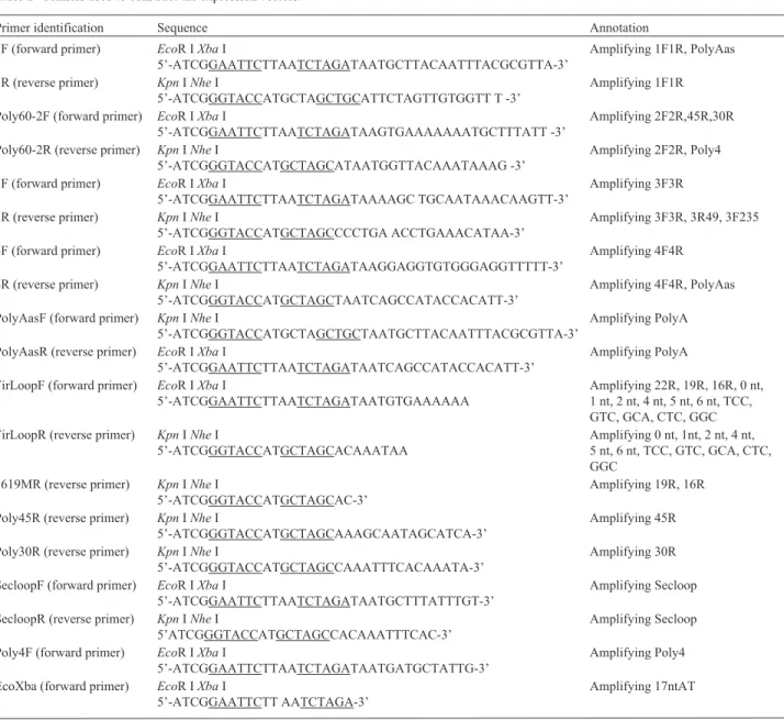
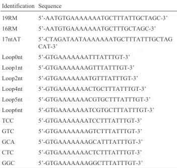
The deletion of selected nucleotides was used to es-tablish the downstream boundary for GFP gene activation by fragment 3F38 (Figure 7A). Northern blotting showed that 3F38d1-3F38D6, 3F38d8 and 3F38d9 activated the
GFP gene (Figure 7B, lanes 1-8 vs. lane 14) whereas
3F38d10-3F38d13 did not (Figure 7B, lanes 9-12vs. lane
14). These results identified 3F38d9 as the critical sequence of 3F38 for GFP gene activation.
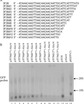
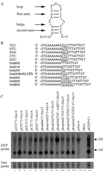
The effects of mutations in 22R DNA on GFP gene expression
Analysis of the DNA sequence of 22R indicated that this fragment may form an incomplete stem-loop structure that included a loop (3 nt), an initial stem (3 bp), a bulge (2 nt) and a second stem (3 bp) (Figure 8A). We examined the influence of loop base type and loop length on the abil-ity of 22R to influence GFP gene activation by introducing mutations in these regions and inserting the fragment into the vector pAlu14 for transfection in HeLa cells. The loop base combination TGC (22R*2, wild type) induced the strongest GFP gene expression (Figure 8C, lane 9vs.lanes 1-5), whereas changing the loop base number from 0 nt to 6 nt showed that a 3 nt loop (22R*2, wild type) produced
the strongest GFP gene expression (Figure 8C, lane 9vs.
lanes 6, 7, 8, 10, 11 and 12). Although most of the loop mu-tants were able to enhance GFP gene expression they were
generally less effective than the wild type fragment (22R) (Figure 8C, lanes 2, 4, 5, 6, 7, 8, 11 and 12vs.lane 13). This finding indicates that if there are no changes in the palin-dromes flanking the loop then many types of loops can en-hance GFP gene expression.
Discussion
SV40PolyA activates luciferase reporter gene expres-sion in HeLa cells (Zhi-Liet al., 2001). In this study, the in-sertion of sense or antisense PolyA between the GFP gene
andAlurepeats orORF2in the vectors pAlu14 or pORF2
resulted in partial recovery of GFP gene expression re-pressed byAlurepeats orORF2. This finding indicated that sense and antisense PolyA enhanced GFP gene expression, with PolyAas causing greater induction than PolyA. Nolan
et al.(1996) found that reversing the orientation of DRE (a 27-bp enhancer) dramatically decreased growth hormone gene expression, indicating that the binding of transfactors to the DRE and the interaction of this complex with the TATA region are directional.
The wild-type SV40 enhancer contains a double tan-dem duplication (72-bp repeat). The single 72-bp repeat contains three functional elements (A, B and C) that range in size from 15 to 22 bp (Shepardet al., 1988). Although Figure 6- The effects of 3F46 deletions on GFP gene expression. (A)
Nu-cleotide sequences of 3F46 and its deleted fragments. (B) The sequences of 3F46 and deleted sequences were inserted downstream of the GFP gene in pAlu14. HeLa cells were transfected with the expression vectors and GFPRNA was detected by northern blotting.
PolyA is a short sequence (240 bp) it contains various re-gions that may differ in their ability to activate genes. To examine this hypothesis, we produced four segments of PolyAas (1F1R, 2F2R, 3F3R and 4F4R) (Figure 2A) and inserted them separately downstream of the GFP gene in pAlu14. 2F2R and 3F3R abolished the inhibition of GFP
gene expression induced by tandemAlurepeats. To
deter-mine which portions of 2F2R activated the GFP gene, we deleted bases from the 3’ end of this segment and found that fragment 22R activated the GFP gene, with double tandem sequences having a stronger effect than the corresponding single sequences. None of the other 2F2R fragments (19R, 16R, Secloop and Poly4) significantly activated the GFP gene.
The 5’ and 3’ UTRs of viral genomes are highly struc-tured and are critical for controlling viral biological pro-cesses. The stem-loop structure is important for gene activation (Daiet al., 1997) and Bio-software predicts vari-ous stem-loop structures in these regions. Most of the stem-loop structures in viral genomes show bulge se-quences in their stems (Yu and Markoff, 2005; Rosskopfet al., 2010; Nickens and Hardy, 2008). To explain the results obtained with the 2F2R fragments, we hypothesized that 22R contained an imperfect palindrome and formed an in-complete stem-loop structure that included a loop (3 nt), an initial stem (3 bp), a bulge (2 nt) and a second stem (3 bp). Fragment 19R [22R with three downstream bases (TGT) deleted] produced fewer transcripts (Figure 4B, lane 3vs.
lane 5), whereas 17ntAT [22R with three upstream bases (GTG) and two downstream bases (GT) deleted] activated the GFP gene when inserted into pAlu14, indicating that the third base (T) downstream of 22R is important for GFP gene activation. 17ntAT was the smallest sequence in 22R to form an incomplete stem-loop structure. The stem-loop structures were destroyed in 19R and 16R, and neither frag-ment activated the GFP gene significantly, which sug-gested that an incomplete stem-loop structure (Figure 8A) was important for GFP gene activation. Examination of the 17ntAT sequence (5’-AAAAAAATGCTTTATTT) sug-gested that it was capable of forming a variety of incom-plete, unstable stem-loops. Figure 8A shows one of the presumed stem-loop structures.
To determine the 3F3R sequences involved in GFP gene activation we produced four overlapping fragments (3F46, 3R49, 3F135 and 3F235) of this segment (Figure 5A). The four fragments were inserted separately between the GFP gene and theAlurepeats in the pAlu14 vector that was then used to transfect HeLa cells. Only 3F46 activated the GFP gene. Sequential (one by one) deletion of bases up-stream of 3F46 showed that removal of the first eight bases (3F46d8) had little influence on GFP gene activation, whereas elimination of the ninth base (3F46d9) markedly attenuated this activation, indicating a critical role for this base (Figure 6B, lane 8). Sequential (one by one) deletion of the downstream bases of 3F38 (fragment 3F46 in which eight bases were deleted) showed that removal of the first nine bases did not markedly affect GFP gene activation whereas the removal of bases 10-13 eliminated the activa-tion of this gene. Together, these findings indicated that the critical sequence in 3F3R for GFP gene activation was 3F38d9 (5’-ATAAACAAGTTAACAACAACAATTGC ATT-3’). This sequence contained 29 bases (A = 15, C = 5, G = 2, T = 7), with the fragment from A12 to C20 contain-ing three AAC repeats that were flanked by GTT and TTG sequences which formed stem-loop structures with the AAC repeats. Fragment 3F38d9 was thus similar to 17ntAT in that both of them formed unstable stem-loop structures. Base mutations and variations in the number of bases (from 0 nt to 6 nt) in the 22R loop showed that the TGC Figure 8- The effects of 22R mutants on GFP gene activation. (A) The
loop (22R, wild type) induced the strongest gene expres-sion, although most of the loop mutants showed some abil-ity to induce this gene (Figure 8C). This finding suggested that an unstable stem-loop structure was required for GFP gene activation by 22R, with many loops partly satisfying this criterion. Changes in loop bases influence the stability
of stem-loop structures (Lamoureux et al., 2006), and
stem-loop structures with 3-4 base loops may be specifi-cally stabilized and have lower folding times (Kuznetsovet al., 2001, 2008). These findings may help to explain the im-portance of 3nt loops in GFP gene activation.
DNA cruciform structures can be formed when intra-strand pairing occurs between complementary bases of in-verted repeat sequences in double-stranded DNA. Cruci-form Cruci-formation is energetically less favorable than B-Cruci-form DNA so that the extrusion of these structures from duplex DNA requires the driving energy provided by negative supercoiling (Seanet al., 2009). Hairpin structures in the cruciform promoter for the bacteriophage N4 virion RNA polymerase are extruded at physiological superhelical
den-sity (Chou et al., 1999). The palindromes in
double-stranded DNA may form incomplete stem-loop structures within small scope (Darlow and Leach, 1998). For this rea-son, the structures formed by these palindrome sequences may play an important role in regulating gene expression in cells.
The critical sequences of 2F2R and 3F3R involved in gene activation have two characteristics in common, na-mely, (1) they can form various stem-loop structures that increase the probability of creating stem-loops by random impact and (2) the stem-loop structures are incomplete and unstable, which ensures that stem-loops promptly revert to a double helix state. Based on these findings, we propose that sequences containing suitably imperfect palindromes activate gene expression by dynamic fluctuations between unstable stem-loop structures and double-strand forms. Ad-ditional experiments are required to confirm this hypothe-sis.
Acknowledgments
This work was supported by grants from the Hebei
Province Natural Science Foundation of China
(C2008001065 and C2011206043) and the Key Project of Hebei Province (08276101D-90).
References
Andreassi C and Riccio A (2009) To localize or not to localize: mRNA fate is in 3’UTR ends. Trends Cell Biol 19:465-474. Belgnaoui SM, Gosden RG, Semmes OJ and Haoudi A (2006)
Human LINE-1 retrotransposon induces DNA damage and apoptosis in cancer cells. Cancer Cell Int 6:1-10.
Chou SH, Tseng YY and Chu BY (1999) Stable formation of a py-rimidine-rich loop hairpin in a cruciform promoter. J Mol Biol 292:309-320.
Dai X, Greizerstein MB, Nadas-Chinni K and Rothman-Denes LB (1997) Supercoil-induced extrusion of a regulatory DNA hairpin. Proc Natl Acad Sci USA 94:2174-2179.
Darlow JM and Leach DR (1998) Evidence for two preferred hair-pin folding patterns in d(CGG) d(CCG) repeat tractsin vivo. J Mol Biol 275:17-23.
Depken M and Schiessel H (2009) Nucleosome shape dictates chromatin fiber structure. Biophys J 96:777-784.
Dietzel S and Belmont AS (2001) Reproducible but dynamic posi-tioning of DNA in chromosomes during mitosis. Nat Cell Biol 3:767-770.
Eggleston AK (2005) Unraveling chromatin organization. Nat Struct Mol Biol 12:6.
Furey TS and Haussler D (2003) Integration of the cytogenetic map with the draft human genome sequence. Hum Mol Genet 12:1037-1044.
Kuznetsov SV, Shen Y, Benight AS and Ansari A (2001) A semiflexible polymer model applied to loop formation in DNA hairpins. Biophys J 81:2864-2875.
Kuznetsov SV, Ren C, Woodson SA and Ansari A (2008) Loop dependence of the stability and dynamics of nucleic acid hairpins. Nucleic Acids Res 36:1098-1112.
Lamoureux M, Patard L, Hernandez B, Couesnon T, Santini GP, Cognet JA, Gouyette C and Cordier C (2006) Spectroscopic and structural impact of a stem base-pair change in DNA hairpins: GTTC-ACA-GAAC versus GTAC-ACA-GTAC. Spectrochim Acta Part A 65:84-94.
Maeshima K and Eltsov M (2007) Packaging the genome: The structure of mitotic chromosomes. J Biochem 143:145-153. Mao J, Li C, Zhang Y, Li Y and Zhao Y (2010) Human
with-no-lysine kinase-4 3’-UTR acting as the enhancer and being targeted by miR-296. Int J Biochem Cell Biol 42:1536-1543.
Nickens DG and Hardy RW (2008) Structural and functional anal-yses of stem-loop 1 of the Sindbis virus genome. Virology 370:158-172.
Nolan EM, Cheung TC, Burton DW and Deftos LJ (1996) Trans-criptional regulation of the human chromoranin A gene by its 5’ distal regulatory element: Novel effects of orientation, structure, flanking sequences, and position on expression. Mol Cell Endocrinol 124:51-62.
Polak P and Domany E (2006)Aluelements contain many binding sites for transcription factors and may play a role in regula-tion of developmental processes. BMC Genomics 7:1-15. Rosskopf JJ, Upton 3rd JH, Rodarte L, Romero TA, Leung MY,
Taufer M and Johnson KL (2010) A 3’ terminal stem-loop structure in Nodamura virus RNA2 forms an essential cis-acting signal for RNA replication. Virus Res 150:12-21. Satzinger H (2008) Theodor and Marcella Boveri: Chromosomes
and cytoplasm in heredity and development. Nat Rev Genet 9:231-238.
Sean P, Nguyen JH and Semler BL (2009) Altered interactions be-tween stem-loop IV within the 5’ noncoding region of coxsackievirus RNA and poly(rC) binding protein 2: Effects on IRES-mediated translation and viral infectivity. Virology 389:45-58.
Tour BE and Laemmli UK (1988) The metaphase scaffold is heli-cally folded: Sister chromatids have predominantly opposite helical handedness. Cell 55:937-944.
Wang XF, Wang XY, Liu J, Feng JJ, Mu WL, Shi XJ, Yang QQ, Duan XC and Xie Y (2009a) Alu tandem sequences inhibit GFP gene expression by triggering chromatin wrapping. Genes Genom 31:209-215.
Wang XF, Jin X, Wang XY, Liu J, Feng JJ, Yang QQ, Mu WL, Shi XJ and Lu ZJ (2009b) Effects of L1-ORF2 fragments on green fluorescent protein gene expression. Genet Mol Biol 32:688-696.
Yu L and Markoff L (2005) The topology of bulges in the long stem of the flavivirus 3’ stem-loop is a major determinant of RNA replication competence. J Virol 79:2309-2324. Zhi-Li Xu, Mizuguchi H, Ishii AW, Uchida E, Mayumi T and
Hayakawa T (2001) Optimization of transcriptional regula-tory elements for constructing plasmid vectors. Gene 272:149-156.
Associate Editor: Carlos F.M. Menck