BIOFILM FORMATION AND BINDING SPECIFICITIES OF CFA/I, CFA/II AND CS2 ADHESIONS OF
ENTEROTOXIGENIC ESCHERICHIA COLI AND CFAE-R181A MUTANT
Iram Liaqat1,2*
1
Department of Zoology, Government College University, Lahore, Pakistan.
Submitted: August 12, 2011; Returned to authors for corrections: December 01, 2011; Approved: June 07, 2012.
ABSTRACT
Enterotoxigenic Escherichia coli (ETEC) strains are leading causes of childhood diarrhea in developing
countries. Adhesion is the first step in pathogenesis of ETEC infections and ETEC pili designated
colonization factor antigens (CFAs) are believed to be important in the biofim formation, colonization and
host cell adhesions. As a first step, we have determined the biofilm capability of ETEC expressing various
types of pili (CFA/I, CfaE-R181A mutant/ CfaE tip mutant, CFA/II and CS2). Further, enzyme-linked
immunosorbent assay (ELISA) assay were developed to compare the binding specificity of CFA/I, CFA/II
(CS1 - CS3) and CS2 of ETEC, using extracted pili and piliated bacteria. CFA/II strain (E24377a) as well as
extracted pili exhibited significantly higher binding both in biofilm and ELISA assays compared to non
piliated wild type E24377a, CFA/I and CS2 strains. This indicates that co-expression of two or more CS2 in
same strain is more efficient in increasing adherence. Significant decrease in binding specificity of
DH5αF’lacIq/∆cotD (CS2) strain and MC4100/pEU2124 (CfaE-R181A) mutant strain indicated the important contribution of tip proteins in adherence assays. However, CS2 tip mutant strain
(DH5αF’lacIq/pEU5881) showed that this specific residue may not be important as adhesions in these strains. In summary, our data suggest that pili, their minor subunits are important for biofilm formation and
adherence mechanisms. Overall, the functional reactivity of strains co expressing various antigens,
particularly minor subunit antigen observed in this study suggest that fewer antibodies may be required to
elicit immunity to ETEC expressing a wider array of related pili.
Key words: ETEC; CFA/I; CFA/II; CS2; Biofilm formation; ELISA assays; asialo-GM1; R181-cotD;
dsc19CotD(His)6
INTRODUCTION
Enterotoxigenic Escherichia coli (ETEC) are leading
causes of diarrhea in children living in developing countries
and the most common cause of traveler’s diarrhea (4).
Infections with ETEC require proper adhesion of bacteria to
the intestinal epithelium. This first step in the pathogenesis of
ETEC diarrhea is mediated through specific surface structures
970
called pili or fimbriae (11), which allow the bacteria to
colonize the intestinal mucosa of small intestine. Hitherto, a
large number of colonization factor antigens (CFAs) have been
identified (27), which include CFA/I, CFA/II, CFA/III and
CFA/IV, and a number of coli surface antigens (CS). CFA/I
and CFA/III are rod like fimbrial antigens with diameters of 6–
8 nm (12, 15). The CFA/II and CFA/IV are composed of
antigenically distinct structures, of which CS1, CS2, and CS3
belong to CFA/II and CS4, CS5, and CS6 belong to CFA/IV
(5, 15). Whereas CS1 and CS2 are morphologically similar to
CFA/I, CS3 was shown to consist of fine fibrils with a diameter
of 2–3 nm.
CS1 pili are composed almost entirely of the major pilin
called CooA. A minor pilin, CooD, is found only at the pilus
tip contributing only one subunit per pilus.CooD is essential
for the transport of CooA across the outer membrane, and the
level of CooD expression determines the number of assembled
pili on cell surface, hence indicating its significance for CS1
pilus assembly initiation (24). Modeling of CFA/I has placed
the CfaB subunits in a recurring interaction pattern with the tip
protein at the end acting as an adhesion, resulting in a helical
structure (19). While conflicting data exists supporting a role
for CfaB and/or CfaE in mediating specific target cell binding,
convincing results from recent studies indicate CfaE as critical
binding subunit. In vitro studies of CFA/I fimbriae expressed in
DH5α suggested CfaE mediated haemagglutination. When
CFA/I was expressed in DH5α with a single-amino-acid
mutation R181A in CfaE resulted in haemagglutination without
affecting pilus assembly (24).
Hemagglutination has also been proven useful in the
determination of the colonization factor (CF) carbohydrate
receptor specificity. The CF and the receptor molecules act as
logical targets for inhibiting the interaction between pathogen
and host cell. Thus, the use of CF, CF analogs, receptors, or
receptor analogs could prevent adherence of pathogens (26).
Alternatively, antibody to CF could prevent the initial
attachment to the host cell. The glycosphingolipid receptor
candidates for P. aeruginosa adhesions include
gangliotetraosyl ceramide (asialo-GM), gangliotriaosyl
ceramide (asialo-GM2), lactosylceramide and sialic
acid-containing glycosphingolipids (2). However, as for the
majority of the receptors for adhesive factors of the
enterovirulent bacteria, the intestinal receptors for the adhesive
factors of ETEC remain unknown. Little is known about
receptors specific for CS1 and CFA/I strains. Some studies
have shown that the latter bind to sialic acid-containing
glyconjugates (29). In erythrocytes, CFA/I (26), can recognize
sialic acid, sialic acid-containing glycopeptides (8), the
GM2-like glycoconjugate or the asialo-GM1 (2, 3). An analogy
between the carbohydrate specificities of the CFA/I and CFA/II
adhesins was observed, since hemagglutination inhibition is
successfully obtained using same preparations of complex
carbohydrates (20). The ETEC adhesins utilizing these
glycosphingolipids as receptors have not been unambiguously
identified. It has been reported previously that this binding
activity in CFA/I is exclusively mediated by the minor pilin
subunit (24). Colonization factors also enable ETECto adhere
and colonize resulting in biofilm formation (22)
Biofilm-formation in ETEC strains is responsible for many serious
infections in patients with indwelling bladder catheters and
bowel diseases (17). Also intracellular biofilm-like aggregates
formed inside bladder cells by these strains, make them hard to
reach by both hostdefence mechanisms and antibiotics (1).
Hence, in this background, very first objective of our
study was to examine the biofilm-forming capacity of ETEC
strains expressing various types of pili (CS2, CFA/I, CFA/II
(CS1-CS3) onabiotic surfaces. Notably, we have probed the
ability of MC4100/pJGX15W (CFA/I) to compete with
MC4100/pEU2124 (CfaE-R181A) mutant piliated strains
during biofilmformation.
Also we showed that ETEC pili can utilize asialo-GM1, as
a receptor. Hence, we tested the binding of piliated
recombinant bacteria with asialo-GM1. Additionally we
asialo-GM, receptor occur at the tip of the pili. The rationale for this
approach is the known ability of CFA/I and CFA/II expressing
recombinant ETEC bacteria and tip mutants to attach to both
human and rabbit intestinal mucosae (29). Whilst the previous
studies suggested involvement of R181A in the minor,
tip-associated pilin, critical for hemagglutination of bovine
erythrocytes and reported it a conserved feature in CS1 and
CFA/I strains (23, 24), we found that site directed mutagenesis
at R181 point of DH5αF’lacIq/pEU588 (called as DH5αF’lacIq/pEU5881 in this study) minor pilin subunit has resulted in no effect on bovine haemagglutination. Further, to
investigate whether minor pilin subunit of CS2 pili may
contribute amino acid residues to a combined pilin
receptor-binding site, we generated a self complimented CotD using the
same strategy described for CfaE and CfaB from the CFA/I
system (21). Our aim was to compare its binding specificity to
bovine erythrocytes compared to purified CS2 pili. However,
work is in progress in our lab to express it in stable,
naturally-folded form and compare its binding specificity with that of
purified CS2 pili.
MATERIALS AND METHODS
Bacterial strains and growth conditions
ETEC K12 strains MC4100, MC4100/pJGX15W
(cfaABCE), MC4100/pEU2124 (cfaABCE1) (24);
DH5αF’lacIq/pEU588 and DH5αF’lacIq/pBAD33-cotD were grown in Luria–Bertani (LB) medium without or with
appropriate antibiotics at concentrations as follows. Antibiotics
added to liquid or solid media were ampicillin (Ap) 100 μg
ml−1, chloramphenicol (Cm) 50 μg ml−1, or tetracycline (Tc) 20
μg ml−1. CF expression of ETEC E24377a (CS1+CS3+LT+ST+) was obtained by growing in CFA medium (Becton Dickinson,
Franklin Lakes, NJ). For solid media, 2% agar was added.
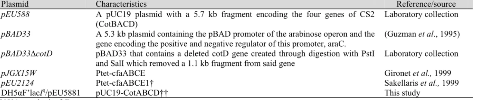
Plasmids used in this study are listed in Table1.
Table 1. Plasmids used in this study.
Plasmid Characteristics Reference/source
pEU588 A pUC19 plasmid with a 5.7 kb fragment encoding the four genes of CS2 (CotBACD)
Laboratory collection
pBAD33 A 5.3 kb plasmid containing the pBAD promoter of the arabinose operon and the gene encoding the positive and negative regulator of this promoter, araC.
(Guzman et al., 1995)
pBAD33∆cotD pBAD33 that contains a deleted cotD gene created through digestion with PstI and SalI which removed a 1.1 kb fragment from said gene
Laboratory collection
pJGX15W Ptet-cfaABCE Gironet et al., 1999
pEU2124 Ptet-cfaABCE1† Sakellaris et al., 1999
DH5αF’lacIq/pEU5881 pUC19-CotABCD†† This study
†R181A mutation in cfaE. †† R181A mutation in CotD.
Antisera
Rabbit anti CFA/I pili antibody was custom made by
immunizing a rabbit (Millipore Corporation, Millipore)with
crude CFA/I pili extracted from MC4100/pJGX15W
(cfaABCE). Nonspecific antibodies were removed by
adsorption with sonicated MC4100/pJGX15W. Antisera
against CS2 pili was available from previous studies in the lab.
Biofilm assay in microtitre plates
Biofilm formation of ETEC strains was monitored
following Liaqat et al. (16). Briefly, Cells were grown for 18 h
in respective media and two hundred fifty micro-litres were
transferred to 25 ml fresh medium, transferred to 96-well
flat-bottommicroplates (Beckton Dickinson, USA), and incubated
at 37°C for 62, 117 and 170 h. Adhered cellswere then stained
with 0.1 % crystal violet for 30 min.Crystal violet wasthen
solubilized by the addition of 33% glacial acetic acid and the
OD563 wasmeasured. Each strain was assayed in eight wells on
972 Hemagglutination and piliation assays
For slide hemagglutination, 18 h old cultures were
resuspended in PBS (0.24% Tris-HCl, 0.88% NaCl) pH 7.4, to
give an OD600 of 10. In glass slides, 20 μl of bacterial suspension
was mixed with 20 μl of TBS containing 0.1 M D-mannose and 20 μl of washed bovine erythrocyte suspension. For detection of pili by slide agglutination, 25 μl of bacterial suspension in TBS (OD600
of 10) was incubated for 1 min. at room temperature with 25 μl of
anti-CS2 or anti CFA/I serum in glass slides (24). The degree of
hemagglutination was observed visually based on the clump size
and time of agglutination.
Extraction of pili, SDS-PAGE gel electrophoresis and Immunoblotting
CS2 pili from 18 h old LB cultures of DH5αF’lacIq/pEU588,
CFA/I from MC4100/pJGX15W (cfaABCE), MC4100/pEU2124
(cfaABCE1) and CFA/II from E24377A were extracted using the
method as described previously ( Sakellaris et al.24). Crude pili
preparations from the extraction steps and normalized whole-cell
lysates or heat extract were separated by SDS-PAGE gels. Samples of 10 μl were loaded on a 15% SDS polyacrylamide gel and electrophoresed at 160 V. Immunoblotting was carried out
using polyclonal anti CS2 or anti CFA/I antibody at a dilution of
1:1,000 and goat anti-rabbit immunoglobulin G whole molecule
(1:1,000) as secondary antibody. Blots were developed with
BCIP/NBT (Roche) substrate according to manufacturer
instructions.
ELISA assay for whole cell and pili binding
Polyvinyl chloride plates (Falcon; Becton Dickinson) were
coated with washed erythrocytes (OD600=1) and incubated for 16 h
at 4°C. For asialo-GM1 (GgO4Cer) binding, solvent was suspended
in methanol (5 μgml-1), added to wells of microtiter plates and
allowed to evaporate for 18 h at room temperature. Wells were
blocked with gelatin blocking buffer (Sigma, USA) for 1 h at RT
and were washed five times with TBS. Blocking was followed by washing and 100 μl of two fold serially diluted bacterial suspensions (OD600=30 in the first well) or crude pili solution (50
μg of pili in the first well) was added. After wells were washed as previously described, goat pAb to E. coli in gelatin blocking buffer
(1:5,000) (or for wells with fimbriae, anti-CS2/CFA/I [2nd bleed/Ist bleed respectively; 1:1,000) was added for 1 h at RT. Following
another washing step, alkaline phosphatase-conjugated rabbit
goat immunoglobulin G whole molecule (Sigma) or goat
anti-rabbit immunoglobulin G whole molecule (Sigma) in gelatin
blocking buffer (1:1,000) was added for 1 h at 37°C. Wells
without erythrocytes, or without asialo-GM1 were used as
controls. The bound enzyme was detected by the addition of
alkaline phosphatase substrate (PNPP, Sigma 71768) diluted in
diethanolamine buffer per well for 5 to 30 min at 37°C. The
control wells were treated in the same manner except that blank
control wells had no bacteria or pili. Absorbance was measured at
OD405 nm with a microplate reader (POLAR star Omega). Each
experiment was repeated twice with six replicates per plate.
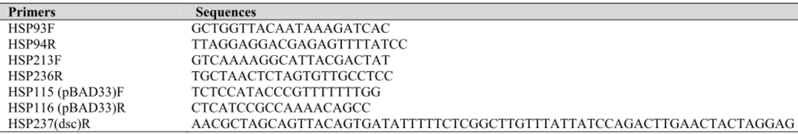
Site directed mutagenesis of CotD
Site-directed mutagenesis of changing Arg-181 of CotD to
Ala was performed with the template plasmid pEU588, and
mutagenic primers (HSP213F, 236R). Plasmid pEU588 was
digested with DpnI and purified using PCR purification kit (Roche
Diagnostic, Germany), self ligated and transformed to DH5αF’lacIq
. The presence of R181A mutation was confirmed by
nucleotide sequencing of the entire CotD gene and of the region
surrounding the directed mutation for R181 allele. Primers used
are listed in Table 2.
Table 2. Primer sequences.
Primers Sequences
HSP93F GCTGGTTACAATAAAGATCAC HSP94R TTAGGAGGACGAGAGTTTTATCC HSP213F GTCAAAAGGCATTACGACTAT HSP236R TGCTAACTCTAGTGTTGCCTCC
HSP115 (pBAD33)F TCTCCATACCCGTTTTTTTGG
HSP116 (pBAD33)R CTCATCCGCCAAAACAGCC
Construction and expression of donor strand compliment CotD
Complementary primers (top strand) AACGCTAGCAGT
TACAGTGATATTTTTCTCGGCTTGTTTATTATCCAGACTT
GAACTACTAGGAG and bottom strand GATCCAACTAT
CGATCTGATGCAACATCATCATCATCATCATTAATGCGG
GAGACTTTATCTGC which contain the coding sequence for a
hairpin linker (DNKQ) followed by the first 19 residues of mature CotA, were used to amplify DH5αF’lacIq
/pBAD33-cotD. The
amplicon was digested with DpnI, ligated and transformed to DH5αF’lacIq
following methods as described previously (24).
Sequencing of entire DH5αF’lacIq/dsc19CfaE(His)6 was done to confirm the correct construction.
The strains expressing His6-tagged CotD was grown in LB
medium with Cm50 at 37°C to late logarithmic phase (OD600=0.8),
followed by induction for 3 h by addition of 0.1-0.5%
L-arabinose. Heat extracts were prepared by boiling at 65°C for 20
minutes. After transfer to nitrocellulose, Immuno blot analysis was
performed by chemiluminescence using monoclonal
antipolyhistdine, antibody produced in mouse (1:2,000 dilution)
and HRP labeled anti-mouse IgG as secondary antibody (1:5,000).
RESULTS
Biofilm formation studies
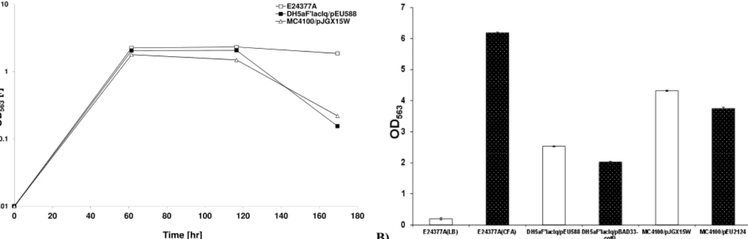
The biofilm-forming capacity of ETEC strains was assessed
inmicrotitre plates by quantitative crystal violet staining. Arange
of ETEC strains including E24377a (CFA/II), DH5aF'lacIq/ pEU588 (CS2), DH5aF'lacIq/pEU588∆CotD, MC4100/pJGX15W (CFA/I) and MC4100/pEU2124 (CfaE-R181A) were includedfor
comparison. It transpired that among tested E. coli isolates,
E24377a produced maximum biofilm after 60 hours, followed by
DH5aF'lacIq/pEU588 and MC4100/pJGX15W. Significant
decrease (P<0.05) in biofilm formation of MC4100/pJGX15W
was observed compared to E24377a and DH5aF'lacIq/pEU588 after 117 hours (Figure 1a). The decreasing rate of biofilm
formation observed in DH5aF'lacIq/pEU588 and
MC4100/pJGX15W might correspond to presence of single type
of fimbriae hence lowering their ability to adhere and form
biofilm.
Having analysed the biofilm-forming capacity of a range of
differentpiliated ETEC isolates, we proceeded with investigating
whether after 60 hours recombinant piliated CFA/II, CS2 and
CFA/I strains could outperform their respective non piliated
CFA/II (E24377a grown in LB), CotD mutant (DH5aF'lacIq/
pBAD33∆cotD) or CfaE-R181 mutant (MC4100/pEU2124)
strains respectively. It was observed that piliated E24337a
produced highly significant (P< 0.001) biofilm compared to non
piliated E24337a strain. Also significant decrease in biofilm
formation was observed in DH5aF'lacIq/pBAD33∆cotD and MC4100/pEU2124 respectively compared to recombinant wild
type strains (Figure 1b).
A) 0.01
0.1 1 10
0 20 40 60 80 100 120 140 160 180
OD
563
[/
]
Time [hr]
E24377A DH5aF'lacIq/pEU588 MC4100/pJGX15W
B)
Figure 1. a) Biofilm quantification of ETEC E. coli strains b). Biofilm quantification of piliated and recombinant piliated ETEC strains monitored after 60 hours. Bacteria were grown in static LB/CFA cultures for 7 days at 37ºC. Significant decrease in biofilm formation was observed in piliated
(E24377a grown in CFA media) versus non piliated (E24377a grown in LB media), CotD and tip mutants compared to CS2 and CFA/I recombinant
974 SDS-PAGE and Immunoblotting of CS2, CFA/I and CFA/II
pili
About 6.8, 3.1, 1.7 and 7.5 mgml-1 of crude pili from MC4100/pEU2124 (cfaABCE1); MC4100/pJGX15W (cfaABCE); DH5αF’lacIq/pEU588 and E24377a were obtained from 1 L of culture fluid. SDS-PAGE of the extracted ETEC pili showed band
that migrated at a position corresponding to a molecular mass of
17 kDa (CFA/II and CS2) and 16kDA (CFA/I). Pili concentration
was measured using Imaje J. Immunoblotting of the crude CFA/I
recombinant and tip mutant pili fractions employing anti CFA/I
serum revealed reacting protein bands at 16 kDa. Interestingly,
CFA/II crude pili were found to be cross reacted with anti-CS2
serum (Data not shown).
Haemagglutination
CS2 pili expression on DH5αF’lacIq/pEU588 ETEC mediate specific adherence to bovine erythrocytes in a mannose-resistant
manner. However, DH5αF’lacIq/pEU5881 clones bearing CotD-R181A mutants exhibited either strong or no haemagglutination
activity, indicating that R 181A is not critical for formation of a
receptor binding epitope on CotD of CS2 strains.
Expression of DH5αF’lacIq/dsc19CotD(His)6
Following Poole et al. (21), we constructed a plasmid that
expresses a CotD variant containing a C-terminal extension
consisting of a hairpin tetrapeptide linker followed by the 19
residue donor strand from the N terminus of mature CotA, and a
terminal hexahistidine affinity tag. Restriction digestion and
sequencing confirmed the correct sequence and validated accuracy
of dsc19CotD(His)6. Immunoblotting of DH5αF’lacIq/dsc19CotD (His) showed an obvious band upon induction with 0.4%
L-arabinose (Data not shown).
Mannose-resistant haemagglutination assay (MRHA) of
DH5αF’lacIq/dsc19CotD(His) showed strong agglutination of bovine erythrocytes as well as anti CFA/I serum without induction
on CFA media however on LB media agglutination was observed
when induced with 0.4% L-arabinose.
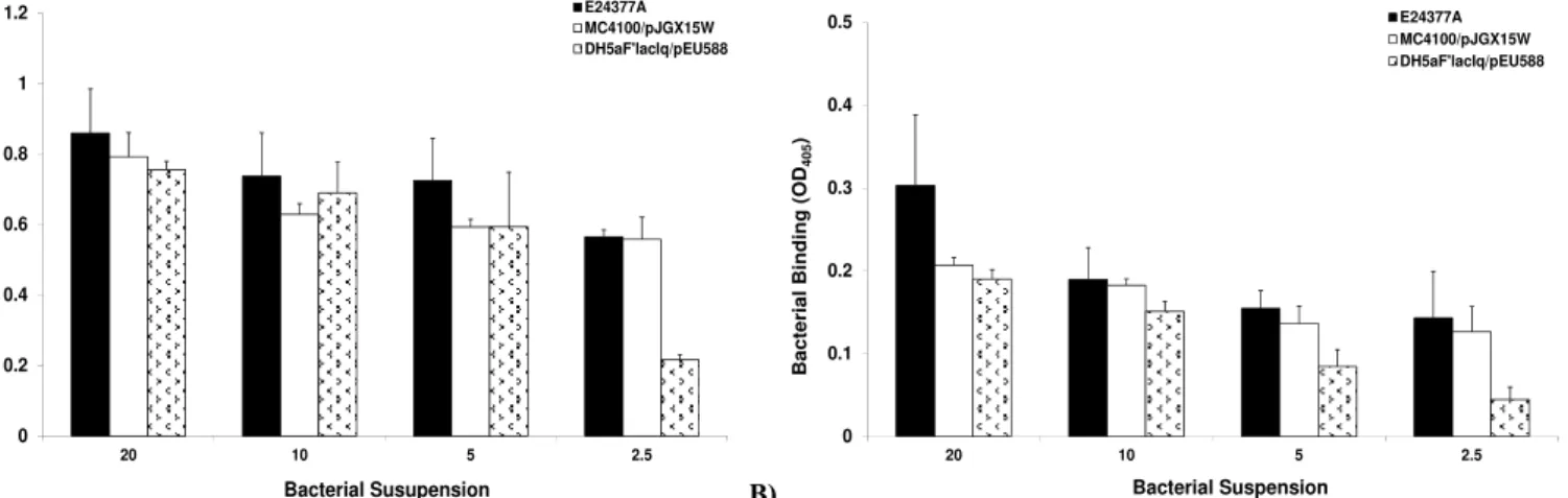
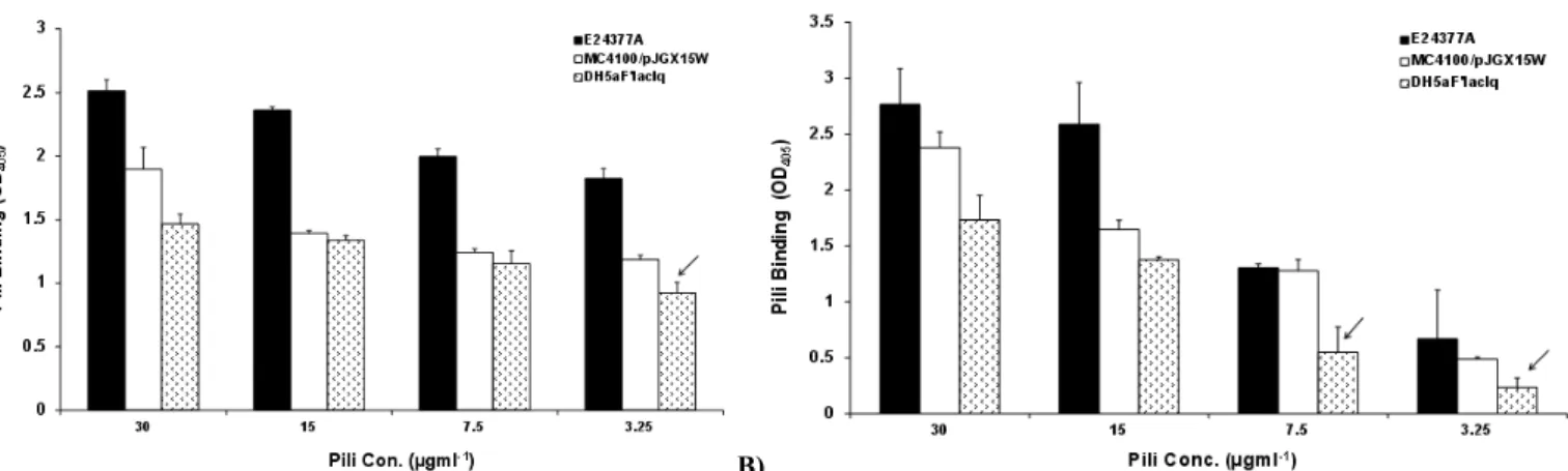
Adherence of CS2, CFA/I and CFA/II pili and piliated bacteria with bovine erythrocytes and asialo-GM1
The ability of purified pili and piliated bacteria to bound with
bovine erythrocytes and asialo-GM1 was studied in microtiter plate
assay. Both pili and bacteria bound to erythrocytes and asialo-GM1
in a dose-dependent manner (Figure 2a, b). We found that
wild-type E24377a bound abundantly to bovine erythrocytes and
asialo-GM1. In contrast, non significant difference in binding capacity of
MC4100/pJGX15W and DH5αF’lacIq/pEU588 was observed at OD600=5. However, highly significant decrease in binding capacity
of DH5αF’lacIq/pEU588 was observed at OD600=2.5.
Binding assay of purified fimbriae and fimbriated bacteria
with asialo-GM1 indicated binding in a concentration dependent
manner with maximum binding observed at 30 μgml-1
in E24377a.
DH5αF’lacIq/pEU588 and crude pili of this strain exhibited the lowest binding capacity (Figure 3a, b). Together, this data strongly
suggest that the carbohydrate residues of glycolipids may be the
binding site for piliated bacteria.
A) 0 0.2 0.4 0.6 0.8 1 1.2
20 10 5 2.5
Bacter ial Bind in g (O D405 ) Bacterial Susupension E24377A MC4100/pJGX15W DH5aF'lacIq/pEU588 B) 0 0.1 0.2 0.3 0.4 0.5
20 10 5 2.5
B a c ter ial B inding (OD 405 ) Bacterial Suspension E24377A MC4100/pJGX15W DH5aF'lacIq/pEU588
Figure 2. Binding of whole cell piliated recombinant ETEC E. coli strains to (a) bovine erythrocytes (b) asialo-GM1, Bacterial strains were incubated with bovine erythrocytes and asialo-GM1 coated microtitre plates. Whole cell binding was assessed using goat pAb to E. coli andalkaline phosphatase-conjugated rabbit anti-goat immunoglobulin G whole molecule as secondary antibody. Wells without erythrocytes, or without asialo-GM1 were used as
A) B)
Adherence of CFA/I and CfaE-R181A mutant piliated
bacteria and pili with bovine erythrocytes and asialo-GM1
The adherence assays were performed employing
MC4100/pJGX15W (CFA/I) and Mc4100/pEU2124 (CfaE–
R181A) strains and pili as mentioned above. According to our
preliminary data, there was significant decrease (P<0.001) in
binding capacity of MC4100/pEU2124 to bovine erythrocytes
compared to MC4100/pJGX15W. However, binding to
asialo-GM1 was significantly low only at maximum OD600=20,
afterwards non significant decrease was observed (Figure 4a,
b).
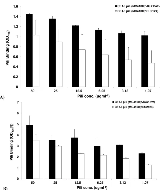
The adherence assays using crude pili of
MC4100/pJGX15W and MC4100/pEU2124 showed the same
trend as observed for piliated and CfaE –R181A mutant
piliated bacteria. Significant (P< 0.05) and highly significant
(P<0.001) decrease in binding of MC4100/pEU2124 pili to
bovine erythrocytes and asailoGM1 was observed respectively
(Figure 5a, b).
A) 0 0.2 0.4 0.6 0.8 1 1.2 1.4
20 10 5 2.5
Bac
ter
ial Binding
(O
D405
)
Bacterial Suspension
MC4100 MC4100/pJGX15W MC4100/pEU2124
B) 0 0.2 0.4 0.6 0.8 1 1.2
20 10 5 2.5
Ba
cte
rial Binding
(OD
40
5
)
Bacterial Suspension
MC4100 MC4100/pJGX15W MC4100/pEU2124 Figure 3. Binding of extracted crude pili of ETEC strains to (a) bovine erythrocytes (b) asialo-GM1. Pili, incubated with bovine erythrocytes
and asialo-GM1 coated microtitre plates. Binding was assessed using anti-CS2/CFA/I [2nd bleed/Ist bleed respectively; 1:1,000) as primary
antibody following another washing step, with goat anti-rabbit immunoglobulin G whole molecule as secondary antibody. Wells without
erythrocytes, or without asialo-GM1 were used as controls. The bound enzyme was detected with alkaline phosphatase conjugate method. The
OD405 as a measure of binding was recorded.
Figure 4. Binding of CFA/I strain (MC4100), CFA/I recombinant (MC4100/pJGX15W) and CfaE-R181 mutant strains (MC4100/pEU2124) to
(a) bovine erythrocytes (b) asialo-GM1, Bacterial strains were incubated with bovine erythrocytes and asialo-GM1 coated microtitre plates Whole
cell binding was assessed using goat pAb to E. coli andalkaline phosphatase-conjugated rabbit anti-goat immunoglobulin G whole molecule as
976
A)
0 0.2 0.4 0.6 0.8 1 1.2 1.4 1.6
50 25 12.5 6.25 3.13 1.07
P
ili
B
ind
in
g
(
O
D40
5
)
Pili conc. (ugml-1)
CFA/I pili (MC4100/pJGX15W) CFA/I pili (MC4100/pEU2124)
B)
0 1 2 3 4 5 6 7
50 25 12.5 6.25 3.13 1.07
Pili
Bin
d
ing (OD
40
5
[/])
Pili conc. (ugml-1)
CFA/I pili (MC4100/pJGX15W)
CFA/I pili (MC4100/pEU2124)
Figure 5. Binding of CFA/I pili from recombinant (MC4100/pJGX15W) and CfaE-R181 mutant (MC4100/pEU2124) strains to
(a) bovine erythrocytes (b) asialo-GM1. Crude pili were incubated with bovine erythrocytes and asialo-GM1 coated microtitre
plates Binding was assessed usinganti CFA/I [Ist bleed respectively; 1:1,000) as primary antibody following another washing
step, with goat anti-rabbit immunoglobulin G whole molecule as secondary antibody. Wells without erythrocytes, or without
asialo-GM1 were used as controls. The bound enzyme was detected with alkaline phosphatase conjugate method (Arrows shows
significant decrease in binding ability of tip mutant compared to recombinant MC4100/pJGX15W strain). The OD405 as a measure
of binding was recorded.
DISCUSSION
This study was undertaken to obtain insights into
adherence mechanisms of ETEC strains. In ETEC, cell surface
structures such as fimbriae/pili have been shown to be
necessary for initial colonization on biotic and abiotic surfaces
resulting in well-established biofilms (6). In our study, all
may be due to greater production of glycocalyx in these strains
in biofilm mode (14). Also significantly higher biofilm
production by E24377a compared to MC4100/pJGX15W and
DH5aF'lacIq/pEU588 may be related to presence of two antigenic types (i.e., CS1 and CS3), hence contributing towards
greater adherence and or/glycocalyx production. Also
significantly high biofilm produced by piliated and
recombinant piliated strains may be due to the fact that piliated
E. coli organisms are more hydrophobic compared to their
non-piliated counterparts (14). A comparison between
MC4100/pJGX15W (CFA/I) and (CfaE-R181 mutant) showed
significantly decreased biofilm formation in
MC4100/pEU2124. Likewise decreased biofilm formation
observed in DH5aF'lacIq/pEU588∆cotD compared
DH5aF'lacIq/pEU588 can be justified with the fact that biofilm formation may be a minor pili/tip-associated event as noticed
for PAK pili of P. aeruginosa (28).
It has previously been reported that R181A mutation in
CFA/I pili manifest MRHA inhibition of bovine erythrocytes
so we tested that whether CS2 bearing the same mutation
affected the agglutination of bovine erythrocytes in MRHA
assay. Using strain DH5αF’lacIq/pEU588, we found that R181 residue is not essential for the agglutination of bovine
erythrocytes since tested clones having R181A mutation
showed either very strong or no agglutination at all. This
indicates R181 which specifies binding to both bovine and
human erythrocytes in CFA/I and CS1 pili (23, 24), is not a
conserved feature in CS2 pili and minor pilins of CS2 does not
inhibit pilus-mediated hemagglutination.
Adapting a strategy used by Poole et al. (21), we produced
dsc19CotD, a stabilized variant of the CS2 pilin minor subunit,
extending the C terminus with the N-terminal β-strand from the
CotA major subunit. This modification facilitated folding of
CotD into an erythrocyte-binding-ready conformation in the
absence of its chaperone, CotB. In the Chaperon-usher (CU)
pathway the chaperone is necessary for transit of proper
subunit and catalysing incorporation into a filament (25),
except where subverted by providing the in cis missing donor
β-strand (10). Immunoblotting showed expression at 0.4% L-arabinose induction. MRHA assay exhibited rapid
agglutination on CFA medium without induction.
Demonstration that donor strand complementation is common
to the CU and AC pathways reopens the evolutionary question
about whether these assembly processes have arisen along
convergent lineages or by divergent routes in which the
ancestral relationship is so remote that primary sequence
similarity has been lost. Purification and expression in a stable,
naturally-folded will further determine relative binding of
purified CotD to bovine and porcine blood mirrors that of
whole CS2 pili. It will suggest us whether that CotA plays no
role in determining erythrocyte binding specificity. The
ongoing work in our lab is continued to prove the validity of
above hypothesis.
The present study suggests that biofilm production and
adherence mechanisms of ETEC isolates are associated with
colonization factors and multiple CF genes located on the
plasmid (CFA/I, CFA/II), as well as on its chromosome (CS2).
The fact that biofilm formation and adherence was observed in
mutant/tip mutant indicates that other adhesins may act
simultaneously or at distinct steps of the adherence process
(18). Also this study provides corroborative evidence that in
contrast to CS1 and CFA/I, R181A mutation in CotD of CS2 is
not necessary for receptor binding moiety.
In conclusion, it is clear that pili are important structures
in adhesion by ETEC strains. Several questions about the
assembly of these unusual covalently linked structures remain
to be addressed. Efforts have been made in this study to study
pili role in vitro using recombinant pilin subunits and strains.
In vitro adhesion studies using bovine erythrocytes and
asailoGM1 have clarified the role of the pili in these processes
and strengthen the rationale for using pilus proteins as vaccine
components. An additional advantage suggested by the
functional reactivity of minor subunit antigen is that fewer
978
expressing a wider array of related pili. Finally, genome
sequencing and comparison will lead to a better understanding
of the evolution of the pilus-encoding pathogenicity islands and
how they have spread through Gram-negative pathogens
considering E. coli as model organism.
ACKNOWLEDGEMENTS
This work was supported by Endeavour Award
(Australian scholarships), 2010.
REFERENCES
1. Anderson, G.G.; Palermo, J.J.; Schilling, J.D. et al. (2003). Intracellular bacterial biofilm-like pods in urinary tract infections. Science., 301:105– 107.
2. Baker, N.; Hansson, G.C.; Leffler, H. et. Al. (1990). Glycosphingolipid receptors for Pseudomonas aeruginosa. Infect. Immun., 58:2361-2366. 3. Buhler, T.; Hoschutzky, H.; Jann, K. (1991). Analysis of colonization
factor antigen I, an adhesin of enterotoxigenic Escherichia coli O78:H11 fimbrial morphology and location of the receptor-binding site. Infect.
Immun., 59:3876–3882.
4. Clavijoa, A.P.; Baia, J.; Gómez-Duarte, O.G. (2010). The Longus type IV pilus of enterotoxigenic Escherichia coli (ETEC) mediates bacterial self-aggregation and protection from antimicrobial agents. Microbial.
Pathogenesis., 48:230-238.
5. de Lorimier, A.J.; Byrd, W.; Hall, E.R. et al. (2003). Murine antibody response to intranasally administered enterotoxigenic Escherichia coli
colonization factor CS6. Vaccine., 21:2548–2555.
6. Domka, J.; Lee, J.; Bansal, T. et al. (2007). Temporal gene-expression in
Escherichia coli K-12 biofilms. Environ. Microbiol., 9:332-346.
7. Evans, D.G.; Evans, D.J.; Clegg, S. et al. (1979). Purification and characterization of the CFA/I antigen of enterotoxigenic Escherichia
coli. Infect. Immun., 25:738–748.
8. Gonzales, E.A.; Blanco, J. (1985). Comparative study of inhibition of mannose-resistant hemagglutination caused by CFA/I, CFA/II, K88, and K99-positive Escherichia coli strains. FEMS. Microbiol. Lett., 29:115– 121.
9. Jones, C.H.; Jacob-Dubuisson, F.; Dodson, K. et al. (1992). Adhesin presentation in bacteria require molecular chaperones and ushers. Infect.
Immun., 60:4445-4451.
10. Jedrzejczak, R.;Dauter, Z.;, Dauter, M. et al. (2006).Structure of DraD invasin from uropathogenic Escherichia coli: a dimer with swapped
beta-tails. Acta. Crystallogr. D. Biol. Crystallogr.,62:157–164.
11. Jonson, A.B.; Normark, S.; Rhen, M. (2005). Fimbriae, pili, flagella and bacterial virulence. Contrib. Microbiol., 12:67–89.
12. Knutton, S.; Lloyd, D.R.; Candy, D.C.;et al. (1985). Adhesion of enterotoxigenic Escherichia coli to human small intestinal enterocytes.
Infect. Immun., 48:824–831.
13. Lee, K.K.; Sheth, H.B.; Wong, W.Y. et al. (1994). The binding of
Pseudomonas aeruginosa pili to glycosphingolipids is a tip-associated
event involving the C-terminal region of the structural pilin subunit. Mol.
Microbiol., 11:705-713.
14. Leung, J.W.; Liu, Y.L.; Desta, T. et al. (1998). Is there a synergistic effect between mixed bacterial infection in biofilm formation on biliary stents? Gastrointest. Endosc., 48:250-257.
15. Levine, M.M.; Ristaino, P.; Marley, G. et al. (1984). Coli surface antigens 1 and 3 of colonization factor antigen II-positive enterotoxigenic Escherichia coli: morphology, purification, and immune responses in humans. Infect. Immun., 44:409–420.
16. Liaqat, I.; Bachmann, R.T.; Sabri, A.N. et al. (2010). Isolate-specific effects of patulin, penicillic Acid and EDTA on biofilm formation and growth of dental unit water line biofilm isolates. Curr. Microbiol., 61:148-156.
17. Macfarlane, S.; Dillon, J.F. (2007). Microbial biofilms in the human gastrointestinal tract. J. Appl. Microbiol., 102:1187–1196.
18. Mazariego-Espinosa, K.; Cruz, A.; Ledesma, M.A. et al. (2010). Longus, a type IV pilus of enterotoxigenic Escherichia coli, is involved in adherence to intestinal epithelial cells. J. Bacteriol., 192:2791-800. 19. Mu, X.Q.; Savarino, S.J.; Bullitt, E. (2008). The three-dimensional
structure of CFA/I adhesion pili: traveler's diarrhea bacteria hang on by a spring. J. Mol. Biol., 376:614–620.
20. Orø, H.S.; Kolstø, A.B.; Wennerås, C. et al. (1990). Identification of asialo-GM1 as a binding structure for Escherichia coli colonization
factors antigens. FEMS. Microbiol. Lett., 72:289–292.
21. Poole, ST, McVeigh AL, Anantha RP et al (2007). Donor strand complementation governs intersubunit interaction of fimbriae of the alternate chaperone pathway. Mol Microbiol 63:1372-1384.
22. Proft, T.; Baker, E.N. (2009). Pili in Gram-negative and Gram-positive bacteria - structure, assembly and their role in disease. Cell. Mol. Life. Sci., 66:613-635.
23. Sakellaris, H.; Balding, D.P.; Scott, J.R. (1996). Assembly proteins of CS1 pili of enterotoxigenic Escherichia coli. Mol. Microbiol., 21:529-541.
24. Sakellaris, H.; Penumalli, V.R.; Scott, J.R. (1999). The level of expression of the minor pilin subunit, CooD, determines the number of CS1 pili assembled on the cell surface of Escherichia coli. J. Bacteriol., 181:1694-1697.
priming of pilus subunits facilitates a topological transition that drives fiber formation. Cell.,111:543–551.
26. Sharon, N.; Lis, H. (1993). Carbohydrates in cell recognition. Sci. Amer., 268:82-89.
27. Thomas, L.V.; McConnell, M.M.; Rowe, B. et al. (1985). The possession of three novel coli surface antigens by enterotoxigenic Escherichia coli
strains positive for the putative colonization factor PCF8775. J. Gen.
Microbiol., 131:2319–2326.
28. van Schaik, E.J.; Giltner, C.L.; Audette, G.F. et al. (2005). DNA binding: a novel function of Pseudomonas aeruginosa type IV pili. J.
Bacteriol., 187:1455-1464.
29. Wennerås, C.; Holmgren, J.; Svennerholm, A.M.; (1990). The binding of colonization factor antigens of enterotoxigenic Escherichia coli to intestinal cell membrane proteins. FEMS. Microbiol. Lett., 54:107-112.
São Paulo, 10 de janeiro de 2013.
ERRATA
Referente ao artigo “
Biofilm formation and binding specificities of CFA/I, CFA/II and CS2
adhesions of Enterotoxigenic
Escherichia coli
and
CfaE
-R181A mutant”
publicado no
Brazilian Journal
of Microbiology
volume 43(3)12 – edição de Julho-Setembro.
Página 969
Linha 7:
retirar o nome do autor Harry Sakellaris
2Onde se lê:
Iram Liaqat
1,2*,
Harry Sakellaris
2Leia se:
Iram Liaqat
1,2*Linha 9 e 10:
retirar a instituição
2The University of Western Australia, School of Biomedical,
Biomolecular & Chemical Sciences Microbiology M502, 35 Stirling Hwy Crawley WA 6009.
Onde se lê:
1Department of Zoology, Government College University, Lahore, Pakistan;
2The University of
Western Australia, School of Biomedical, Biomolecular & Chemical Sciences Microbiology M502, 35
Stirling Hwy Crawley WA 6009.
Leia se:
1Department of Zoology, Government College University, Lahore, Pakistan.
Atenciosamente,
p/
Prof. Adalberto Pessoa Junior
Editor of Brazilian Journal of Microbiology
Av. Prof. Lineu Prestes, 2415 – ICB III – Sala SBM