Ana I. P. G. Pereira
Dissertation presented to obtain the Ph.D degree in Neuroscience
Instituto de Tecnologia Química e Biológica António Xavier | Universidade Nova de Lisboa
Oeiras,
Ana I. P. G. Pereira
Dissertation presented to obtain the Ph.D degree in Neuroscience
Instituto de Tecnologia Química e Biológica António Xavier | Universidade Nova de Lisboa
Oeiras, September, 2015
Auditory Cues in Social
Transmission of Fear
Research work coordinated by: Marta Moita, PhD
!
!
!
!
!
!
!
!
!
!
!
!
!
!
! !
!
!
!
!
!
!
!
!
! !
!
!
TABLE OF CONTENTS
Acknowledgements iii Abbreviation list vii
Resumo 1
Abstract 5 Chapter I – General Introduction 9
Part I – Neuronal Pathways Underlying Innate and Learned
Defense Behaviors I.I Defense Responses 15 I.II Neuronal Circuits of Innate and Learned Defense Behaviors 17 a) Innate Defense Responses 19 b) Learned Defense Responses 26 References 32 Part II – Social Transmission of Fear 43 II.I Sensory Cues underlying Social Transmission of Fear 46 II.II Sensory Stimuli in Intraspecies Communication in Rodents 50 II.III Social Transmission of fear in the lab 54 References 59 Chapter II - Fearful Silence: Rats use the Cessation of
Results 69 Discussion 83 Materials and Methods 85 References 90 Chapter III – Neuronal Pathways underlying Defense
Responses triggered by the Cessation of
Acknowledgments
First I would like to thank my supervisor, Marta Moita PhD, for accepting me in her lab and for all the support along these years. For all the discussions, ideas and advices. For guiding experiments, promote critical thinking and give space for new ideas. For above all trying to make each member of her team a better scientist, which is more than accomplishing a PhD. And for teaching me the most valuable lesson in science: always keep close to the data. For being a source of inspiration as a scientist and as a human being.
I would also like to thank my co supervisor, Susana Lima PhD, for all the critical input along these years and support in the experimental design and data analysis. Also for her precious guiding and teachings in molecular biology. And for her boldness and strength as a scientist, which are truly inspiring.
A great acknowledge to my thesis committee, Luisa Vasconcelos PhD and Rui Costa PhD, for the valuable discussions and guidance along the project. Their input was very important in guarantying the completion of the projects I proposed to develop and above all, it made me look at my project and results in a different and challenging perspective.
who was always helpful and that contributed with valuable input to the writing of the present thesis.
I must also thank the entire Champalimaud Neuroscience Programme (CNP) for being such an amazing community always ready to face new challenges. The years spent with this group made me remember the importance of questioning and wondering, and that there is no such thing as a “crazy idea” as long as it is carefully explored. In its’ heart it keeps the most fundamental thing for a scientist, the sense of wonder. In this group I have to particularly acknowledge the INDP 2008, with whom I shared my first year of classes and that nowadays are some of my closest friends at the CNP.
I must also express my great gratitude to Instituto Gulbenkian Ciência and to the amazing team of scientists working there. It was a privilege to spend my first years of PhD in this institute.
To Alexandre Estrela, my most sincere acknowledge. His input into this work was crucial since one of the most significant discoveries achieved during this PhD resulted from an interaction between his and our work.
I cannot forget my friends and closest family for always supporting me. They were a great help in rationalizing the bad moments and celebrating the good ones.
ABBREVIATION LIST
AFC auditory fear conditioning AOS acessory olfactory system BL basolateral amygdala BM basomedial amygdala CD conditioned demonstrator CeA central amygdala
CeL lateral portion of the central amygdala CeM medial portion of the central amygdala ChR2 Channelrhodopsin2
CoA cortical amygdala CR conditioned response CS conditioned stimulus EO experienced observer ICC inferior culliculus IEG immediate early gene LA lateral amygdala MeA medial amygdala MGB medial geniculate body
MGm medial division of the medial geniculate body MOS main olfactory system
ND naïve demonstrator NO naïve observer PAG periaqueductal gray
SCR skin conductance response SI social interaction
SSDR species-specific defense responses STF social transmission of fear
RESUMO
Quando um individuo detecta uma ameaça no seu ambiente, são desencadeadas alterações tanto a nível fisiológico como comportamental. Estas alterações podem ser detectadas por outros indivíduos que se encontram nas proximidades, alterando o comportamentos deste últimos. Esta transferência de informação relativa a um perigo iminente é denominada Transmissão Social de Medo. A utilização de informação social que sinaliza a presença de uma ameaça pode ter benefícios a curto prazo (como seja evitar um perigo eminente), ou estar na base da aprendizagem social sobre novas ameaças. Apesar da prevalência deste fenómeno no reino animal, bem como a sua importância para a sobrevivência, pouco se sabe sobre os mecanismos neuronais subjacentes.
sensoriais que suportam a transmissão de medo, demonstrámos que os observadores não necessitam de contacto, informação visual ou auditiva conferida por gritos de alarme para expressarem uma resposta de medo desencadeada pela imobilidade dos seus conspecificos. Utilizam no entanto informação auditiva que sinaliza a transição súbita de movimento para imobilidade. Durante a fase inicial da interação social, os ratos movem-se na caixa produzindo sons característicos do seu movimento. Estes sons diminuem drasticamente quando o demonstrador fica imóvel. Foi assim hipotetizado que esta transição do som do movimento para silêncio é necessária para a imobilidade dos observadores. De forma a testar esta hipótese, reproduzimos o som de um rato a mover-se durante a fase da interação social em que ambos os ratos estão imóveis, abolindo o silêncio. Verificámos que a reprodução deste som aboliu a imobilidade dos observadores, e ainda que a sua terminação desencadeou de novo imobilidade nestes últimos.
De forma a testar se a cessação do som do movimento é suficiente para desencadear imobilidade, realizámos um outro conjunto de experiências em que colocámos ratos com experiência previa com choques na mesma caixa de interação social, mas desta vez sozinhos. Durante a sessão de teste o mesmo som proveniente do movimento de um rato foi reproduzido dentro da caixa de forma continua, com exceção de dois períodos de um minuto de silêncio. Os sujeitos estiveram mais tempo imóveis durante esses períodos de silêncio em comparação com os períodos de mesmo duração anteriores à cessação do som. Estas experiências confirmam que a cessação do som do movimento é necessária e suficiente na transmissão social de medo.
Condicionamento Auditivo de Medo. Estes estudos demonstraram que a Amígdala lateral é necessária para a aprendizagem, armazenamento e expressão de medo em resposta a estímulos sonoros. Porem, na maior parte destes estudos a resposta de medo é desencadeada pela apresentação de um som, enquanto que no nosso caso é decorrente da sua cessação. De forma a investigar quais os mecanismos neuronais subjacentes à expressão de medo desencadeado pela súbita cessação do som do movimento, começámos por averiguar se a atividade na amígdala lateral também é importante para a expressão de medo desencadeada pelo nosso estimulo.
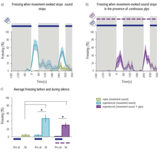
Para tal, recorremos a uma manipulação optogenética para inibir a Amígdala lateral especificamente durante o intervalo de silêncio introduzido no som do movimento. Nesta experiência utilizámos 2 grupos, ArchT (que expressa a bomba de protões ArchT na Amígdala lateral permitindo a sua inibição) e Controlo. Comparando a percentagem de tempo que os ratos estiveram imóveis durante o período de silêncio e o período antecedente a este no grupo ArchT, não foram encontradas diferenças estatisticamente significativas. Pelo contrário, os animais controlos estiveram imóveis significativamente mais tempo durante o silêncio comparado com o período que o precede. Verificou-se ainda uma diferença significativa na imobilidade entre os animais do grupo ArchT e Controlo durante o silêncio, mostrando que atividade neuronal na Amígdala lateral é importante para a expressão de respostas de medo desencadeadas pela cessação do som do movimento.
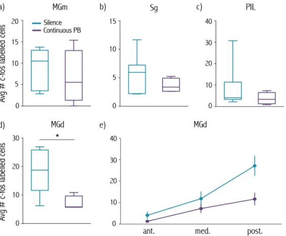
de células que respondem à terminação de diferentes sons em vários subnúcleos desta região. Estudos anatómicos revelaram ainda que existem projeções diretas destes subnúcleos para a Amígdala lateral.
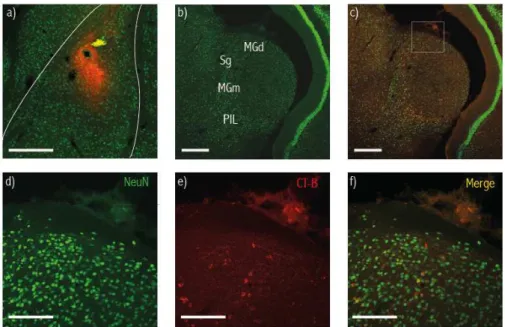
A análise da expressão de c-fos revelou um aumento significativo no numero de células marcadas pela proteína c-fos no núcleo dorsal do Corpo Geniculado Medial em animais expostos ao som do movimento com intervalos de silêncio, quando comparado com animais sujeitos a este som de forma contínua. Este aumento foi particularmente marcado na zona mais posterior deste subnúcleo. Uma vez que esta região tem projeções diretas para a Amígdala lateral (confirmadas no presente estudo), sugerimos que a ativação de células neste subnúcleo, desencadeada pela cessação do som do movimento, leva à ativação de células pos-sinapticas na Amígdala lateral desencadeando imobilidade.
ABSTRACT
When an animal faces a threat, both behavioral and physiological changes occur that promote the avoidance of the menace. Individuals in the surroundings of the fearful animal (both con and heterospecifics) may detect some of these changes, that become cues that signal an impending danger. The detection of such cues can therefore trigger defense behaviors in observers, in a phenomenon called Social Transmission of Fear.
The use of social information to signal danger can have both immediate benefits like the avoidance of the menace, or underlie social learning about threats. Despite its prevalence and importance for survival, very little is known about the neuronal mechanism underlying it.
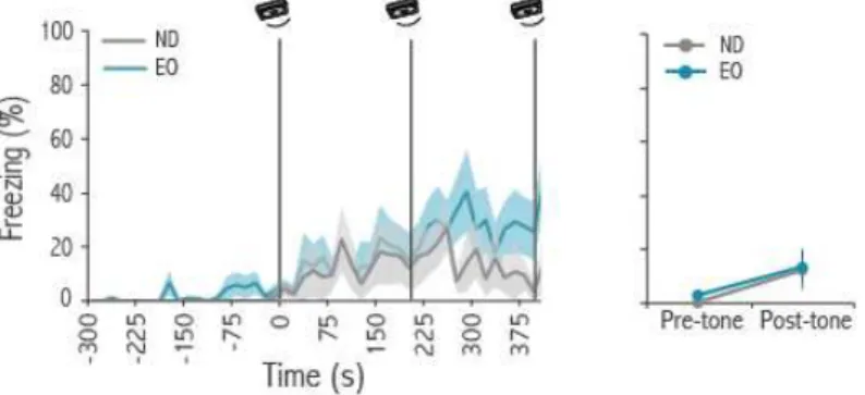
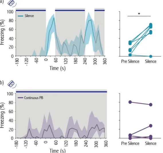
likely to signal the sudden transition from motion to immobility. During the baseline period rats move around in the social interaction chamber producing rustling sounds, which decreased dramatically when the demonstrator rat started freezing. We then hypothesized that the transition from the sound of movement to silence is necessary to trigger freezing in observers. In order to test this hypothesis we played back the sound of a rat moving while both rats were immobile, disrupting silence. We found that the playback of the movement-evoked sound disrupted freezing by observers and that freezing resumed immediately after the sound playback re-instated by silence.
In another set of experiments we placed experienced rats alone in the social interaction chamber. During the test session the same movement-evoked sound used in the previous experiment was played continuously, except for two one-minute periods of silence. Experienced rats significantly increased their levels of freezing during the periods of silence compared with baseline. These experiments confirm that the absence of movement-evoked sound is necessary and sufficient to induce fear in observer rats.
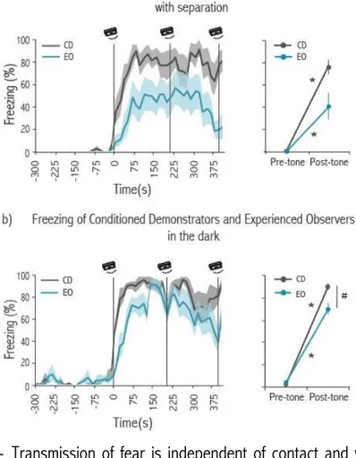
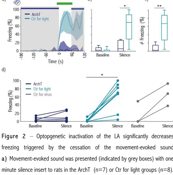
For this purpose, we optogenetically inhibited the Lateral Amygdala specifically during a silence gap introduced during the playback of the movement-evoked sound. For this experiment we had two groups: ArchT (expressing the ArchT proton pump in Lateral Amygdala which when stimulated by light allows neuronal inhibition) and Control (with fiber implants but no ArchT expression in Lateral Amygdala). We didn’t found any significant increase in freezing between baseline and silence in the ArchT group. On the contrary, such an increase was found in the Control group. Moreover, there was a significant difference in percentage of freezing between the ArchT and the Control group during the period of silence, showing that activity in Lateral Amygdala is important for the display of defense responses triggered by the cessation of movement-evoked sounds.
triggering freezing.
CHAPTER I – GENERAL INTRODUCTION
“The most common mutual service in the higher animals is towarn one another of danger by means of the united senses of all.Every sportsman knows, as Dr. Jaeger remarks (7. “Die Darwin’sche Theorie”, s.101.), how difficult it is to approach animals in a herd or troop. Wild horses and cattle do not, I believe, make any danger-signal; but the attitude of any of them who first discover an enemy warns the others”
Chapter I - General Introduction
Although fear is an emotion that most likely has been felt by the majority of humans, its definition is far from being unanimously agreed upon. Fear can be defined as the conscious feeling of being afraid of an impending danger. However, behavioral neuroscientists have classically defined and studied “fear” and “fear responses” as the set of physiological and behavioral changes that occur in response to stimuli that signal a potential threat. The main debate arises from how related the mechanisms underlying the last are with the ones that generate the conscious feeling of being afraid (LeDoux JE 20141, Gross C
20122, Adolphs R 20133). This debate is of particular importance when
studying the feeling of fear in humans, and whether it can be extended to other animals. However, the study of the neuronal mechanisms underlying defense responses, the classically called fear system, is of major importance not only to understand the emotion of fear, but also the survival and adaptation of different species to their environment.
In the context of this thesis, the fear system will be approached based on the circuits which underlie the display of defense responses, including its behavior output as well as the physiologic and autonomic changes concomitant to it.
Defense responses can be triggered when an individual directly recognizes a threat. Fear can be expressed right from the first encounter with a threat or with a cue that signals it, being presumably innate (Veen T 20004, Du Y 20125,
Goth A 20016, Gross C 20122). Fear responses can also be learned; in this
such threat will lead to the display of fear. Several years of research in the fear system has led to extensive literature regarding both innate and learned fear behaviors triggered by the direct detection of a threat (for reviews see Gross C 20122, Adolphs R 20133, Herry C 20147).
Importantly, information conveyed by others can also be used to detect threats in the environment. Social transmission of fear takes place when cues provided by an individual (demonstrator) trigger the display of defense responses in other individuals, either con- or heterospecifics (observers). These cues are the result of changes in physiological and behavioral responses of demonstrator individuals upon the direct detection of a threat. The responses of observers to such social cues can be either innate or learned (Hollen LI 20098, Enjin A 20139). Although currently there are several known
examples of transmission of fear in wild populations (Seyfarth RM 198010,
Zuberbuhler K 200111, Ito R 200912, Wilson DR 200413, Ono M 200314, Hingee
M 200915, Coleman SW 200816, Curio E 197817), the mechanisms underlying it
are still largely unknown. Only more recently have studies started being performed under laboratory settings (Masuda A 200918, Kim EJ 201019, Jeon D
201020, Sanders J 201321, Chen Q 200922, Atsak P 201123, Bruchey AK
201024, Olsson A 200725, Jones CE 201426, Church RM 195927).
The work developed during the following thesis focused on social transmission of fear using rats as an experimental model, and attempted to unravel the neuronal mechanisms underlying it. Our main goals were:
1) To establish a behavioral paradigm for the study of social transmission of fear under laboratory settings;
3) To investigate the brain regions involved in the response to sensory cues provided by others upon the perception of a threat;
As previously mentioned, very little is known about the neuronal mechanisms underlying social transmission of fear. The first part of the following introduction will thus focus on the characterization of defense behaviors and on the description of the neuronal pathways underlying both innate and learned fear responses when they are triggered by the direct recognition of the threat. A review of this literature may provide important insight into the potential brain regions and neuronal mechanisms underlying social transmission of fear.
Part I - Neuronal Pathways Underlying Innate and Learned Defense Behaviors
I.I Defense responses
The fear system can be considered a survival circuit that responds to information about a potential threat with endocrine, autonomic and behavioral changes. The adopted behavior should be the one that enhances the prospect of avoiding or escaping the threat, minimizing injury, and increasing the likelihood of survival. The activation of this survival circuit may also lead to the formation of memories of this encounter, which may be useful in the future (Ledoux JE 20141).
Defense behaviors are species specific, and presumably innate.
Bolles’ (Bolles RC 197028) outlined in his species-specific defense
reactions (SSDR) theory that when an animal faces a threat, its behavioral repertoire becomes restricted to a set of prepackaged behaviors. Freezing, fight and flight are examples of these behaviors that are common to most animal species. According to this theory, SSDR can be rapidly acquired as avoidance responses in tasks where an animal has to perform an action in order to avoid an aversive stimulus. On the other hand, other behaviors of the animal’s repertoire (e.g. grooming) may need extensive training or may never be learned as avoidance responses. Namely, a subject learns very fast to run away from an alley to avoid a shock because fleeing is part of his SSDR. However, extensive training is needed if the animal must bar press to avoid a shock, since this action is not in his defensive prepackaged repertoire.
the biologically important features of the threat source and the characteristics of the environment determine the expression of a given defense behavior (Blanchard RJ 196929). In particular, if an escape path is available, flight is
usually the best response, however if no such escape exists, then freezing (characterized by complete immobility) is more advantageous (Blanchard RJ 197130).
The above referenced studies also concluded that the environment might become associated with the threat itself (Blanchard RJ 196929). Thus, although
there are predefined defense behaviors, their expression is flexible depending on the characteristics of the surroundings. Moreover, it is influenced by the expectation of the threat that is provided by cues in the environment (Bolles RC 197631). Subsequent studies by Fanselow and Lester lead to the
development of the predatory eminence theory, which proposed that the prey’s perceived likelihood of being caught by the predator is what determines the displayed defense behavior. This likelihood is influenced by the distance to, or thetemporalprobability of encountering the threat (e.g. if an animal is hidden, it would take longer for a predator to detect it than if it is in a open arena), and not only by the characteristics of the environment (Fanselow MS 199432).
Freezing, fight and flight are examples of such defense responses and have been reported as part of the coping strategies of almost all vertebrates (Mirza RS 200333, Oliveira R 201134, Gabrielsen GW 198535, Ellison K 201236,
Blanchard RJ 197137, Mateo JM 199638, Roelofs K 201239). These different
coping strategies are generally categorized as active or passive. This categorization is based on changes in both the motor output and in the patterns of autonomic activity (Bandler R 200040, Bittencourt AS 200441).
Following the classical categorization, fight and flight are considered active coping strategies characterized by increased motor activity, hypertension, tachycardia and non-opioid mediated analgesia. On the other hand, passive coping strategies are characterized by reduced somatomotor activity, sometimes hypotension and bradycardia, and opioid-mediated analgesia. Freezing, characterized by immobility, body tenseness, shallow breathing, exophtalamus and absence of sniffing, has been proposed as a passive coping strategy. However, it has also been proposed that freezing is a defense response during which the animal is highly attentive to the environment, being its classification as a passive response arguable.
Innate defense responses are hence believed to result from the activation of developmentally programmed neuronal circuits, and the stimulus that triggers them is intrinsically threatening.
On the other hand, learned defense responses are triggered by a previously neutral cue that was associated with an innate aversive stimulus.
The Amygdala, located in the most ventral part of the mammalian brain, has been widely implicated in both learned and innate fear responses (reviewed in Gross C 20122, Pape H 201042, Maren S 200443, Herry C 20147).
Based on anatomy and cellular properties, it is subdivided into several subnuclei. The basolateral complex encompasses the lateral (LA), basolateral (BL) and basomedial (BM) subnuclei that receive most of the sensory inputs to amygdala. This complex is a cortex like structure and the most common cell types are multipolar, pyramidal-shaped or stellate projection neurons. These projection neurons mainly use the neurotransmitter glutamate, and they contribute to most of the projections to other amygdala nuclei and the rest of the brain.
One important projection site of this complex is the central nucleus of the amygdala (CeA) that in turn projects to brainstem nuclei responsible for the generation of different aspects of defense responses. It is believed that most neurons in the CeA are GABAergic, being that most of the projections arising from this nucleus are inhibitory (Pape H 201042).
The medial (MeA) and cortical (CoA) amygdala are also important for the display of defense behaviors. They receive strong inputs from olfactory sensory areas, namely the main and accessory olfactory bulbs, processing information about predator odors and pheromones (Takahashi L 201444, Meredith M
elevated number of glutamic acid decarboxylase (GAD) positive neurons (both interneurons and projection neurons (Keshavarzi S 201447)), whereas most
cells in CoA are glutamatergic (Sah P 200348).
Other brain regions like the Hypothalamus and the Periaqueductal Gray (PAG) are also part of the so called “fear circuit”, being densely interconnected with the amygdala. The later is classically viewed as an output station downstream of amygdala, important for the coordination of the behavioral manifestations of the defense responses (freezing, fight or flight). In the further sections, we will review the contributions of these different structures and their subnuclei in the display of both innate and learned defense responses. The majority of these studies were performed in rodents.
a) Innate Defense Responses
Innate defense behaviors are mostly displayed in response to painful stimuli, predators, aggressive conspecifics or cues of different sensory modalities that signal the previous. Namely, a looming visual stimulus, which resembles an avian predator, triggers both freezing and escape responses in mice (Yilmaz M 201349, Shang C 201550, Wei P 201551). It has also been
shown that auditory stimuli like a broad band white noise (Xiong XR 201552) or
a train of 17-20KHz frequency sweeps (Mongeau R 200353) delivered at high
intensities, as well as a noxious somatosensory stimulus like a footshock, can equally trigger innate defense behaviors (Gross C 20122).
CeA, shown to be necessary for the expression of learned freezing, do not affect innate fear responses to cat exposure (Martinez RC 201154). This
suggests the existence of a pathway dedicated to the display of coping strategies towards predators. The posterior ventral MeA (pvMEA), a region that receives strong inputs from both the Main Olfactory System (MOS) and Accessory Olfactory System (AOS) seems to be particularly important in the response to cat odor. The LA and pBM, which are strongly interconnected and receive inputs from both auditory and visual sensory processing areas, are proposed to integrate non-olfactory predator-derived cues (Gross C 20122).
The pvMEA and the pBM project to the dorsalmedial part of the ventralmedial Hypothalamus (dmVMH), proposed to be part of a predator-responsive circuit in the hypothalamus. Pharmacogenetic inhibition of the dmVMH significantly decreases defense responses towards a predator in comparison with controls, but has no significant effect when the threat is an aggressive conspecific or a noxious footshock (Silva BA 201355). This network targets the dorsolateral
part of the PAG (dlPAG), and pharmacological inactivation of the dPAG (including the medial and lateral part) is sufficient to significantly reduce defense responses in mice to the presentation of a predator rat (Gross C 20122, Silva BA 201355). These results suggest that the dlPAG is an important
site for the orchestration of defense responses towards predators. One interesting aspect of the dlPAG is that it does not receive direct inputs from the spinal chord that receives cutaneous, deep somatic and visceral primary afferents that provide information about noxious stimulation. This suggests that this subnucleus might respond to stressors other than physical (Bandler R 200040), which may be important to orchestrate coping behaviors towards
defense responses towards conspecifics. Pheromonal and olfactory information from conspecifics activate the posterior dorsal MeA (pdMeA) that projects to the ventrolateral VMH (vlVMH) (Gross C 20122). In mice, optogenetic activation
of the vlVMH triggers aggressive behaviors towards both male and female intruders, as well as towards inanimate objects (Lin D 201156). The vlVMH and
the dorsal medial premammilary nucleus (dmPMD) are part of the conspecific-responsive circuit in the hypothalamus. The introduction of an intruder mouse in the homecage of another mouse, leads to the display of several defense reactions that can be either passive (freezing, on-the back position) or active (upright standing, boxing…). It has been found that this interaction increases c-fos expression in the dmPMD of the intruder but not of the resident. Also, intruders with lesions in this subnucleus showed a major deficit in passive defense behaviors, while keeping certain key active responses (Motta SC 200957). The dPAG also seems to be important for the display of coping
strategies towards conspecifics, given that pharmacological inhibition of this area in mice reduced defense responses when facing an aggressive mouse (Silva BA 201355). The segregation between predator and conspecific defense
neuronal circuits may also be kept in the PAG, since the dmPMD projects mainly to the dorsomedial PAG (dmPAG) (Gross C 20122).
the axonal terminals of the corticofugal neurons of the Auditory Cortex, targeting the cortical ICC, is sufficient to induce flight. The escape response is mediated by the inputs from the cortical ICC to the dPAG (Xiong XR 201552).
It has also been shown that a looming stimulus, an expanding dark disc, which simulates an approaching threat from above the animal, can induce visually innate fear responses such as escape and freezing (Yilmaz M 201349).
Recent work focusing on the display of freezing triggered by this stimulus in mice, revealed that a subcortical pathway from the medial inferior layer of the Superior Culliculus (SC) to the lateral posterior nucleus of the Thalamus and forward to the LA, mediates visually evoked innate freezing (Wei P 201551).
Interestingly, another study reported that optogenetic activation of parvalbumin positive neurons in the SC (SC PV+) triggers impulsive escape
followed by long lasting freezing in mice. The authors report that light induced activation of SC PV+ axonal terminals in the parabigeminal nucleus is sufficient
to trigger the defense behaviors. Given the projections from the parabigeminal nucleus to the amygdala, in particular to the central nucleus, the authors propose that SC PV+ neurons form a subcortical visual pathway that transmits
threat relevant information to the Amygdala (Shang C 201550). Although these
two works report different pathways underlying visually evoked innate defense responses, they provide evidence that the Amygdala is necessary for the display of innate defense behaviors triggered by visual stimuli.
Blair H 200559). These initial motor responses are in general followed by
stretch positions and immobility in enclosed spaces, defense behaviors that are conditioned to the environment. Both the spinothalamic and the spino-parabrachial tracts transmit nociceptive information from the periphery to the forebrain including the Amygdala. This information can be sent both directly through the spinothalamic and the spino-parabrachial tract (Kruger L 199860,
Han S 201561, Cliffer KD 199162) and indirectly through the paraventricular
nucleus of the Thalamus (Penzo MA 201563) to the lateral portion of the CeA
(CeL). The LA also receives nociceptive information indirectly through the somatosensory thalamus and cortex (Lanuza E 200464, Bubser M 199965).
Bilateral electrolytic lesions of the posterior intralaminar thalamic complex destroy fibers from both these tracts, leading to the disruption of freezing after footshock delivery (Lanuza E 200464). Moreover, pretraining lesions of both
the posterior intralaminar thalamic nucleus (PIN) and the insular cortex significantly attenuated the magnitude of shock-induced activity in lesioned rats compared to controls (Shi C 199966).
Nociceptive information about footshocks can also be conveyed to different subnuclei of the amygdala through the paraventricular nucleus of the Thalamus. Footshock stimulation leads to a significant increase in c-fos expressing neurons in this region (Penzo MA 201563). In addition, a study
combining immunohistochemistry with retrograde tracing revealed that c-fos expressing cells in this thalamic nucleus, in response to footshocks, project to the CeA and BL subnuclei, as well as to the Prefrontal Cortex and Nucleus Accumbens (Bubser M 199965).
The LA is also proposed to be an important site for processing aversive nociceptive information. Footshock stimulation in an enclosed box leads to an increase of c-fos expression in all subnuclei of the LA, when compared to animals just exposed to the box. Unilateral electrolytic lesions of the PIN and the medial division of the MGB (MGm) significantly decreased the number of c-fos labeled cells in LA after footshock when compared with intact animals. This result suggests that the LA is involved in the processing of footshocks and that the somatosensory information is provided by the MGm and PIN (Lanuza E 200867). It has also been shown that electrolytic lesions and muscimol
inactivation of the LA reduced the unconditioned response of head movement to the presentation of an eyelid shock (Blair H 200559). In a different study
using a similar paradigm, it was also shown that cells in the LA respond to eyelid shock delivery. The authors also report that muscimol inactivation of the PAG greatly reduced these responses, and it was found that cells in different columns of the PAG also respond to this aversive nociceptive stimulus. These results suggest that the PAG may participate in relaying information about the aversive stimuli to the LA (Johansen J 201068).
the control of a c-fos promoter showed that footshock stimulation triggers neuronal activity in the BL resulting in the expression of the fused protein in 3% of the neurons. ChR2 is a light gated channel, whose activation by light leads to neuronal depolarization (Nagel G 200369). Optical excitation of the
footshock responsive neurons expressing ChR2/YFP decreased both heart and respiration rate, and increased the levels of freezing compared with controls where ChR2/YFP was expressed in a random population of neurons (Gore F 201570).
Together, these results suggest that information about nociceptive aversive stimulus like a footshock is transmitted to both BL and CeA.
b) Learned Defense Responses
As previously mentioned, coping strategies can be evoked by a stimulus that is not intrinsically aversive, as long as this stimulus was previously paired with an innate threat. One paradigm that has been extensively used in laboratory studies to assess the mechanisms of learned fear responses is Auditory Fear Conditioning (AFC). In this paradigm, an initial neutral stimulus, like a pure tone, is paired with an innately aversive stimulus like a footshock (termed unconditioned stimulus US). An association is made between the two, and the animal learns that the presentation of the sound (now the Conditioned Stimulus CS) predicts the US. The presentation of the CS alone is now sufficient to trigger defense behaviors.
Behavioral, anatomical and physiological studies based on this paradigm revealed cellular mechanisms as well the neuronal circuits underlying aversive learning towards an auditory cue. There is extensive literature showing that the Amygdala is a key structure for fear learning and memory (reviewed in Maren S 200443, Pape H 201042, Herry C 20147). In what concerns the
neuronal pathways providing auditory information to the Amygdala, it has been shown that several subnuclei of the auditory thalamus project directly to the LA (Neot DD 199971), with the exception of the ventral MGB (MGv) (the primary
input to Auditory Cortex area 1). These nuclei also project to the Auditory Cortex (Smith PH 201272, Kimura A 200373), and cortical projections to LA
originate in the secondary auditory and perirhinal cortex (Romanski LM 199374, McDonald AJ 199875). Hence, information about an auditory stimulus
201080). Importantly, is has been reported that after AFC, CS-evoked
responses are enhanced in cells both in the MGm of the auditory thalamus and in auditory cortical areas, showing that plasticity occurs in the pathways that provide CS information to the LA (for review see Maren S 200443, Herry C
20147, Ehrlich I 200981).
Several studies have demonstrated that the LA is necessary for learning, storage and expression of defense behaviors triggered by sounds (Hitchcock J 198682, Hitchcock J 198783, LeDoux JE 199084, Romanski LM 199374, Quirk GJ
199585, Schafe GE 200586, Rumpel S 200587, Han JH 200988, Johansen J.
201068, Gouty-Colomer LA 201589). Some of the first evidence that LA is an
important site for the acquisition of auditory fear learning was provided by studies performing electrolytic lesions in this nucleus (Hitchcock J 198682,
Hitchcock J 198783, LeDoux JE 199084). These studies showed that animals
with LA lesions have impaired auditory fear learning, since both potentiated startle and freezing responses to the presentation of the conditioned CS are decreased when compared with intact animals. Its role in integrating information of different sensory modalities has been elucidated by electrophysiology studies showing that cells in LA receive convergent inputs from both the CS and the US (Romanski LM 199374). Both pharmacological
manipulations and electrophysiology recordings provided evidence of synaptic plasticity in this nucleus. It has been reported that AFC increases CS-evoked responses in LA (Quirk GJ 199585, Rogan MT 199790, Repa JC 200191,
Johansen J 201068), which is consistent with conditioning induced changes in
auditory responses. Pharmacological blockade of N-methyl-D-aspartate (NMDA) receptors (Rodrigues SM 200192, Miserendino MJ 199093) or
consolidation (Schafe GE 200586, Schafe GE 200094). Together, these results
show that synaptic plasticity in this nucleus underlies auditory fear memory. The role of LA in fear memory storage has also been shown by both lesion and molecular studies (Maren S 199695, Han JH 200988). Taking advantage of
previous findings (Han JH 200796) showing that LA neurons overexpressing
cyclic adenosine monophosphate response element–binding protein (CREB) were preferentially activated during fear conditioning compared with neurons with non-altered CREB expression, the authors used an inducible diphtheria-toxin strategy to specifically ablate CREB overexpressing neurons after fear learning. This manipulation significantly blocked expression of the fear memory and this loss was persistent over time, suggesting that the ablation of a specific neuronal subpopulation in LA is sufficient to permanently abolish an aversive memory (Han JE 200988).
The US-evoked depolarization of pyramidal cells in LA is thought to underlie hebbian plasticity, by favoring synaptic association between neurons that respond to the US and afferents with a concomitant, although weaker, CS-evoked response. This hypothesis has been tested by expressing ChR2 in LA pyramidal cells of rats that were subjected to an AFC task where the CS coterminated with light activation of these neurons instead of a footshock. This pairing was sufficient to support fear learning (Johansen J 201068).
Changes in firing rate due to fear learning have also been shown in other nuclei in amygdala. The LA sends strong projections to the BL and a study performed in mice showed that cells in this nucleus showed increased initial phasic responses to the presentation of the CS after FC (Herry C 200897). A
even outlasting, the presentation of the CS. These results suggest that neurons in the BM nucleus are not passive relays of the phasic responses seen in LA. Importantly, inactivation of both BM and BL decreased fear expression in a testing session, revealing the importance of basal nucleus for the display of acquired defense behaviors in response to an initial neutral cue (Amano T 201198). Importantly, a recent study reported that footshock stimulation
triggers neuronal activity in the BL in mice. Optogenetic stimulation of ChR2/YFP protein expressed in neurons activated by the footshock was sufficient to trigger defensive behaviors as well as drive auditory fear learning (Gore F 201570).
The expression of conditioned defense behaviors to noxious stimuli such as footshocks is believed to be under the control of CeA. Interestingly, lesions of this nucleus disrupt aversive learning supported by footshocks, but don’t interfere with conditioned defense responses to a predator (Gross C 20122,
Martinez M 201154).
The CeA is classically viewed as a relay between the basolateral complex and the hypothalamic, midbrain, and brainstem systems. Electrolytic lesions of its downstream targets, the lateral hypothalamic nucleus and the PAG, reduce respectively the increase in mean arterial pressure and freezing to the CS (LeDoux JE 199899). The basolateral complex of the Amygdala project directly
activity dependent neuronal plasticity in CeL is necessary for fear memory acquisition (Ciocchi S 2010100). This and other studies (Duvarci S 2011101,
Han S 201561, Herry C 20147) contributed to the idea that CeL is mostly
involved in fear acquisition, while activity in CeM is closely related with fear expression (but see Penzo MA 2013102, Penzo MA 201563). Given that most of
the brainstem projecting cells is concentrated in CeM, this subnulceus is thought to be the main output to downstream effector targets.
In support of this view, distinct neuronal populations in CeM differentially affect the physiological and behavioral components of a defensive response. Namely, neuronal activity in cells that project to the dorsal vagal complex modulate changes in heart rate when an animal is exposed to a previously learned threatening environment. On the other hand, intermingled cells in CeM that project to the vPAG affect the expression of freezing. Inhibition of these later cells by oxytocin significantly decreases freezing but has no effect in the cardiovascular component (Viviani D 2011103).
Interestingly, the classical view that the PAG is just an output station downstream of Amygdala has been recently challenged. Pairing dPAG stimulation with an auditory CS is sufficient to support AFC; however, if BL is inhibited, conditioning does not occur. This data suggests that BL may be downstream target of dPAG in aversive auditory learning (Kim E 2013104).
Also, pharmacological inactivation of PAG reduces shock-evoked responses in LA and the acquisition of aversive learning (Johansen J 201068). This data
REFERENCES
1. LeDoux, J.E. Coming to terms with fear. Proc. Natl. Acad. Sci. USA. 111, 2871–2878 (2014).
2. Gross, C. & Canteras, N. The many paths to fear. Nature Rev. Neurosci. 13, 651–658 (2012).
3. Adolphs, R. The biology of fear. Curr. Biol. 23, 79–93 (2013).
4. Veen, T., Richardson, D., Blaakmeer, K. & Komdeur, J. Experimental evidence for innate predator recognition in the Seychelles warbler. Proc. R. Soc. B: Biol. Sci. 267, 2253-2258 (2000).
5. Du, Y. et al. Innate Predator Recognition in Giant Pandas. Zoolog. Sci. 29, 67–70 (2012).
6. Göth, A. Innate predator-recognition in Australian brush-turkey (Alectura lathami, Megapodiidae) hatchlings. Behaviour 138, 117–136 (2001).
7. Herry, C. & Johansen, J. Encoding of fear learning and memory in distributed neuronal circuits. Nature Neurosci. 17, 1644–1654 (2014). 8. Hollen, L.I. & Radford, A.N. The development of alarm call behaviour in mammals and birds. Anim. Behav. 78, 791-800 (2009).
9. Enjin, A. & Suh, G. Neural mechanisms of alarm pheromone signaling. Mol. Cells 35, 177-181 (2013).
10. Seyfarth, R., Cheney, D. & Marler, P. Vervet monkey alarm calls: Semantic communication in a free-ranging primate. Anim. Behav. 28, 1070-1094 (1980).
eavesdropping on heterospecific alarm calls in a non-vocal lizard Oplurus cuvieri cuvieri (Reptilia: Iguania). Proc. R. Soc. B: Biol. Sci. 277, 1275-1280 (2009).
13. Wilson, D. & Hare, J. Animal communication: Ground squirrel uses ultrasonic alarms. Nature 430, 523–523 (2004).
14. Ono, M., Terabe, H., Hori, H. & Sasaki, M. Components of giant hornet alarm pheromone. Nature 424, 637–638 (2003).
15. Hingee, M. & Magrath, R. Flights of fear: a mechanical wing whistle sounds the alarm in a flocking bird. Proc. R. Soc. B: Biol. Sci. 276, 4173–4179 (2009).
16. Coleman, S. W. Mourning dove (Zenaida macroura) wing-whistles may contain threat-related information for con- and hetero-specifics. Naturwissenschaften 95, 981–986 (2008).
17. Curio, E., Ernst, U. & Vieth, W. Cultural Transmission of Enemy Recognition: One Function of Mobbing. Science 202, 899–901 (1978).
18. Masuda, A. & Aou, S. Social Transmission of Avoidance Behavior under Situational Change in Learned and Unlearned Rats. PLoS ONE 4, e6794 (2009).
19. Kim, E., Kim, E., Covey, E. & Kim, J. Social Transmission of Fear in Rats: The Role of 22-kHz Ultrasonic Distress Vocalization. PLoS ONE 5, e15077 (2010).
20. Jeon, D. et al. Observational fear learning involves affective pain system and Cav1.2 Ca2+ channels in ACC. Nature Neurosci. 13, 482–488 (2010).
Background in Mice. PLoS ONE 4, e4387 (2009).
23. Atsak, P. et al. Experience modulates vicarious freezing in rats: a model for empathy. PLoS ONE 6, e21855 (2011).
24. Bruchey, A., Jones, C. & Monfils, M.-H. Fear conditioning by-proxy: social transmission of fear during memory retrieval. Behav. Brain Res. 214, 80–4 (2010).
25. Olsson, A., Nearing, K. & Phelps, E. Learning fears by observing others: the neural systems of social fear transmission. Soc. Cogn. Affect. Neur. 2, 3–11 (2007).
26. Jones, C., Riha, P., Gore, A. & Monfils, M.-H. Social transmission of Pavlovian fear: fear-conditioning by-proxy in related female rats. Anim. Cogn. 17, 827–834 (2014).
27. Church, R. Emotional reactions of rats to the pain of others. J. Comp. Physiol. Psychol. 52, 132 - 134 (1959).
28. Bolles, R. Species-specific defense reactions and avoidance learning. Psychol. Rev. 77, 32–48 (1970).
29. Blanchard, R. J. & Blanchard, D. C. Passive and active reactions to fear-elicitaing stimuli. J. Comp. Physiol. Psychol. 68, 129–135 (1969).
30. Blanchard, R. J. & Blanchard, D. C. Defensive reactions in the Albino Rat. Learn. Motiv. 2, 351–362 (1971).
31. Bolles, RC & Collier, AC. The effect of predictive cues on freezing in rats. Anim. Learn. Behav. 4, 6-8 (1976).
32. Fanselow, M. S. Neural organization of the defensive behavior responsible for fear. Psychon. Bull. Rev. 1, 429–438 (1994).
(2003).
34. Oliveira, R., Silva, J. & Simões, J. Fighting zebrafish: characterization of aggressive behavior and winner-loser effects. Zebrafish 8, 73–81 (2011). 35. Gabrielsen, GW, Blix, AS & Ursin, H. Orienting and freezing responses in incubating ptarmigan hens. Physiol. Behav. 34, 925-934 (1985).
36. Ellison, K & Ribic, C. Nest Defense-Grassland Bird Responses To Snakes. (2012).
37. Blanchard, R. & Blanchard, D. Crouching as an index of fear. J. Comp. Physiol. Psychol. 67, 370–375 (1969).
38. Mateo, J. M. The development of allarm-call response behaviour in free-living juvenile Belding’s ground squirrels. Anim. Behav. 52, 489–505 (1996).
39. Hagenaars, M., Oitzl, M. & Roelofs, K. Updating freeze: Aligning animal and human research. Neurosci. Biobehav. Rev 47, 165-176 (2014).
40. Bandler, R., Keay, K., Floyd, N. & Price, J. Central circuits mediating patterned autonomic activity during active vs. passive emotional coping. Brain Res. Bull. 53, 95–104 (2000).
41. Bittencourt, A., Carobrez, A., Zamprogno, L., Tufik, S. & Schenberg, L. Organization of single components of defensive behaviors within distinct columns of periaqueductal gray matter of the rat: role of N-METHYL-d-aspartic acid glutamate receptors. Neuroscience 125, 71-89 (2004).
42. Pape, HC & Pare, D. Plastic synaptic networks of the amygdala for the acquisition, expression, and extinction of conditioned fear. Physiol. Rev. 90, 419-463 (2010).
44. Takahashi, L. Olfactory systems and neural circuits that modulate predator odor fear. Front. Behav. Neurosci. 8(72) (2014).
45. Meredith, M & Westberry, JM. Distinctive responses in the medial amygdala to same-species and different-species pheromones. J. Neurosci. 24, 5719-5725 (2004)
46. Root, C., Denny, C., Hen, R. & Axel, R. The participation of cortical amygdala in innate, odour-driven behaviour. Nature 515, 269–273 (2014). 47. Keshavarzi, S., Sullivan, R., Ianno, D. & Sah, P. Functional Properties and Projections of Neurons in the Medial Amygdala. J. Neurosci. 34, 8699–8715 (2014).
48. Sah, P, Faber, E. & Armentia, D. M. The amygdaloid complex: anatomy and physiology. Physiol. Rev. 83, 803-834 (2003).
49. Yilmaz, M. & Meister, M. Rapid innate defensive responses of mice to looming visual stimuli. Curr. Biol.! 23, 2011–2015 (2013).
50. Shang, C. et al. A parvalbumin-positive excitatory visual pathway to trigger fear responses in mice. Science 348, 1472–1476 (2015).
51. Wei, P. et al. Processing of visually evoked innate fear by a non-canonical thalamic pathway. Nature Commun. 6, (2015).
52. Xiong, X. et al. Auditory cortex controls sound-driven innate defense behaviour through corticofugal projections to inferior colliculus. Nature Commun. 6, (2015).
53. Mongeau, R., Miller, G., Chiang, E. & Anderson, D. Neural correlates of competing fear behaviors evoked by an innately aversive stimulus. J. Neurosci. 23, 3855–3868 (2003).
55. Silva, B. et al. Independent hypothalamic circuits for social and predator fear. Nature Neurosci. 16, 1731–1733 (2013).
56. Lin, D. et al. Functional identification of an aggression locus in the mouse hypothalamus. Nature 470, 221–226 (2011).
57. Motta, S. et al. Dissecting the brain’s fear system reveals the hypothalamus is critical for responding in subordinate conspecific intruders. Proc. Natl. Acad. Sci. USA 106, 4870–4875 (2009).
58. De Oca, B. M., DeCola, J. P., Maren, S. & Fanselow, M. S. Distinct regions of the periaqueductal gray are involved in the acquisition and expression of defensive responses. J. Neurosci. 18, 3426–3432 (1998). 59. Blair, H. T., Sotres-Bayon, F., Moita, M. A. & Ledoux, J. E. The lateral amygdala processes the value of conditioned and unconditioned aversive stimuli. Neuroscience 133, 561–569 (2005).
60. Kruger, L., Sternini, C., Brecha, N. & Mantyh, P. Distribution of calcitonin gene‐related peptide immunoreactivity in relation to the rat central somatosensory projection. J. Comp. Neurol. 273, 149–162 (1988).
61. Han, S., Soleiman, M., Soden, M., Zweifel, L. & Palmiter, R. Elucidating an Affective Pain Circuit that Creates a Threat Memory. Cell 162, 363-374 (2015).
62. Cliffer, K.D., Burstein, R. & Giesler, G.J. Distributions of spinothalamic, spinohypothalamic, and spinotelencephalic fibers revealed by anterograde transport of PHA-L in rats. J. Neurosci.11, 852-868 (1991). 63. Penzo, M. et al. The paraventricular thalamus controls a central amygdala fear circuit. Nature 519, 455-459 (2015).
65. Bubser, M. & Deutch, A. Y. Stress Induces Fos Expression in Neurons of the Thalamic Paraventricular Nucleus that Innervate Limbic Forebrain Sites. Synapse 32, 13–22 (1999).
66. Shi, C. & Davis, M. Pain pathways involved in fear conditioning measured with fear-potentiated startle: lesion studies. J. Neurosci. 19, 420–430 (1999).
67. Lanuza, E., Moncho-Bogani, J. & Ledoux, J. Unconditioned stimulus pathways to the amygdala: effects of lesions of the posterior intralaminar thalamus on foot-shock-induced c-Fos expression in the subdivisions of the lateral amygdala. Neuroscience 155, 959–968 (2008).
68. Johansen, J., Tarpley, J., LeDoux, J. & Blair, H. Neural substrates for expectation-modulated fear learning in the amygdala and periaqueductal gray. Nature Neurosci. 13, 979–986 (2010).
69. Nagel, G. et al. Channelrhodopsin-2, a directly light-gated cation-selective membrane channel. Proc. Natl. Acad. Sci. USA. 100, 13940–13945 (2003).
70. Gore, F., Schwartz, E.C., Brangers, B.C. & Aladi, S. Neural Representations of Unconditioned Stimuli in Basolateral Amygdala Mediate Innate and Learned Responses. Cell 162 134-145 (2015).
71. Doron, N.N. & Ledoux, J.E. Organization of projections to the lateral amygdala from auditory and visual areas of the thalamus in the rat. J. Comp. Neurol. 412, 383–409 (1999).
72. Smith, P., Uhlrich, D., Manning, K. & Banks, M. Thalamocortical projections to rat auditory cortex from the ventral and dorsal divisions of the medial geniculate nucleus. J. Comp. Neurol. 520, 34–51 (2012).
117, 1003–1016 (2003).
74. Romanski, L.M. & LeDoux, J.E. Information cascade from primary auditory cortex to the amygdala: corticocortical and corticoamygdaloid projections of temporal cortex in the rat. Cereb. Cortex 3, 515–532 (1993). 75. McDonald, A.J. Cortical pathways to the mammalian amygdala. Prog. Neurobioll. 55, 257–332 (1998).
76. Romanski, L.M. & LeDoux, J.E. Equipotentiality of thalamo-amygdala
and thalamo-cortico-amygdala circuits in auditory fear conditioning. J. Neurosci. 12, 4501–4509 (1992).
77. Campeau, S. & Davis, M. Involvement of subcortical and cortical afferents to the lateral nucleus of the amygdala in fear conditioning measured with fear-potentiated startle in rats trained concurrently with auditory and visual conditioned stimuli. J. Neurosci. 15, 2312–2327 (1995).
78. Jarrell, T.W., Gentile, C.G., Romanski, L.M., McCabe, P.M. & Schneiderman, N. Involvement of cortical and thalamic auditory regions in retention of differential bradycardiac conditioning to acoustic conditioned stimuli in rabbits. Brain Res. 412, 285–294 (1987).
79. Johnson, L., Hou, M., Prager, E. & LeDoux, J.E. Regulation of the Fear Network by Mediators of Stress: Norepinephrine Alters the Balance between Cortical and Subcortical Afferent Excitation of the Lateral Amygdala. Front. Behav. Neurosci. 5, (2011).
80. Antunes, R. & Moita, M.A. Discriminative auditory fear learning requires both tuned and nontuned auditory pathways to the amygdala. J. Neurosci. 30, 9782–9787 (2010).
82. Hitchcock, J.M. & Davis, M. Lesions of the amygdala, but not of the cerebellum or red nucleus, block conditioned fear as measured with the potentiated startle paradigm. Behav. Neurosci. 100, 11-22 (1986).
83. Hitchcock, J.M. & Davis, M. Fear-potentiated startle using an auditory conditioned stimulus: effect of lesions of the amygdala. Physiol. Behav. 39, 403-408 (1987).
84. LeDoux, J.E., Cicchetti, P. & Xagoraris, A. The lateral amygdaloid nucleus: sensory interface of the amygdala in fear conditioning. J. Neurosci. 10, 1062-1069 (1990).
85. Quirk, G.J., Repa, C. & LeDoux, J.E. Fear conditioning enhances short-latency auditory responses of lateral amygdala neurons: parallel recordings in the freely behaving rat. Neuron 15, 1029–1039 (1995).
86. Schafe, G.E., Doyère, V. & LeDoux, J.E. Tracking the fear engram: the lateral amygdala is an essential locus of fear memory storage. J. Neurosci. 25, 100-104 (2005).
87. Rumpel, S., LeDoux, J.E., Zador, A. & Malinow, R. Postsynaptic receptor trafficking underlying a form of associative learning. Science 308, 83–88 (2005).
88. Han, J.-H. H. et al. Selective erasure of a fear memory. Science 323, 1492–1496 (2009).
89. Jaarsma, D., Elgersma, Y. & Kushner, S.A. Arc expression identifies the lateral amygdala fear memory trace. Molecul. Psychiatr. 1-12 (2015). 90. Rogan, M.T., Stäubli, UV & LeDoux, J.E. Fear conditioning induces associative long-term potentiation in the amygdala. Nature 390, 604-607 (1997).
724–731 (2001).
92. Rodrigues, S.M. & Schafe, G.E. Intra-amygdala blockade of the NR2B subunit of the NMDA receptor disrupts the acquisition but not the expression of fear conditioning. J. Neurosci. 21, 6889-6896 (2001).
93. Miserendino, M. & Davis, M. NMDA and non-NMDA antagonists infused into the nucleus reticularis pontis caudalis depress the acoustic startle reflex. Brain research 632, 215-222 (1993).
94. Schafe, G.E. & LeDoux, J.E. Memory consolidation of auditory pavlovian fear conditioning requires protein synthesis and protein kinase A in the amygdala. J. Neurosci. 20, 1-5 (2000).
95. Maren, S., Aharonov, G. & Fanselow, M.S. Retrograde abolition of conditional fear after excitotoxic lesions in the basolateral amygdala of rats: absence of a temporal gradient. Behav. Neurosci. 110, 718-726 (1996). 96. Han, J.H., Kushner, S.A., Yiu, A.P., Cole, C.J. & Matynia, A. Neuronal competition and selection during memory formation. Science 316, 457-460 (2007).
97. Herry, C. et al. Switching on and off fear by distinct neuronal circuits. Nature 454, 600–606 (2008).
98. Amano, T., Duvarci, S., Popa, D. & Paré, D. The fear circuit revisited: contributions of the basal amygdala nuclei to conditioned fear. J. Neurosci. 31, 15481–15489 (2011).
99. LeDoux, J.E., Iwata, J., Cicchetti, P. & Reis, D.J. Different Projections of the Central Amygdaloid Nucleus Mediate Autonomic and Behavioral Correlates of Conditioned Fear. J. Neurosci. 8, 2517–2529 (1998).
100. Ciocchi, S. et al. Encoding of conditioned fear in central amygdala inhibitory circuits. Nature 468, 277–282 (2010).
Conditioning. J. Neurosci. 31, 289–294 (2011).
102. Li, H. et al. Experience-dependent modification of a central amygdala fear circuit. Nature Neurosci. 16, 332–339 (2013).
Part II - Social Transmission of Fear
“Neither the mouse nor the gazelle can afford to learn to avoid; survival is too urgent, the opportunity to learn is too limited, and the parameters of the situation make the necessary learning impossible”
Robert C. Bolles “Species-specific defence reactions and avoidance learning”
The above statement by Bolles R.C. points out that in natural populations the costs of learning by self-experience about the threats in the environment can be too high, risking the survival of the individual. The existence of hardwire neuronal pathways that allow the innate recognition of predators is then extremely advantageous since they underlie the display of defense responses towards the threat. Importantly, even if a given individual is able to recognize a threat, it may not detect it until the threat is in close proximity. Social information that signals impending danger has been shown to both increase alertness (Wilson DR 20041, Ito R 20112) and trigger defense behaviors in
observer animals (Seyfarth RM 19803). This phenomenon is described as
social transmission of fear and most likely is of major importance in threat avoidance. Given that it does not require the direct detection of the threat but the use of information conveyed by others, it is likely to decrease the risk of direct encounters with a predator. However, it implies that at least one of the individuals in the environment is able to recognize the threat, either innately or subsequently to learning.
survival of indigenous preys since these species may fail to recognize cues or hunting strategies from novel invasive species (Gomez-Mestre I 20014, Polo
Cavia N 20105, Berger J 20116). The ability to learn to associate novel cues to
threatening situations is then advantageous in dynamic environments. Currently, there are several pieces of evidence that learning plays an important role in developing defense responses towards threats. For the particular case of predator avoidance this learning may rely on social information (reviewed in Griffin AS 20047).
Defense responses towards predators can therefore be acquired through social learning. According to Heyes’, “Social learning refers to learning about other agents or the inanimate world that is influenced by observation of, or interaction with, another individual or its products. These products can include deposits, such as scent marks, and the effects of actions on objects and environments” (Heyes C 20128). For the particular case of social learning
about threats, many authors pointed out that the mechanisms underlying it are similar to those of Pavlovian Stimulus-Stimulus association (Mineka S 19939,
Griffin AS 20047, Heyes C 20128). In the case of observational fear
snake in a model monkey. Importantly, observer monkeys didn’t respond fearfully to the snake prior to the experiment. The authors found a strong relationship between the models’ fear of snakes and the acquired fear of the observers tested posteriorly. Interestingly, during the conditioning session there was a very high correlation between the disturbance triggered by the snake in the model and the disturbance in the observer monkey. This is in agreement with the idea that the fear responses of a demonstrator act as an US that triggers UR in the observer.
An essential part of observational fear is then the perception of social information that can be accessed through different sensory modalities. Whether there are mechanisms and neuronal circuits devoted to social information is still a matter of debate (Adolphs R 201010).
In the context of the following thesis we focused on the mechanisms of social transmission of fear. On one hand, studying this behavior addresses an important ecological phenomenon that may deeply influence survival. A better understanding of how information is transferred between individuals in the context of fear will also shed light into the mechanisms of social learning about threats. Studying this phenomenon in laboratory settings will also contribute to unravel the neuronal mechanism underlying it and increase the knowledge about how social information is processed in the brain. Finally, it creates a framework to study how individuals integrate information acquired by self-experience with information conveyed by others, which is of major importance to understand group dynamics.
In the next sections we will review the literature on social transmission of fear in several species and in both natural or laboratory settings. To avoid confounds given the different nomenclature in these studies, we will designate by demonstrator the animal that display the fear responses triggered by a direct recognition of either an innate or learned aversive cue. The witnesses of such display will be designated observers.
II.I Sensory Cues underlying Social Transmission of Fear
Transmission of fear between conspecifics has been reported in many species, ranging from invertebrates to birds, fish and mammals (Ono M11, Hingee M
200912, Cornell HN 201113, Coleman SW 2008 14, Mirza RS 200315,
Zuberbuhler K 200116, Wilson DR 20041, Hollen LI 200917, Enjin A 201318).
Chemical communication is quite pervasive and a wide range of products like pheromones (intraspecies cues), kairomones (interspecies cues that benefit the recipient) and allomones (interspecies cues that benefit the emitter) have been mentioned in animal communication. The secretion of pheromones can convey information about the releaser, namely sex, social status and reproductive state. These chemicals can be detected through the olfactory and gustatory system and can influence the physiology and behavior of the individual that encounters them in a variety of ways. As an example, many species secrete chemicals in threatening situations termed alarm pheromones that trigger defense behaviors like freezing, attack or disperse in conspecifics (Ferrero DM 201019, Beni Y 201420).
Chen and Li found that the weaver ant O. smaragdina preys giant honeybees A. dorsata while the latter are foraging for nectar under flowers. When the honeybees perceive the threat, they fly away from the risky plants leaving an alarm pheromone that prevents other bees to visit the flowers were the attacks took place (Chen & Li 201221).
Defense responses triggered by chemical cues have also been shown in fish. The exposure of juvenile yellow perch, Perca flavescens, to damage-released alarm cues from injured conspecifics triggered antipredator responses like an increased use of a shelter and freezing (Mirza RS 200315).