531 531 531 531 531 Mem Inst Oswaldo Cruz, Rio de Janeiro, Vol. 93(4): 531-537, Jul./Aug. 1998
Parasite Genotypically Related to a Monoxenous
Trypanosomatid of Dogs Flea Causing Opportunistic
Infection in an HIV Positive Patient
Raquel S Pacheco /
+, Mauro CA Marzochi*, Marize Q Pires, Célia MM Brito*,
Maria de Fátima Madeira*, Elizabeth GO Barbosa-Santos*
Departamento de Bioquímica e Biologia Molecular, Instituto Oswaldo Cruz, Av. Brasil 4365, 21045-900 Rio de Janeiro, RJ, Brasil *Departamento de Ciências Biológicas, Escola Nacional de Saúde Pública, Fiocruz,
Rio de Janeiro, RJ, Brasil
An HIV positive patient presenting a clinical picture of visceral leishmaniasis co-infection was sub-mitted to a bone marrow aspiration after admission to hospital. Amastigotes forms were seen in the bone marrow aspirate and the parasite grew in culture as promastigotes. Molecular analyses showed that the flagellates isolated did not belong to the genera Leishmania, Trypanosoma or Sauroleishmania. It was not possible to establish infection in laboratory animals. In vitro culture of mouse peritoneal macroph-ages revealed the invasion of the host cells by the flagellates and their killing 48 hr after infection. Opportunistic infection with an insect trypanosomatid was suspected. Further hybridization analyses against a pannel of different monoxenous and heteroxenous trypanosomatids showed kDNA cross-ho-mology with Leptomonas pulexsimulantis a trypanosomatid found in the dog’s flea.
Key words: opportunistic infection - AIDS - monoxenous trypanosomatid - minicircle - hybridization
The family Trypanosomatidae of the order Kinetoplastida is characterized by the presence of a kinetoplast-mitocondrial complex rich in DNA. This family includes parasite flagellates that un-dergo cyclical development in both vertebrate and invertebrate hosts. These parasites are best known as agents of important diseases in humans, domes-tic animals and plants. In addition to these heteroxenous organisms, several genera such as
Crithidia, Herpetomonas, Blastocrithia and Lep-tomonas are restricted to a single host (monoxenous). Usually they are found infecting different insect orders such as Diptera, Hemiptera and Siphonaptera (Wallace 1966, Vickerman 1976, 1994). Such monoxenous or “lower trypano-somatids” have never been confirmed as patho-genic for human beings. Five species of the genus
Leptomonas hosted by fleas (Siphonaptera) have been listed (McGhee & Cosgrove 1980). These parasites exist as attached and free promastigotes and amastigotes in the gut of the dog fleas. Amastigote forms are resistent to external enviroment and are deposited via feces of infected fleas (McGhee & Cosgrove 1980). More recently, Beard et al. (1989) reported the isolation of a new
species of Leptomonas (Leptomonas pulex-simulantis, ATCC 50186) from the dog’s flea Pulex simulans. Giemsa-stained preparations of the flea feces colected from the scalp of a dog demonstrated the presence of round aflagellate forms.
In the past few years, new pathological aspects of protozoan infections caused mainly by Leishma-nia and Trypanosoma cruzi have emerged in immunosupressed patients. Particularly in cases of HIV co-infection the clinical symptoms are fre-quently peculiar. Cases of central nervous system tumor-like lesions (Ferreira et al. 1991, Pittella 1993, Rocha et al. 1994, Pacheco et al. 1998) and cutane-ous lesions manifestations (Amato et al. 1997) re-lated to the reactivation of Chagas’ disease have been reported in association with immunosuppression. Distinct Leishmania isoenzymatic patterns and spe-cific dermotropic zymodemes have also been found causing visceral leishmaniasis (Campino et al. 1994, Jimenez et al. 1995, Alvar et al. 1997).
A recent publication reported (Dedet et al. 1995) a case of diffuse cutaneous infection caused by a presumed monoxenous trypanosomatid in a patient infected with HIV. The patient developed a diffuse leishmaniasis-like syndrome with numer-ous amastigotes in the skin nodules. The parasite isolated differed isoenzymatically from Leishma-nia, Trypanosoma and Sauroleishmania. Another interesting paper (Jimenez et al. 1996) reported the finding of an “unusual Leishmania-like parasite” in a case of visceral leishmaniasis/HIV co-infec-tion. No kDNA sequence homology was observed with Leishmania nor with other genera of lower
This work received financial support from the Interna-tional Atomic Energy Agency (IAEA, Vienna, Austria), PAPES (Fiocruz) and Fundação Nacional de Saúde.
+Corresponding author. Fax: +55-21-590.3495
532 532 532 532
532 Opportunistic Infection in an HIV Positive Patient Raquel S Pacheco et al.
trypanosomatids in cross-hybridization experi-ments. The authors stressed that immunocompro-mised patients could be vulnerable to other non-human trypanosomatids.
The present paper describes the isolation and the genotypic and phenotypic characterization of a monoxenous trypanosomatid found in the bone marrow of an HIV positive patient presenting a visceral leishmaniasis-like syndrome.
MATERIALS AND METHODS
Patient medical history - A 35-year-old hetero-sexual male, living in the district of Jacarepaguá, Rio de Janeiro, Brazil was admitted to hospital in 1994 complaining of weight loss and weakness. The physical examination revealed enlargement of cer-vical lymph nodes, splenomegaly and fever. Labo-ratory analyses showed basically a hypochromic anemia and the hematocrit was 36%. The white blood cells and platelets count was normal. Serum chemistry values were within normal limits except for a serum alkaline phosphatase level of 9.0 U/100 ml [normal range 1.5 to 4 U (Bodansky)]. Serologi-cal test for hepatitis (A, B and C), toxoplasmosis and syphilis were negative. HIV infection was later diagnosed using enzyme-linked immunoabsorbent assay (ELISA) and Western blot techniques but un-fortunately a CD4 count was not obtained.
The patient presented tatoo marks on his body and denied belonging to the intravenous drug user group or to other risk groups. He is resident in an area considered as endemic for cutaneous leish-maniasis. Although no cutaneous scar had been found, the first clinical suspicion was that of visceralizing L. (Viannia) braziliensis infection. Immunofluorescence antibody test (IFAT) for leish-maniasis was high with a titer of 1/320. Search for amastigotes forms in the bone marrow aspirate was positive. The patient was treated for 20 days with N-methyl-glucantime antimoniate (20 mg/kg per day) and recovered completely. No recurrence was detected over a period of two years after treatment.
Parasite isolation and stocks of trypanosomatids used as reference - A sample from the patient’s bone marrow aspirate was inoculated in biphasic medium containing penicilin (1000 U/ml, Gibco) and streptomicin (1000 U/ml, Gibco). Parasites were subsequently propagated in Schneider’s Drosophila
medium (Gibco) supplemented with heat-inactivated 10% fetal bovine serum and 1% glutamine. The same culture was utilized to prepare the cell mass for phenotypic and genotypic analyses and cryopreserved in liquid nitrogen.
Trypanosomatids used as reference in this study were as follow: L. (Viannia) braziliensis (MHOM/ BR/75/M2903), L. (V) guyanensis (MHOM/BR/ 75/M4147), L. (Leishmania) amazonensis (IFLA/ BR/67/PH8), L. (L) chagasi (MHOM/BR/74/
PP75), S. tarentolae (ATCC 30267), T. cruzi (Y strain), Endotrypanum schaudinni (IM217),
Phytomonas davidi (ATCC 30287), Blastocrithidia culicis (ATCC 30268), Crithidia deanei (ATCC 30255), Herpetomonas samuelpessoai (ATCC 30252), L. lactosovorans (ATCC 30970) and L. pulexsimulantis (ATCC 50186).
Parasite study
Mouse peritoneal macrophage culture - The methodology used has been previously reported (Eslami & Tanner 1994). The peritoneal cavity of outbread Swiss-Webster mice was each washed with 5 ml of RPMI medium (Sigma). The resident peri-toneal cells were pooled, counted with a haemocytometer and made up to 2 x 10⁶ viable cells/ ml in RPMI medium. The cell suspension was placed onto several sterile coverslips within a culture flask and incubated at 37oC. Culture promastigotes were centrifuged prior to the inoculation at 900 G for 10 min at 4oC, counted and adjusted at a ratio of 1:5 (macrophage/parasite) in RPMI. After different times (day 0, 1, 2 and 5), the coverslips were fixed in methanol and stained with Giemsa in order to observe the parasites in the macrophages.
Multilocus enzyme electrophoresis - Isoenzyme electrophoresis was performed according to Momen et al. (1985) and Pacheco et al. (1994) in 1% agarose gel using the following enzymes: ac-onitase (ACON, E.C.4.2.1.3), phosphoglucomutase (PGM, E.C.1.4.1.9), peptidase 3 (PEP-3, E.C.3.4.11), nucleotidase (NH, E.C.3.2.2.1), malate dehydrogenase (MDH, E.C.1.1.1.37), isocitrate dehydrogenase (IDH, E.C.1.1.1.42), peptidase D (PEP-D, E.C.3.4.13.9), peptidase 2 (PEP-2, E.C.3.4.11.1), malic enzyme (ME, E.C.1.1.1.40), glucose-6-phosphate isomerase (GPI, E.C.5.3.1.9), 6-phosphogluconate dehydrogenase (6PGD, E.C.1.1.1.43) and glucose-6-phosphate dehydro-genase (G6PD, E.C.1.1.1.49).
kDNA analysis and Southern blot hybridiza-tion - The technique of extraction and analysis of kDNA restriction profiles was carried out accord-ing to Pacheco et al. (1986). Purified kDNA prepa-rations (1mg) were digested with the restriction
533 533533 533533 Mem Inst Oswaldo Cruz, Rio de Janeiro, Vol. 93(4), Jul./Aug. 1998
amazonensis (IFLA/BR/67/PH8), L. (L) chagasi
(MHOM/BR/74/PP75) and total kDNA from the patient’s isolate were radiolabelled with a 32P
dATP using the random primer method (KIT-Fiocruz, Brazil) to an specific activity of about 109 dpm/mg according to a modified protocol (Pacheco
et al. 1994). Hybridizations were carried out at 65oC overnight, membranes were washed in 0.1X SSC/ 0.5% SDS at 65oC 3 times 30 min each and exposed to X-ray films overnight with an inten-sifying screen at -70oC.
RESULTS
Giensa-stained preparations of the parasite from Schneider’s Drosophila medium showed pleomor-phic flagellate forms with predominance of slen-der and shorter promastigotes. Round flagellate
forms, although less frequent, were also observed. The size range of the promastigotes appeared to be larger than Leishmania promastigotes. Parasites grew profusely in the culture medium. Ultrastruc-ture was examined by transmission and scanning electromicroscopy and excluded Trypanosoma in-fection (not shown).
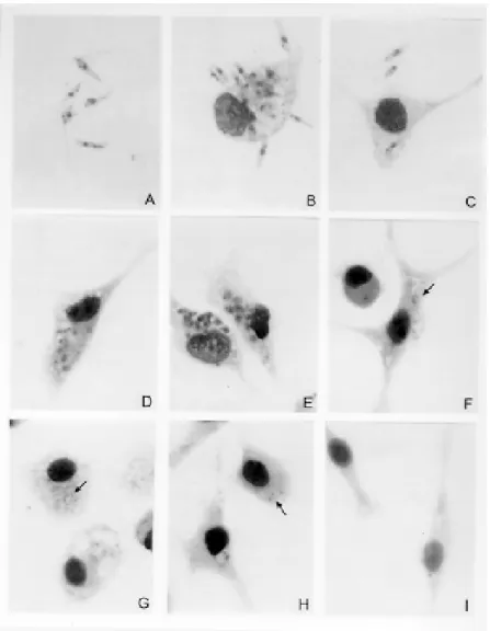
Macrophage culture - Peritoneal macrophages infected in vitro with promastigotes forms revealed internalized and degenerated parasites after 24 hr of infection. Nucleous and kinetoplast could hardly be observed. After 48 hr of infection, macroph-ages contained several vacuoles and small bodies. After five days, macrophages were found free of infection and presenting small inclusions, suggest-ing non-patogenicity or low virulence of the para-site (Fig. 1).
534 534 534 534
534 Opportunistic Infection in an HIV Positive Patient Raquel S Pacheco et al.
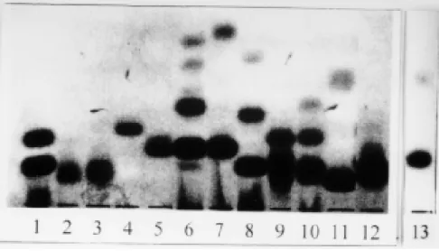
Isoenzyme electrophoresis - Results obtained using 12 enzymatic systems have allowed us to exclude any phenotypic identity of the isolate with
Leishmania, Sauroleishmania, Trypanosoma,
Endotrypanum, Phytomonas, Crithidia,
Blastocrithidia, Leptomonas and Herpetomonas. Fig. 2 shows electromorphs for the enzyme MDH detected in the different genera.
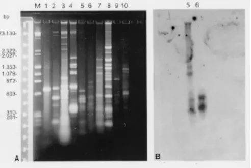
Genotypic analysis - The kDNA restriction pro-file of the isolate after digestion with the restric-tion endonuclease MspI showed no genotypic
simi-larity with L. (V) braziliensis, L. (L) amazonensis
and L. (L) chagasi (Fig. 3A). Southern blot hy-bridization of maxi and minicircles fragments with specific cloned minicircles of L. (V) braziliensis,
L. (L) amazonensis and L. (L) chagasi used as probes revealed no sequence homology with the kDNA from the parasite isolated. Homologous sequences could only be detected with the respec-tive homologous kDNAs (Fig. 3B, C, D).
KDNAs from distinct species of trypano-somatids were further digested with the restriction
Fig. 2: MDH electrophoretic patterns detected among different species of trypanosomatids. 1: patient’s isolate; 2: Leishmania (V) braziliensis; 3: L. (V) guyanensis; 4: L. (L) amazonensis; 5: L.(L) chagasi; 6: Crithidia deanei; 7: Blastocrithidia culicis; 8: Herpetomonas samuelpessoai; 9: Phytomonas davidi; 10: Leptomonas pulexsimulantis; 11: Endotrypanum schaudinii; 12: Trypa-nosoma cruzi; 13: Sauroleishmania tarentolae.
535 535535 535535 Mem Inst Oswaldo Cruz, Rio de Janeiro, Vol. 93(4), Jul./Aug. 1998
enzyme HaeIII. Results showed in Fig. 4A revealed that each species displayed a different genotypic pattern. Nevertheless, when the radiolabelled kDNA from the human isolate was used as probe against the repertoire of trypanosomatids, homolo-gous sequences in other minicircles fragments could only be found in the L. pulexsimulantis
kDNA (Fig. 4B).
DISCUSSION
The presence of intracellular amastigote forms in Giemsa-stained preparations of bone marrow aspirates, the association with clinical and sero-logical findings as well as the growth of the para-site predominantly as promastigotes have firstly led us to conclude that the patient was experienc-ing a visceral leishmaniasis/HIV co-infection. In immunocompetent individuals, even in spite of treatment and aparent cure of the disease, Leish-mania parasites can persist and reactivate (Saravia et al. 1990, Guevara et al. 1993, Aebischer 1994, Schubach et al. 1998) raising the question on mo-lecular determinants of virulence in some parasite strains. No relapse of the symptoms was observed in our immunocompromised patient over a period of two years. Evidence for low virulence of the parasite, in this case, was also supported by ab-sence of cutaneous manifestations or visceral in-volvement after inoculation in the hind paws or via intraperitoneal in three golden hamsters. No histopathological alteration was observed in the skin, spleen and liver over a period of one year of
observation (not shown). Likewise, reduced viru-lence was also evident from the parasite behavior within macrophages 48 hr after infections.
The second step was to characterize the para-site isolated. Some areas in the district of Jacarepaguá in Rio de Janeiro, where the patient is resident, are considered endemic for tegumentar leishmaniasis and cutaneous lesions are found in dogs and humans (Lopes et al. 1984, Pacheco et al. 1986). L. (V) braziliensis has been recorded ever since. In other foci near the metropolitan region of Rio de Janeiro both forms (tegumentar and/or vis-ceral) of the disease are present in humans and domestic animals, being L. (V) braziliensis and L. (L) chagasi the unique species detected until now (Oliveira-Neto et al. 1986, Marzochi et al. 1994, Barbosa-Santos 1994). In such areas is not excep-tional to find dogs and human beings living to-gether in the same dwelling. Isoenzyme electro-phoresis using 12 enzymatic loci and restriciton fragment length polymorphisms of kDNA analy-sis revealed patterns different from the most preva-lent species of Leishmania causing tegumentar or visceral leishmaniasis in Brazil. Comparison with phenotypic and genotypic profiles of other genera of heteroxenous and monoxenous trypanosomatids also showed no identity.
Hybridization of kDNA restriction fragments is frequently used to distinguish between strains and related species (Barker 1987). By using kDNA probes is possible to show that kDNA minicircles from organisms belonging to the principal
536 536 536 536
536 Opportunistic Infection in an HIV Positive Patient Raquel S Pacheco et al.
mania species complexes do not share extensive sequence homologies (Barker 1987). However, intraspecific variations are frequently detected using homologous kDNA probes (Pacheco et al., unpublished results). In the present case, no kDNA cross-homology could be detected between the parasite isolated and three different type-species representative of the Leishmania species com-plexes found in the New World. Although kDNAs from both human isolate and L. pulexsimulantis
have displayed dissimilar restriction profiles, they share homologous sequences in minicircle frag-ments ranging from 603 to 310 bp. Whether the observed divergence is attributable to intraspecific variations or the in vivo generation of new poly-morphisms under specific conditions (Pacheco et al. 1995) is difficult to ascertain. However, such homology seems to be specific and conserved in some minicircle classes in both kDNAs as no ho-mologous sequences could be detected in minicircles from other species or even in another related Leptomonas species. This finding allows us to authenticate the close genotypic relationship between both parasites.
The immune system depression in the patient might explain the opportunistic parasitism by this trypanosomatid. The presence of Leptomonas of the dog’s flea in an HIV positive patient reinforces the idea that humans under immunossupression conditions may be vulnerable to other insect trypanosomatids giving rise to clinical manifesta-tions similar to leishmaniasis. On the other hand it is not also so difficult to explain the mechanism of transmission being men and dogs living together. Feces deposited by infected adult fleas are usually well supplied with amastigotes which retain their infectiousness after drying at 25, 37 and 45oC (McGhee & Cosgrove 1980).
The literature reported in 1980 a possible, whereas not proved, case of human infection by
Herpetomonas (McGhee & Cosgrove 1980). The present article sustains that the possibility of lower trypanosomatids infecting humans exists and should be considered by attending physicians.
ACKNOWLEDGMENTS
To Prof. Maria Auxiliadora de Sousa (Núcleo de Tripanosomatídeos, Instituto Oswaldo Cruz) for the do-nation of monoxenous trypanosomatids, Dr Maurílio J Soares (Departamento de Ultraestrutura e Biologia Celular, Instituto Oswaldo Cruz) for the electromicros-copy studies and Drs Claudio Siqueira, Tania Amaral and Luiz Franco (Setor de Hematologia do Hospital da Lagoa, Rio de Janeiro) for the hematological and clini-cal follow up of the patient.
REFERENCES
Aebischer T 1994. Recurrent cutaneous leishmaniasis: a role of persistent parasites? Parasitol Today 10:
25-28.
Alvar R, Canavete C, Gutierrez-Solar B, Jimenez M, Laguna F, Lopez-Velez R, Molina R, Moreno J 1997. Leishmania and human immunodeficiency virus co-infection: the first 10 years. Clin Microbiol Reviews 10: 298-319.
Amato JCP, Amato-Neto V, Amato VS, Duarte MIS, Uip DE, Boulos M 1997. Lesões cutâneas como únicas manifestações de reativação da infecção pelo Try-panosoma cruzi em receptora de rim por transplante. Rev Soc Bras Med Trop 30: 61-63.
Barbosa-Santos EGO, Marzochi MCA, Urtado W, Queiroz F, Chicarino J, Pacheco RS 1994. Leishmaniasis dis-seminated by Leishmania braziliensis in a mare (Equus cabalus): immunotherapy and chemotherapy assays. Mem Inst Oswaldo Cruz 89: 217-220. Barker DC 1987. DNA diagnosis of human
leishmania-sis. Parasitol Today 3: 177-184.
Beard CB, Butler JF, Orshar EC 1989. In vitro growth characterization and host-parasite relationship of Leptomonas pulexsimulantis n. sp., a trypanosomatid flagellate of the flea Pulex simulans. J Parasitol 75: 658-668.
Campino L, Santos-Gomes G, Pratlong F, Dedet JP, Abranches P 1994. HIV-Leishmania co-infection in Portugal: isolation of Leishmania infantum MOM-24. Trans R Soc Trop Med Hyg 88: 394.
Dedet JP, Roche B, Pratlong F, Cales-Quist D, Jouannelle J, Benichou JC, Heurre M 1995. Diffuse cutaneous infection caused by a presumed monoxenous trypanosomatids in a patient infected with HIV. Trans R Soc Trop Med Hyg 89: 644-646.
Eslami Z, Tanner CE 1994. Time course and intensity of infection in vitro in the resident peritoneal macroph-ages of resistent and suspectible mice exposed to different doses of Leishmania donovani pro-mastigotes. Int J Parasitol 24: 743-747.
Ferreira MS, Nishioka AS, Rocha A, Silva AM, Ferreira RG, Olivier W, Tostes S 1991. Acute fatal Trypano-soma cruzi meningoencephalitis in a human immu-nodeficiency virus positive hemophiliac patient. Am J Trop Med Hyg 45: 723-727.
Guevara P, Ramirez JL, Rojas E, Scorza JV, Gonzalez N, Anez N 1993. Leishmania braziliensis in blood 30 years after cure. Lancet 341: 1341.
Jimenez M, Ferrer-Dufol M, Canavete C, Gutierrez-So-lar B, Molina R, Laguna F, Lopez-Velez R, Cercenado E, Dauden E, Blazquez J, Guevara CL, Gomez J, De La Torre J, Barros C, Altes J, Serra T, Alvar J 1995. Variability of Leishmania (Leishma-nia) infantum among stocks from immunocompro-mised, immunocompetent patients and dogs in Spain. FEMS Microbiol Letters 131: 197-204.
Jimenez M, Lopez-Velez R, Molina R, Canavete C, Alvar J 1996. HIV co-infection with a currently non-patho-genic flagellate. Lancet 347: 264-265.
Lopes UG, Momen H, Grimaldi Jr G, Marzochi MCA, Pacheco RS, Morel CM 1994. Schizodeme and zymodeme characterization of Leishmania in the investigation of foci of visceral and cutaneous leish-maniasis. J Parasitol 70: 89-98.
537 537537 537537 Mem Inst Oswaldo Cruz, Rio de Janeiro, Vol. 93(4), Jul./Aug. 1998
Visceral leishmaniasis in Rio de Janeiro. Parasitol Today 10: 37-40.
McGhee RB, Cosgrove WB 1980. Biology and physiol-ogy of the lower trypanosomatidae. Microbiol Re-views 44: 140-173.
Momen H, Grimaldi Jr G, Pacheco RS, Jaffe CL, McMahon-Pratt D, Marzochi MCA 1985. Brazilian Leishmania stocks phenotypically similar to Leish-mania major. Am J Trop Med Hyg 34: 1076-1084. Oliveira-Neto MP, Marzochi MCA, Grimaldi Jr G,
Pacheco RS, Toledo LM, Momen H 1986. Concur-rent human infection with Leishmania donovani chagasi and Leishmania braziliensis braziliensis. Ann Trop Med Parasitol 80: 587-592.
Pacheco RS, Brandão AA, Sibajev A, Cupolillo E, Momen H, Degrave W 1994. The Genus Crithidia: Genotypic diversity among species. J Protozool Res 4: 71-82.
Pacheco RS, Ferreira MS, Machado MI, Brito CMM, Pires MQ, Da-Cruz AM, Coutinho SG 1998. Chagas’ disease and HIV co-infection: Genotypic character-ization of the Trypanosoma cruzi strain. Mem Inst Oswaldo Cruz 93: 165-169.
Pacheco RS, Lopes UG, Morel CM, Grimaldi Jr G, Momen H 1986. Schizodeme analysis of Leishma-nia isolates and comparison with some phenotypic techniques, p. 57-65. In JA Riou, Leishmania Taxonomie et Phylogenese. Applications Eco-epidemiologiques, IMEEE, Montpellier.
Pacheco RS, Martinez JE, Valderrama L, Momen H, Saravia NG 1995. Genotypic polymorphisms in ex-perimental dermal leishmaniasis. Mol Biochem Parasitol 69: 197-209.
Pittella JEH 1993. The central nervous system involve-ment in Chagas’ disease: un updatting. Rev Inst Med Trop São Paulo 35: 111-116.
Rocha A, Meneses ACO, Silva AM, Ferreira MS, Nishioka AS, Burgarelli MKN, Almeida E, Turcatto Jr G, Metze K, Lopes ER 1994. Pathology of pa-tients with Chagas’ disease and acquired immuno-deficiency syndrome. Am J Trop Med Hyg 50: 261-268.
Saravia NG, Weigle K, Segura I, Giannini SH, Pacheco RS, Labrada LA, Gonçalves AM 1990. Recurrent lesions in human Leishmania braziliensis infection: reactivation or reinfection? Lancet 18: 398-402. Schubach A, Marzochi MCA, Cuzzi-Maya T, Oliveira
AV, Araujo ML, Oliveira ALC, Pacheco RS, Momen H, Coutinho SG, Marzochi KBF 1998. Cutaneous scars in american tegumentar leishmaniasis patients: a site of Leishmania (Viannia) braziliensis persis-tence and viability eleven years after antimony therapy and clinical cure. Am J Trop Med Hyg 58: in press.
Southern EM 1975. Detection of specific sequences among DNA fragments separated by gel electro-phoresis. J Mol Biol 98: 503-517.
Vickerman K 1976. The diversity of the kinetoplastid flagellates, p. 1-34. In WH Lumsden, DA Evans (eds), Biology of the Kinetoplastida, Academic Press, London.
Vickerman K 1994. The evolutionary expansion of trypanosomatid flagellates. Int J Parasitol 24: 1317-1331.
538 538 538 538