33 3333 3333 Mem Inst Oswaldo Cruz, Rio de Janeiro, Vol. 100(1): 33-38, February 2005
Programmed cell death in
Trypanosoma cruzi
induced by
Bothrops jararaca
venom
Poliana D eolindo, André S. Teixeira-Ferreira, Edésio JT M elo* ,
Andrea Cristina Veto Arnholdt* * , Wanderley de Souza* * * , Elias W Alves/+, Renato A D aM atta*
Laboratório de Química e Função de Proteínas e Peptídeos *Laboratório de Biologia Celular e Tecidual **Laboratório de Biologia do Reconhecer, Centro de Biociências e Biotecnologia, Universidade Estadual do Norte Fluminense, Av. Alberto Lamego 2000, 28013-600 Campos dos Goytacazes, RJ, Brasil ***Laboratório de Biologia Celular Hertha Meyer, Instituto de Biofísica Carlos
Chagas Filho, Universidade Federal do Rio de Janeiro, Rio de Janeiro, RJ, Brasil
Cells die through a programmed process or accidental death, know as apoptosis or necrosis, respectively.
Bothrops jararaca is a snake whose venom inhibits the growth of Trypanosoma cruzi epimastigote forms causing mitochondrion swelling and cell death. The aim of the present work was to determine the type of death induced in epimastigotes of T. cruzi by this venom. Parasite growth was inhibited after venom treatment, and 50% growth inhibition was obtained with 10 µg/ml. Ultrastructural observations confirmed mitochondrion swelling and kine-toplast disorganization. Furthermore, cytoplasmic condensation, loss of mitochondrion membrane potential, time-dependent increase in phosphatidylserine exposure at the outer leaflet plasma membrane followed by permeabilization, activation of caspase like protein and DNA fragmentation were observed in epimastigotes through-out a 24 h period of venom treatment. Taken together, these results indicate that the stress induced in epimastigote by this venom, triggers a programmed cell death process, similar to metazoan apoptosis, which leads to parasite death.
Key words: Trypanosoma cruzi - programmed cell death -Bothrops jararaca - snake venom
In multicellular organisms, programmed cell death (PCD), also known as apoptosis, is important to control cell number for proper development and tissue homeo-stasis, removal of unwanted cells and functional control of the immune, haemopoietic and nervous systems (Welburn et al. 1997). The process of PCD is activated by genetically controlled cell suicide machinery (Ameisen 1996). In vertebrate cells, PCD can be activated by exter-nal stimuli (ethanol, reactive oxygen species, and recep-tor ligands) and internal processes (mitotic catastrophe, replication failures, developmental programmed cell death) (Fröhlich & Madeo 2000). PCD is also a common response to cell stress caused by different toxins (Vaux 2002). Inde-pendent of the stimulus, PCD usually involves alteration in the mitochondrial membrane permeability, caspase ac-tivation, phosphatidylserine (PS) exposure, nuclear and cytoplasmic condensation, DNA fragmentation and break-age of the cell into apoptotic bodies, which are engulfed by the surrounding cells (Vaux & Strasser 1996). PCD is a process found in virtually all nucleated metazoan cells, and it has been recently associated with several species of unicellular eukaryotes, notably kinetoplastids (Ameisen et al. 1995, Welburn et al. 1996, Moreira et al. 1996, Ridgley
Financial support: CNPq, Faperj, Pronex
+Corresponding author. E-mail: [email protected]
Received 27 July 2004 Accepted 15 December 2004
et al. 1999, Arnoult et al. 2002, Lee et al. 2002, Zangger & Xu 2002), yeast (Madeo et al. 1999), apicomplexans (Picot et al. 1997, Peng et al. 2003), and amitochondrion para-sites (Chose et al. 2002, Mariante et al. 2003).
Snake venoms have been identified as the richest source of enzymes among poisonous animal, displaying a variety of biological activities (Tan & Ponnudurai 1992).
Bothrops jararaca is a snake of the viperidae family, largely
distributed in Brazil and responsible for 90% of the ophid-ian envenomations in humans (Bochner & Struchiner 2003). Trypanosoma cruzi is the agent of Chagas disease
affecting 16-18 million people in Latin America. We have previously demonstrated that B. jararaca venom
inhib-ited the growth of epimastigote forms of T. cruzi, causing
mitochondrion swelling and kinetoplast disorganization (Gonçalves et al. 2002). Because of the ultrastructural alteration observed in the mitochondrion after venom treat-ment it was suggested that impairtreat-ment of this organelle was probably responsible for growth inhibition (Gonçalves et al. 2002). However, how epimastigotes were demising was not analyzed. Thus, the aim of the present work was to identify if PCD or necrosis was being induced in T. cruzi epimastigote forms during B. jararaca venom
treat-ment.
MATERIALS AND METHODS
Parasite and venom - Epimastigote forms of T. cruzi
3 4 3 4 3 4 3 4
3 4 Cell death by snake venom • Poliana D eolindo et al.
Lyophilized B. jararaca venom was purchased from
the Instituto Butantan (São Paulo, Brazil). Venom stock solutions (2.5 mg/ml) were prepared in 20 mM Tris, 300 mM NaCl, pH 8.0, esterilized in a 0.22 µm filter (Millipore) and kept at 4°C. Crude venom solutions were diluted in the cell culture medium before use.
Treatment of cultured parasites - T. cruzi
epi-mastigotes were seeded (1.5 x 10⁶ cell/ml) into flasks (final volume 4 ml) containing medium supplemented with 0 (con-trol), 5, 10, 25 or 50 µg/ml of venom. Parasite growth was monitored, up to 7 days post-seeding, by the counting of formalin fixed parasites in a hemacytometer chamber daily. The inhibitory K0.5 (50% growth inhibition) was estimated
by plotting the parasite numbers from day 4 as a percent-age over venom concentration; cultures without any venom were used as 100% growth. For most of the fol-lowing characterizations, parasites were treated with double of the amount of the inhibitory K0.5 of the venom
(20 µg/ml) for a maximum of 24 h. For mitocondrial mem-brane potential and PS exposure assays cells were treated at different time points, as below. And for detection of caspase-3 like activity cells were treated with 10 µg/ml for 24 h. Control parasites were cultured in parallel without venom and analyzed at the same time.
Cell viability and PS exposure assays - Epimastigotes
were treated with venom and the viability assay was per-formed in a flow cytometer (Coulter Elite, SP) with propidium iodide (PI) (Sigma) as a vital stain. Cells were washed with phosphate buffer saline(PBS) and 1.5 x 10⁶ cells in 500 µl of PBS were incubated with 10 µg/ml of PI for 30 min and analyzed. For PS exposure assay, epi-mastigotes were treated with venom for 4, 8, and 24 h. PS exposure on the outer leaflet of the plasma membrane was measured by incubating cells with 1 µg/ml of annexin V-FITC (Santa Cruz Biotechnology Inc., US) for 20 min in a buffer containing 140 mM of sodium chloride, 10 mM of HEPES, 2.5 mM calcium chloride at pH 7.4. Viability was also assayed in parallel using PI as before. Cells were analyzed in a flow cytometer. Data, histograms and dot plot were obtained using WinMDI 2.8 software.
Transmission electron microscopy - Epimastigotes
treated with venom were washed three times with PBS, fixed for 2 h with 2.5% glutaraldehyde, 2% recently pre-pared formaldehyde in 0.1 M phosphate buffer, pH 7.2. Cells were washed in the same buffer, post-fixed for 1 h with 1% osmium tetroxide in 0.1 M phosphate buffer, de-hydrated in graded acetone and embedded in epoxy resin. Ultrathin sections were stained with uranyl acetate and lead citrate, and observed in a ZEISS EM900 transmission electron microscope.
Mitochondrion membrane potential (Dym) analy-sis - Alterations in Dym were analyzed in epimastigotes
treated for 4 h with venom. After treatment, cells were centrifuged at 1500 g for 10 min and adhered to cover slips coated with poly-L-lysine. Cells attached to cover slips were incubated in LIT medium containing 50 µg/ml of rhodamine 123 during 20 min, washed and observed in a laser scan confocal microscopy Zeiss SM 410 using a 488 nm and 543 nm argon laser.
Detection of caspase-3 like activity - The CM1
anti-body used was a gift from Dr Marlene Benchimol (Universidade Santa Úrsula, Brazil) and Dr Robert C Armstrong (Idun Pharmaceuticals, US), and a detailed description was provided by Namura et al. (1998). Treated epimastigotes were immunolabeled following a modifica-tion of the method used by Mariante et al. (2003). Cells were fixed for 30 min in 3% recently prepared formalde-hyde in PBS, washed, incubated for 10 min in 50 mM amonium chloride in PBS, washed and attached to cover slips coated with poly-L-lysine. Cells were washed and incubated for 10 min in a blocking buffer containing 2% bovine serum albumin, 0.2% non-fat milk powder and 0.8% Triton X-100 in PBS at room temperature. After that, cells were incubated overnight in CM1 primary antibody di-luted (1:5000) in the blocking buffer at 4°C. Cells were washed with PBS, incubated for 10 min in the blocking buffer and further incubated for 40 min with a FITC la-beled goat anti-rabbit IgG (Sigma) diluted 1:100 at 4°C. Cells were washed with PBS, mounted in N-propyl-gallate and observed in the Confocal microscope.
In situ nick-end labeling - Treated epimastigotes were
washed with PBS, fixed with 3% recently prepared formal-dehyde in PBS and attached to cover slips coated with poly-L-lysine. In situ DNA nick-end labeling was per-formed using a DNA fragmentation detection kit (FragEL, Oncogene) according to manufacturer’s specifications.
RESULTS
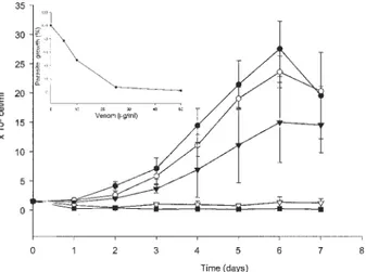
The effect of crude venom treatment on epimastigotes was dose-dependent. No growth was detected when para-sites were treated with 25 or 50 µg/ml of venom (Fig. 1). The inhibitory K0.5 (50% growth inhibition) at 4th day
was 10 µg/ml (Fig. 1, inset).
Flow cytometry analysis of epimastigotes treated with venom showed cytoplasmic condensation (Fig. 2A) and 97% of dead cells indicated by propidium iodide staining
Fig. 1: growth inhibition of Trypanosoma cruzi epimastigote forms by Bothrops jararaca venom. Cells were cultured for 7 days with
increasing venom concentrations (! 0; " 5; ! 10; " 25; # 100 µg/ml) and counted daily. The inhibitory K0.5 (50% growth
3 5 3 53 5 3 5 3 5 Mem Inst Oswaldo Cruz, Rio de Janeiro, Vol. 100(1), February 2005
(Fig. 2B). Only 3% of the cell population of untreated para-sites showed cell death.
Ultrastructural observations of untreated epimastigote showed all the typical ultrastructural characteristics of the trypanosomatids. The flagellar pocket and the flagel-lum close to the kinetoplast (large condensed mitochon-drion DNA), and a normal mitochonmitochon-drion next to the cell periphery were seen (Fig. 3A). Epimastigotes treated with venom presented mitochondrion swelling and kinetoplast disorganization (Fig. 3B). Treated parasites also exhibited a rounded shape instead of the normal elongated appear-ance of untreated cells (Fig. 3B). Margination and con-densation of nuclear chromatin was rarely observed after venom treatment (not shown).
Due to the swelling of the mitochondria observed af-ter treatment of parasites with venom, alaf-terations in the Dym were analyzed. After incubation with rhodamine 123, untreated epimastigotes showed intense labeling of the mitochondrion as expected (Fig. 4A, B). Parasites treated with venom showed a decrease in fluorescence, indicat-ing lost of Dym (Fig. 4C, D).
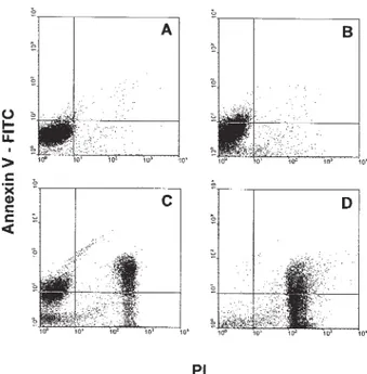
PS exposure in the outer leaflet of the plasma mem-brane is a common feature of cells undergoing apoptosis. Using annexin V-FITC and PI, PS exposure and membrane permeability of parasites treated with venom were simul-taneously accessed. Untreated parasites were annexin V and PI negative (Fig. 5A). After 4 h of venom treatment, 7% of the parasite population exposed PS and exhibited no plasma membrane permeability (Fig. 5B). After 8 h of treatment, the population exposing PS and preserving membrane integrity increased to 21%. However, another 26% of treated cells exposed PS and were also positive for PI (Fig. 5C). After 24 h, 94% of the treated parasite popu-lation was PI positive and 50% of this popupopu-lation exposed PS (Fig. 5D).
Caspase-3 activity is a hallmark of cells dying by PCD. By immunofluorescence it was possible to detect a caspase-like protein in treated epimastigotes. Untreated parasites presented an elongated morphology and did not label for the CM1 antibody (Fig. 6A, B). Treated epimastigotes presented round (cytoplasmic shrinkage) and elongated forms (Fig. 6C). Only the rounded shape parasites had caspase-like protein activity (Fig. 6D).
Fig. 2: cytoplasmic condensation and viability of Trypanosoma cruzi epimastigote forms after Bothrops jararaca venom
treat-ment. Parasites were treated for 24 h with 20 µ g/ml of venom. Untreated (white area) and treated parasites (gray area) can be distinguished in the overlay histograms; parasite size (A) and propidium iodide (PI) intercalation (B) are shown. Note the smaller size and higher PI positive fluorescence (97% as defined by the M1 region) in parasites treated with venom. This experiment is repre-sentative of five performed.
Fig. 3: transmission electron microscope images of Trypanosoma cruzi epimastigote forms treated with Bothrops jararaca venom.
Parasites were incubated for 24 h with 20 µg/ml of venom. A: untreated parasite showing mitochondrion (m), Golgi apparatus (g), kinetoplast (k), flagellar pocket (p), flagellum (f) and nucleus (n); B: treated parasite exhibiting a rounded shape, mitochondrion swelling (arrow), and kinetoplast disorganization. Bars = 0.5 µm. This experiment is representative of three performed.
3 6 3 6 3 6 3 6
3 6 Cell death by snake venom • Poliana D eolindo et al.
DISCUSSION
In a previous work we have shown that epimastigote forms of T. cruzi had their growth arrested after B. jararaca
venom treatment (Gonçalves et al. 2002). Here we con-firmed that this treatment induced growth arrest and mito-chondrion alteration. Furthermore, venom treatment caused cytoplasmic condensation, loss of mitochondrion membrane potential, exposure of PS on the outer leaflet of the plasma membrane, caspase 3 like protein activation Fig. 4: Trypanosoma cruzi epimastigote forms treated with Bothrops
jararaca venom show reduction in the mitochondrion membrane
potential (Dym). Parasites were treated with 20 µg/ml of venom for 4 h and incubated with rhodamine 123. A: differential interference contrast (DIC) image of untreated parasites; B: confocal laser scan-ning microscopy (CLSM) image of untreated parasites showing mitochondrion labeling with rhodamine 123 (arrows); C: DIC im-age of treated parasite; D: CLSM imim-age of treated parasite. Note the decrease in fluorescence of rhodamine 123 labeling in treated parasites indicating loss of ∆ψm. Bars = 5 µm. This experiment is representative of two performed.
Fig. 5: time-dependent increase in phosphatidylserine (PS) expo-sure by Trypanosoma cruzi epimastigote forms treated with Bothrops jararaca venom. Parasites were treated for 4, 8, and 24 h with 20 µg/ml of venom and incubated with 10 µg of propidium iodide (PI) and 1 µ g/ml of annexin V-FITC. A: untreated parasites did not expose PS and were negative for PI; B: after 4 h of treatment, 7% of the parasites exposed PS and were negative for PI; C: after 8 h, 21% of the parasites exposed PS and were negative for PI, 26% were positive for both markers and 21% were PI positive only; D: parasites treated for 24 h exhibited 94% of the parasite population PI positive. This experiment is representative of three performed.
Fig. 6: Trypanosoma cruzi epimastigote forms treated with Bothrops jararaca venom exhibited caspase-like activity. Parasites were treated for 24 h with 10 µg/ml of venom. Cells were labeled with CM1 as in materials and methods. A: differential interference con-trast (DIC) image of untreated parasites; B: confocal laser scanning microscopy (CLSM) image of untreated parasites, no labeling can be observed; C: DIC image of treated parasite; D: CLSM image of treated parasite. Note that round cells are labeled and that elon-gated epimastigotes are not (arrows). Bar = 10 µm. This experi-ment is representative of two performed.
Fig. 7: Trypanosoma cruzi epimastigote forms treated with Bothrops jararaca venom exhibited DNA fragmentation. Parasites were
3 7 3 73 7 3 7 3 7 Mem Inst Oswaldo Cruz, Rio de Janeiro, Vol. 100(1), February 2005
and DNA fragmentation. Taken together, these results strongly suggest that this venom induces death in epimastigote forms by triggering PCD with features of metazoan apoptosis.
The induction of apoptosis in other cell types by snake venom treatment has already been described. Venom of Crotalus viridis (Suzuki et al. 1997) and other
hemorragic snake venoms (Araki et al. 1993) induce PCD in vascular endothelial cells, resulting in vascular mal-functions and alteration of the haemostatic process. Ven-oms are a complex mixture with different biological ef-fects, thus we can only speculate on the component(s) responsible for the induction of PCD in venom treated T. cruzi epimastigotes. However, Tempone et al. (2001) have
shown that the venom of Bothrops moojeni presents an
L-amino acid oxidase responsible for growth inhibition of different Leishmania species. L-amino acid oxidase uses
amino acids as substrate producing H2O2, which is a known inducer of PCD in metazoans (Hockenbery et al. 1993, Clement & Pervaiz 1999) and also in unicellular or-ganisms (Madeo et al. 1999, Ridgley et al. 1999, Mariante et al. 2002). The venom used in this work is from a snake of the same genus used in Tempone’s work, thus we are now trying to determine if the oxidases from the same family could be responsible for triggering PCD in epimastigotes due to H2O2 production.
In mammalian cells, a common pro-apoptotic stimulus is the release of cytochrome c from mitochondrion due to the outer membrane permeabilization (Brenner & Kroemer 2000). The kinetoplast disorganization, mitochondrion swelling and loss of its membrane potential seen in epimastigotes after venom treatment suggests that the observed PCD might involve the mitochondrion cell death machinery. This is further confirmed by the activity of caspase-like found in treated epimastigotes. The CM1 antibody is able to recognize the cleavage product of the human or mouse caspase-3 (Namura et al. 1998) and also label Tritrichomonas foetus treated with H2O2 (Mariante
et al. 2003). In this report, epimastigotes were treated with the inhibitory K0.5 venom concentration, which was able to maintained some elongated form of the parasites in the period analyzed. Only epimastigotes that suffered cyto-plasmic condensation were labeled by the antibody. Thus, this labeling was specific and shows that a caspase-like was being activated after venom treatment. This is an-other evidence that a PCD process was induced in epimastigotes after venom treatment.
It was observed that PS exposure after venom treat-ment was time-dependent and was followed by an increase in PI signal indicating membrane permeability. Thus, PCD was being induced after treatment with venom and was followed rapidly by secondary necrosis. This indicates that the venom was extremely toxic and killed epimastigote through a PCD mechanism, probably as a response to cell stress (Vaux 2002) caused by the treatment. DNA frag-mentation is a hallmark of apoptosis (Zhang & Xu 2002). In our experiments DNA fragmentation was clearly de-tected by in situ nick-end labeling further supporting that venom treatment was inducing cell death by an apoptotic mechanism. Furthermore, some cells exhibited two labeled regions. This might indicate that during cell death by
venom treatment, nuclei were fragmenting or that kineto-plast could also suffer the action of endonucleases, which would corroborate with the disorganization appearance as seen at the ultrastructural level. Further experiments are necessary to investigate these possibilities.
Multicellular organisms use physiological mechanisms of cell death for development and morphogenesis, to con-trol cell number, as well as a defensive strategy to remove infected, mutated or damaged cells (Vaux & Korsmeyer 1999). Until recently, it was assumed that PCD was a pro-cess confined to metazoan organisms (Vaux et al. 1994). However, new findings indicate that unicellular eukary-otes exhibited a type of cell death similar to typical mam-malian PCD. PCD has been described in several species including the kinetoplastids Trypanosoma cruzi (Welburn
et al. 1996), L. amazonensis (Moreira et al. 1996) T. brucei brucei (Ridgley et al. 1999), L. donovani (Lee et al. 2002), L. mexicana (Zangger et al. 2002), L. major (Arnoult et al.
2002), the amitochondrial parasites Trichomonas vaginalis
(Chose et al. 2002) and Tritrichomonas foetus (Mariante
et al. 2003), Plasmodium falciparum (Picot et al. 1997), Toxoplasma gondii (Peng et al. 2003), yeast (Madeo et al.
1999) and Dictyostelium (Cornillon et al. 1994). However,
the function of PCD in unicellular organisms is under speculation. It has been proposed that the reason of a PCD pathway in unicellular organisms is to control the cell population. Because these organisms are largely clonal populations, an altruistic mechanism of cell death avoid-ing uncontrolled growth would be logical (Welburn et al. 1997). The prediction that a single molecular mechanism of PCD emerging in evolution prior to the postulated mul-tiple emergences of multicellularity indicates that some of the molecules involved in the PCD mechanisms of phylo-genetically distant organisms might be related (Cornillon et al. 1994).
In conclusion, our results show that T. cruzi
epimas-tigote forms exhibited PCD after B. jararaca venom
treat-ment. This result is another example that the PCD pro-cess occurs in unicellular organism and might be the re-sult of cell stress caused by venom treatment. This find-ing indicates that pharmacological and immunological ma-nipulation of the PCD process may lead to new therapeu-tic approaches to chronic parasitherapeu-tic diseases (Barcinski & DosReis 1999). We are now characterizing the component of venom responsible for this effect on parasites.
ACKNOWLEDGMENTS
To Andrèa Carvalho César and Rafael M Mariante for read-ing the manuscript; Rosemary C Maciel, Adriana A Martins, Marcia A Dutra, and Giovana A de Morais for technical assis-tantship, and Dr Marlene Benchimol and Robert C Armstrong for letting us use the MC1 antibody.
REFERENCES
Ameisen JC 1996. The origin of programmed cell death. Science 272: 1278-1279.
3 8 3 8 3 8 3 8
3 8 Cell death by snake venom • Poliana D eolindo et al.
Araki S, Ishida T, Yamamoto T, Kaji K, Hayashi H 1993. In-duction of apoptosis by hemorrhagic snake venom in vas-cular endothelial cells. Biochem Biophys Res Com190: 148-153.
Arnoult D, Akarid K, Grodet A, Petit PX, Estaquier J, Ameisen JC 2002. On the evolution of programmed cell death: apoptosis of the unicellular eukaryote Leishmania major involves cysteine proteinase activation and mitochondrion permeabilization. Cell Death Differ9: 65-81.
Barcinski MA, DosReis GA 1999. Apoptosis in parasites and parasite-induced apoptosis in the host immune system: a new approach to parasitic diseases. Braz J Med Biol Res32: 395-401.
Bochner R, Struchiner CJ 2003. Epidemiologia dos acidentes ofídicos nos últimos 100 anos no Brasil: uma revisão. Cad Saúde Pública 19: 7-16.
Brenner C, Kroemer G 2000. Apoptosis. Mitochondria-the death signal integrators. Science 289: 1150-1151.
Camargo EP 1964. Growth and differentiation in Trypanosoma cruzi. I. Origin of metacyclic trypanosomes in liquid media. Rev Inst Med Trop São Paulo6: 93-100.
Chose O, Noe C, Gerbod D, Brenner C, Viscogliosi E, Roseto A 2002. A form of cell death with some features resembling apoptosis in the amitochondrial unicellular organism Tri-chomonas vaginalis. Exp Cell Res276: 32-39.
Clement MV, Pervaiz S 1999. Reactive oxygen intermediates regulate cellular response to apoptotic stimuli: an hypoth-esis. Free Radic Res30: 247-252.
Cornillion S, Foa C, Davoust J, Buonavista N, Gross JD, Golstein P 1994. Programmed cell death in Dictyostelium. J Cell Sci 107: 2691-2704.
Frölich KU, Madeo F 2000. Apoptosis in yeast – A monocellu-lar organism exhibits altruistic behaviour. FEBS Letters473: 6-9.
Gonçalves AR, Soares MJ, de Souza W, DaMatta RA,Alves EW 2002. Ultrastructural alterations and growth inhibition of Trypanosoma cruzi and Leishmania major by Bothrops jararaca venom. Parasitol Res 88: 598-602.
Hockenbery DM, Oltvai ZN, Yin XM, Milliman CL, Korsmeyer SJ 1993. Bcl-2 functions in an antioxidant pathway to pre-vent apoptosis. Cell75: 241-251.
Lee N, Bertholet S, Debrabant A, Muller J, Duncan R, Nakhasi HL 2002. Programmed cell death in the unicellular proto-zoan parasite Leishmania. Cell Death Differ9: 53-64. Madeo F, Frohlich E, Ligr M, Grey M, Sigrist SJ, Wolf DH,
Frohlich KU 1999. Oxygen stress: a regulator of apoptosis in yeast. J Cell Biol145: 757-767.
Mariante RM, Guimarães CA, Linden R, Benchimol M 2003. Hydrogen peroxide induces caspase activation and pro-grammed cell death in the amitochondrial Tritrichomonas foetus. Histochem Cell Biol120: 129-141.
Moreira ME, Del Portillo HA, Milder RV, Balanco JM, Barcinski
MA 1996. Heat shock induction of apoptosis in promastigotes of the unicellular organism Leishmania ( Leish-mania) amazonensis. J Cell Physiol167: 305-313.
Namura S, Zhu J, Fink K, Endres M, Srinivasan A, Tomaselli KJ, Yuan J, Moskowitz MA 1998. Activation and cleavage of caspase-3 in apoptosis induced by experimental cerebral ischemia. J Neurosci18: 3659-3668.
Peng BW, Lin J, Lin JY, Jiang MS, Zhang T 2003. Exogenous nitric oxide induces apoptosis in Toxoplasma gondii tachyzoites via a calcium signal transduction pathway. Para-sitology 126: 541-550.
Picot S, Burnod J, Bracchi V, Chumpitazi BF, Ambroise-Tho-mas P 1997. Apoptosis related to chloroquine sensitivity of the human malaria parasite Plasmodium falciparum. Trans R Soc Trop Med Hyg91: 590-591.
Ridgley EL, Xiong ZH, Ruben L 1999. Reactive oxygen species activate a Ca2+-dependent cell death pathway in the unicel-lular organism Trypanosoma brucei brucei. Biochem J340: 33-40.
Suzuki K, Nakamura M, Hatanaka Y, Kayanoki Y, Tatsumi H, Taniguchi N 1997. Induction of apoptotic cell death in hu-man endotelial cells treated with snake venom: implication of intracellular reactive oxygen species and protective ef-fects of glutathione and superoxide dismutases. J Biochem 122: 1260-1264.
Tan NH, Ponnudurai G 1992. Comparative study of the enzy-matic, hemorrhagic, procoagulant and anticoagulant activi-ties of some animal venoms. Comp Biochem Physiol103: 299-302.
Tempone AG, Andrade HFJr, Spencer PJ, Lourenço CO, Rogero JR, Nascimento N 2001. Bothrops moojeni venom kills Leishmania spp. with hydrogen peroxide generated by its L-amino acid oxidase. Bichem Biophy Res Commun280: 620-624.
Vaux DL 2002. Apoptosis and toxicology – What relevance? Toxicology181-182: 3-7.
Vaux DL, Korsmeyer SJ 1999. Cell death in development. Cell 96: 245-254.
Vaux DL, Strasser A 1996. The molecular biology of apoptosis. Proc Natl Acad Sci USA93: 2239-2244.
Vaux DL, Haecker G, Strasser A 1994. An evolutionary per-spective on apoptosis. Cell76: 777-779.
Welburn SC, Dale C, Ellis D, Beecroft R, Pearson TW 1996. Apoptosis in procyclic Trypanosoma brucei rhodesiense in vitro. Cell Death Differ 3: 229-236.
Welburn SC, Williams GT, Barcinski MA 1997. Programmed cell death in trypanosomatids. Parasitol Today13: 22-25. Zangger H, Mottram JC, Fasei N 2002. Cell death in
Leishma-nia induced by stress and differentiation: programmed cell death or necrosis? Cell Death Differ9: 1126-1139. Zhang J, Xu M 2002. Apoptotic DNA fragmentation and tissue