Report of the Second Satellite Symposium on Ultrasound
in Schistosomiasis
Joachim Richter/
+, Ana Lúcia Coutinho Domingues*, Cristina H Barata**,
Aluizio R Prata**, José Roberto Lambertucci***
Heinrich-Heine-UniversitaetDuesseldorf, Tropenmedizinische Ambulanz, Geb.11.93, Moorentr. 5, D-40225 Duesseldorf, FR Germany *Universidade Federal de Pernambuco, Recife, PE, Brasil **Faculdade de Medicina do Triângulo Mineiro, Uberaba, MG, Brasil ***Faculdade de Medicina da Universidade Federal de Minas Gerais,
Belo Horizonte, MG, Brasil
A group of experts on schistosomiasis and ultrasonography discussed the experiences and results obtained with the Niamey-Belo Horizonte Protocol on Ultrasonography in Schistosomiasis. A series of recomendations about qualitative and quantitative data obtained by ultrasound in studies performed in Africa and Brazil are presented. Imunological, genetic and epidemiological studies must rely on ultrasound for the identification of patients with periportal thickening/fibrosis.
Key words: schistosomiasis - schistosomiasis mansoni - ultrasound - periportal fibrosis
Ultrasonography (US) is the tool of first choice for assessing liver and spleen changes induced by
Schistosoma spp. in endemic regions; it is the most economic imaging technique which permits the as-sessment of abdominal organs. Another advantage is that US can be performed using portable devices which allow its application in remote areas. In areas without electricity network US devices can be pow-ered by portable generators.
The specificity of the characteristic US features of periportal fibrosis has been proven in several endemic areas like Brazil (Cerri et al. 1984), Sudan (Homeida et al. 1988) and Egypt (Abdel Wahab et al. 1989). Since in these studies different method-ologies have been applied, data obtained were not directly comparable on an international scale. This prompted the World Health Organization to spon-sor and assemble an International Expert Meeting in Cairo in 1991. A standardized protocol was de-veloped and published (Cairo Working Group 1992). This protocol was revised by an expert group in Niamey, Niger in 1996, and during the previous First Satellite Symposium on Ultrasonography in Schis-tosomiasis in Belo Horizonte 1997 (Niamey Work-ing Group 2000). The aim of the present Satellite Symposium was to discuss the practical experi-ences and results obtained with the Niamey-Belo Horizonte US protocol.
ACTUAL EXPERIENCES
The protocol has been applied in Uganda, Senegal, Brazil and Cambodia (to assess Schisto-soma mekongi-associated morbidity (Christoph Hatz, pers. commun., 1999; this latter study is not reported here). Approximative figures are given be-cause data analysis has not yet been completed (Table).
The following criteria of the usefulness of the various parameters of the Niamey-Belo Horizonte protocol were chosen: simplicity, reproducibility, time spent, relation to clinical status, power to dif-ferentiate between patients and control subjects (Minas Gerais study)
PRELIMINARY RESULTS - CORE PROTOCOL
Measurement of segmental portal branch wall thickness
After some training measurements are generally easy to perform by experienced ultrasonographists but this is not the case with less experienced exam-iners (Uganda, Senegal studies).
Reproducibility has been found unacceptably low in sub-samples of patients in Uganda and Senegal. Interobserver variance appears to decrease after training. However, even when performed by experi-enced ultrasonographists, reproducibility of mea-surements was unsatisfactory (Senegal).
Although the measurements of the portal branch walls take relatively long time this would be ac-ceptable if the other criteria above were fulfilled. Measurements were not found to reflect adequately the clinical status (hematemesis due to variceal bleeding, for example) in advanced cases in Senegal.
+Corresponding author. E-mail:
Image patterns (IP)
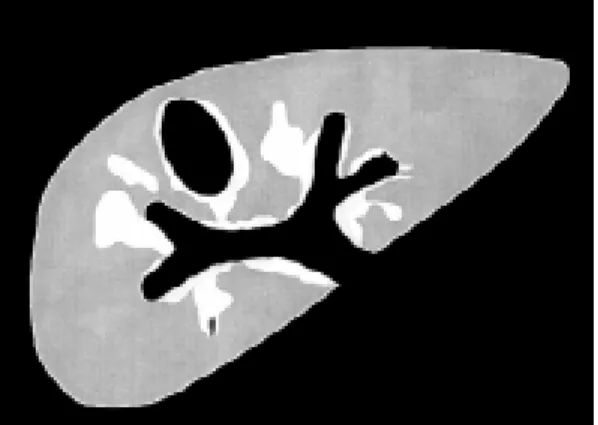
IP (Figures 1-8) are easily understood by experi-enced and less experiexperi-enced examiners.
Interobserver variance was low when training of the different examiners had taken place before (Uganda, Senegal). It is sometimes difficult to discern a pattern D from slight thickening of the wall of the main portal vein. Therefore a measure-ment should be introduced which allows objectivation of the finding. The best way of as-sessing “central periportal thickening” appears to be to measure the wall at portal bifurcation in front of the portal trunk in a oblique liver scan. In the
Minas Gerais study normal values of this measure-ment have been established (up to 5 mm in indi-viduals with a weight up to 40 kg, and 6 mm in subjects weighing more than 40 kg). Data will be reanalyzed in function of body height.
IP assignment does not take much time. Contrary to measurements, when the examiner is looking for IP, he or her concentrates on parenchyma changes. IP A, Dc-F reflected adequately the clinical status of all patients examined. IP B was found to be non-specific as it may be observed in children, after starvation, and in viral infections (e.g. a measle case
Fig. 1: image pattern A (normal sonographic liver appear-ance)
Fig. 2: image pattern B (pronounced peripheral portal branch echogenicity: ‘starry sky pattern’)
Fig. 3: image pattern C (peripheral periportal thickening)
Fig. 4: image pattern D (central periportal thickening) TABLE
Countries and areas where the WHO-Niamey protocol has been applied
Assessment of
Approximate interobserver
Country/area number of patients Patient selection variance
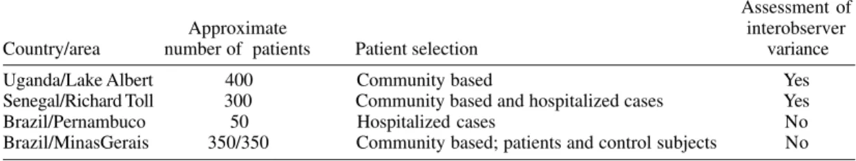
Uganda/Lake Albert 400 Community based Yes
Senegal/Richard Toll 300 Community based and hospitalized cases Yes
Brazil/Pernambuco 50 Hospitalized cases No
in Germany). IP Dc (mild periportal thickening around the portal stem and the peripheral branches) seems to have a worse clinical prognosis than D alone (central periportal thickening alone without peripheral thickening (Gerspacher-Lara et al. 1997). Most of the complicated cases have patterns E, Ec or F (advanced periportal thickening).
Liver measurements
Left liver lobe measurement is easily and rapidly performed. Right liver lobe measurement takes longer because the right liver lobe usually exceeds the dimensions of the US probe and measurement has to be done by adding partial diameters. For the same reason left liver lobe measurement is less subjected to intra-and inter-observer variance than that of the right liver lobe.
Measurement of the right liver lobe takes almost twice the time as compared to left lobe measure-ment.
Relation of liver measurements to clinical status has not yet been assessed systematically in the above study.
Portal hypertension assessment
Whereas the measurement of portal vein diameter at the liver hilus and detection of ascites do not require exceptional skills, the capacity in detecting collaterals largely depends on the experience of the examiner and the preparation of the patient. Reproducibility of portal vein measurements has been found excellent in Senegal and Uganda. Once the ultrasonographist is well trained in taking the measurement neither different experience of the examiner nor different quality of US devices inter-fered with measurement results. In hospitalized pa-tients Doppler examination can be performed. However, reproducibility of Doppler measurements has never been investigated in schistosomiasis patients
Portal vein measurement is rapid. In hospitalized patients in Pernambuco, the measurement of the splenic vein behind the pancreas correlated better with portal and intravariceal pressure than portal vein diameter. Splenic vein measurement be-hind the pancreas requires a good preparation of
Fig. 5: image pattern Dc (peripheral periportal thickening + central periportal thickening)
Fig. 6: image pattern E (central periportal thickening with echogenic patches expanding into the parenchyma)
Fig. 7: image pattern Ec (advanced peripheral periportal thickening + central periportal thickening)
the patient. The high respiratory variation of this vessel (50%) in healthy subjects may be a con-founding factor. It was therefore felt that this mea-surement including the assessment of respiration variation is recommended in hospitalized patients but was less useful in the field. The Niamey-Belo Horizonte Portal Hypertension score was found to reflect more the clinical status of four Senegalese patients with complicated periportal fibrosis than other scores tested in the past (Abdel-Wahab et al. 1993, Richter et al. 1998).
Additional investigations
Gall-bladder wall assessment should be integrated into the core protocol. Measurement is rapidly per-formed and adjustment to other biometric data is simple (up to 3 mm in individuals with a weight <= 30 kg, up to 4 mm in individuals > 30 kg). Character-istic changes (hyperechoic protrusions from the gall-bladder wall into the parenchyma) should be reported. Gall-bladder changes may precede peri-portal changes in a part of infected individuals and may therefore have an independent prognostic sig-nificance (Pinto da Silva et al. 1994).
In non-malarious areas spleen length should be al-ways assessed. Spleen measurements may be also useful in areas of low levels of malaria endemicity. Data obtained in a holoendemic area (Uganda) are yet to be analyzed.
COMPARISON OF DATA OBTAINED BY ULTRA-SONOGRAPHY AND BY OTHER MEANS
The gold standard for the assessment of the speci-ficity of US in vivo is liver biopsy. However, it must be stressed that the way of taking liver biopsies and the volume of the biopsy sample may interfere with biopsy results. A deep wedge liver biopsy of the left liver lobe obtained during surgery or laparoscopy has the highest sensitivity. For ethi-cal reasons this invasive procedure can not be per-formed in patients with early lesions. Blind liver punction does usually not yield enough material and fibrotic material is frequently not easily aspi-rated. Classification criteria have to be uniform dis-cerning between an egg granuloma and periportal fibrosis. Classifications must be standardized
(Coutinho-Domingues 1998). Inter-observer vari-ance in judging the sample must also be assessed.
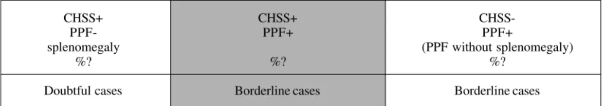
“Hepatosplenic schistosomiasis” is a clinical defi-nition which is not synonymous with periportal thickening as assessed by US. This is crucial when judging any kind of studies (genetic, immunologi-cal, epidemiologic), classifiying patients by one of these or both methods (Lambertucci et al. 2000). 1- Hepatosplenic schistosomiasis is also not syn-onymous with hepato-splenomegaly. Frequently the liver is not enlarged or even shrunken in pa-tients with hepatosplenic schistosomiasis. The clinical definition of hepatosplenic schistosomia-sis adopted by Aluizio Prata is the presence of a firm liver (left liver lobe) and a firm spleen palpable without breathing in a subject with parasitologi-cally proven schistosomiasis. This definition is subject to high inter-observer-variance as it has been observed in Brazil, Uganda and Senegal (Lambertucci et al. 1996).
2 - Periportal fibrosis may occur without splenom-egaly. Hyper-reactive splenomegaly may not be accompanied by ultrasonographically detectable periportal fibrosis. The relation between the differ-ent case definitions are illustrated in Fig. 9. The relation between the two case definitions (ideally maximal overlap percentage) has to be assessed in each endemic region and for each examination team. 3 - If US is applied to assess the value of clinical examination (to allow assessment of a high number of individuals in epidemiological surveys, when resources are limited) other parameters have to be included into the examination protocol. In the first phase, training of clinical examiners should be done with simultaneous ultrasound examination (“clini-cal hepatomegaly” was not associated to organ enlargement in a Senegalese sub-sample). Inter-observer variance should be reduced by intensive group training and ultrasound control in order to eliminate equivocal results.
4 - To allow direct comparison between clinical ex-amination and US results, splenic measurements should include also the extension of the spleen
Fig. 9: relation between the definitions clinical hepatosplenic schistosomiasis (CHSS) and periportal fibrosis (PPF) as assessed by ultrasonography
CHSS+ CHSS+
CHSS-PPF- PPF+ PPF+
splenomegaly (PPF without splenomegaly)
%? %? %?
below the costal margin in patients who do breathe quietly (not in deep inspiration). In huge splenom-egaly palpation using the Hackett classification may be more adequate than ultrasonographic measure-ment, and could be reported on the US sheet.
Improvement of US in detecting early liver pathol-ogy:
1 - Comparative Necropsy-US studies in subjects infected with schistosomiasis but who died of other causes may be helpful in the identification of char-acteristic early US features. Here it is particularly important to use an unequivocal pathological defi-nition of periportal fibrosis and to define and mea-sure macroscopic lesions.
2 - Animal studies.
NOVEL TECHNIQUES FOR THE IDENTIFICATION OF EARLY PATHOLOGY
Actual US technology (portable devices) does not allow a clear-cut definition of early pathology. High-tech US devices (e.g. with harmonic tissue imaging facility, examination of peripheral liver pa-renchyma with high frequency transducers, Dop-pler-US, contrast-Doppler-US)
Computed tomography (Cesmeli et al. 1997) Helicoidal computed contrast-tomography Magnetic resonance imaging (proved useful in neuroschistosomiasis)
INTERFERENCE OF OTHER LIVER DISEASES
Chronic hepatitis and alcoholic diseases are the major confounders when assessing US results in patients with schistosomiasis. Mixed hepatitis C and schistosomiasis is particularly frequent in Egypt, but periportal fibrosis and post-necrotic and/ or alcoholic liver cirrhosis occur frequently also in some areas of Brazil. In epidemiological studies on hepatic morbidity alcoholism must be ruled out by careful medical history. Hepatitis B and C must be ruled out by the determination of at least HBsAg, and anti-HCV antibody .
ACUTE SCHISTOSOMIASIS
Reports on abdominal ultrasound studies in pa-tients with acute schistosomiasis are still scarce and limited data are available on changes of liver texture in this stage of the disease (Lambertucci et al. 1994). More recently, 26 patients with acute S. mansoni
infection were submitted to clinical and ultraso-nographic examination by Barata et al. (1999). For comparison of ultrasound features, each patient was matched by age, gender, weight and height to a non-infected individual. In most patients (21/26) non-spe-cific hepatosplenomegaly was observed without liver texture abnormalities which was frequently
accom-panied by intraabdominal lymph node swelling, es-pecially around the portal hilus. Periportal thick-ening was observed in 5/26 patients. Three of these patients underwent percutaneous liver biopsy, which showed dense inflammatory infiltration of neutro-phils, macrophages and eosinophils in the portal tracts associated with discrete fibrous tissue forma-tion. Periportal thickening regressed six months af-ter chemotherapy. Twenty four months post-therapy liver and spleen volumes had regressed to normal, whereas lymph nodes, although reduced in size were still easily recognized.
Intraabdominal lymph nodes may be seen also in healthy individuals, and lymphadenopathy can be observed in a variety of diseases other than schistosomiasis. The predictive value of particular hilar lymph node morphology changes observed in acute schistosomiasis but not in other disease (acute hepatitis, acute HIV-infection, acute EBV-infection etc.) is underway in Minas Gerais.
CONCLUSIONS AND RECOMMENDATIONS
The preliminary data obtained with the Niamey-Belo Horizonte protocol since 1997 do not yet per-mit to formulate definite recommendations. The pro-tocol needs to be tested further in different endemic areas and by different research groups.
Periportal branch wall measurements appear not to be reliable indicators of periportal fibrosis when analyzed alone, but should still be performed to evaluate this parameter.
It is emphasized that measurements in general must not be abandoned. Measurements increase the objectivity of the results if these fulfill the fol-lowing criteria: (1) measurements must be repro-ducible among different examiners; (2) reflect the clinical status; (3) must be adjusted to individual biometric data (body height and/or weight). Best reproducibility is obtained for measurements be-tween 5 mm and 5 cm, and structures which are not to close to the transducer (because convex trans-ducers distort the image).
Gallbladder assessment should be included into the core protocol.
Adequate training is crucial not only of the ultrasonographist but also of examiners foreseen for clinical assessment in epidemiological studies. Quality assessment and training of palpation should rely also on simultaneous ultrasonography.
ACKNOWLEDGEMENTS
To the Organizing Committee of the VII International Symposium on Schistosomiasis, and in particular Dr Miriam Tendler for giving the opportunity and infrastruc-ture for this Satellite Symposium. Hospitality, logistic and travel costs of Dr Richter were covered by Fiocruz, Rio de Janeiro, and Danish Bilharziasis Laboratory, Den-mark. Dr Prata and Dr Lambertucci wish to thank CNPq, WHO and the National Health Foundation for sponsoring the work that has been developed in Brazil.
REFERENCES
Abdel-Wahab MF, Esmat G, Milad M, Abdel-Razek S, Strickland GT 1989. Characteristic sonographic pat-tern of schistosomal hepatic fibrosis. Am J Trop Med Hyg 40: 72-76.
Abdel-Wahab MF, Esmat G, Farrag A, El-Boracy Y, Strickland GT 1993. Ultrasonographic prediction of esophageal varices in schistosomiasis mansoni. Am J Gastroenterol 88: 560-563.
Barata CH, Pinto-Silva RA, Lambertucci JR 1999. Ab-dominal ultrasound in acute schistosomiasis mansoni. Brit J Radiol 72: 949-952.
Cairo Working Group 1992. The use of diagnostic ultra-sound in schistosomiasis - attempts at standardiza-tion of methodology. Acta Trop 51: 45-63. Cerri GG, Alves VAF, Magalhães A 1984. Hepatosplenic
schistosomiasis mansoni: ultrasound manifestations. Radiology 153: 777-780.
Cesmeli E, Vogelaers D, Voet D, Duyck P, Peleman R, Kunnen M, Afschrift M 1997. Ultrasound and CT changes of liver parenchyma in acute schistosomia-sis. Br J Radiol 70: 758-760.
Coutinho-Domingues AL 1998. Ultra-sonografia na Esquistossomose Mansônica Hépato-esplênica: Avaliação da Intensidade da Fibrose Periportal e da Hipertensão Porta, PhD Thesis, Federal University of Recife, Brazil.
Gerspacher-Lara R, Pinto-Silva RA, Rayes AAM,
Drummond SC, Lambertucci JR 1997. Ultrasonog-raphy of periportal fibrosis in schistosomiasis mansoni in Brazil. Trans R Soc Trop Med Hyg 91: 307-309.
Homeida MA, Abdel-Gadir AF, Cheever AW, Bennett JL, Arbab BMO, Ibrahium SZ, Abdel-Salam IM, Dafalla AA, Nash TE 1988. Diagnosis of pathologi-cally confirmed Symmer’s periportal fibrosis by ul-trasonography: a prospective blinded study. Am J Trop Med Hyg 38: 86-91.
Lambertucci JR, Gerspacher-Lara R, Pinto-Silva RA, Barbosa MM, Teixeira R, Barbosa HF, Serufo JC, Rezende SC, Rayes AM 2000. The Queixadinha Project: a study on morbidity and schistosomiasis control in the northeast of the state of Minas Gerais, in Brazil. Rev Soc Bras Med Trop 29: 127-135. Lambertucci JR, Pinto-Silva RA, Gerspacher-Lara R,
Barata CH 1994. Acute Manson’s schistosomiasis: sonographic features. Trans R Soc Trop Med Hyg 88: 76-77.
Lambertucci JR, Serufo JC, Gerspacher-Lara R, Rayes AA, Teixeira R, Nobre V, Antunes CM 2000. Schis-tosoma mansoni: assessment of morbidity before and after control. Acta Trop 77: 101-109.
Niamey Working Group 2000. Ultrasound in schistoso-miasis.A practical guide to the standardized use of ultrasonography for the assessment of schistoso-miasis-related morbidity. World Health Organization/ TDR/SCH/ULTRASON/document, Geneva, Swit-zerland, in press.
Pinto-Silva RA, Abrantes WL, Antunes CM, Lambertucci JR 1994. Sonographic features of por-tal hypertension in schistosomiasis mansoni. Rev Inst Med Trop São Paulo 36: 355-361.