Magnetic r esonance imaging and ultr asound in
hepatosplenic schistosomiasis mansoni
Ressonância magnética e ultrassonografia na
esquistossomose mansoni hepatoesplênica
José Rober to Lamber tucci
1, Luciana Cr istina dos Santos Silva
1, Luciene Mota Andr ade
2,
Leonar do Campos de Queir oz
3and Rogér io Augusto Pinto-Silva
3ABSTRACT
We re po rt th e f i n d i n gs o f a b d o m i n a l u ltra so u n d a n d m a gn e ti c re so n a n c e i m a gi n g o b se rve d i n a p a ti e n t wi th a d va n c e d
sc histo so miasis manso ni. A 25-ye a r-o ld m a n with he pa to sple nic schisto so m ia sis a nd va rice a l b le e ding co nfirm e d b y uppe r e ndo sco py wa s sub m itte d to a b do m ina l ultra so und a nd m a gne tic re so na nce im a ging. During surge ry fo r po rta l hype rte nsio n, a live r b io psy wa s ta k e n a nd the dia gno sis o f Sym m e rs’ fib ro sis wa s co nfirm e d. Ma gne tic re so na nce im a ging sca ns ga ve m o re pre cise info rm a tio n a b o ut the ga llb la dde r, pe ripo rta l thick e ning a nd a b do m ina l ve no us syste m tha n did the ultra so und.
Ke y-words: He p a to sp le n i c sc h i sto so m i a si s m a n so n i . Ultra so u n d . Ma gn e ti c re so n a n c e i m a gi n g.
RESUMO
Re la ta m o s o s a c h a d o s u ltra sso n o grá f i c o s e à re sso n â n c i a m a gn é ti c a i n tra - a b d o m i n a i s o b se rva d o s e m u m p a c i e n te c o m e sq u i sto sso m o se m a n so n i gra ve . Um ho m e m de 25 a n o s de ida de c o m e sq u isto sso m o se he pa to e splê n ic a e sa n gra m e n to dige stivo de va rize s e so fa gia n a s, c o m dia gn ó stic o c o n firm a do pe la e n do sc o pia , fo i su b m e tido à u ltra so n o gra fia a b do m in a l e re sso n â n c ia m a gn é tic a . Du ra n te a c iru rgia de hipe rte n sã o po rta , u m fra gm e n to de fíga do fo i o b tido e o e xa m e histo ló gic o c o n firm o u o dia gn ó stic o de fib ro se de Sym m e rs. A re sso n â n c ia m a gn é tic a fo rn e c e u in fo rm a ç õ e s m a is pre c isa s so b re a ve síc u la b ilia r, e spe ssa m e n to pe ripo rta l e siste m a ve n o so a b do m in a l do q u e a u ltra sso n o gra fia .
Pal avr as-chave s: Esq u i sto sso m o se m a n so n i h e p a to e sp lê n i c a . Ultra sso n o gra f i a . Re sso n â n c i a m a gn é ti c a .
1 . Se r viç o de Do e nç as Infe c c io sas e Par asitár ias da Fac uldade de Me dic ina da Unive r sidade Fe de r al de Minas Ge r ais, B e lo Ho r izo nte , MG. 2 . Lab o r ató r io s He r me s Par dini, B e lo Ho r izo nte , MG. 3 . Se r viç o de Radio lo gia do Ho spital das Clínic as da Unive r sidade Fe de r al de Minas Ge r ais, B e lo Ho r izo nte , MG.
Addr e ss to: Dr. Jo sé Ro b e r to Lamb e r tuc c i. Se r viç o de Do e nç as Infe c c io sas e Par asitár ias/FM/UFMG. Av. Alfr e do B ale na 1 9 0 , Santa Efigê nia, 3 0 1 3 0 - 1 0 0 B e lo Ho r izo nte , MG, B r asil,
Te l: 5 5 3 1 3 2 4 8 - 9 8 2 1 E- mail: lamb e r @ ne t. e m. c o m. b r Re c e b ido par a pub lic aç ão e m 2 6 /3 /2 0 0 4 Ac e ito e m 5 /5 /2 0 0 4
Diagnosis of hepatosplenic sc histosomiasis and stratific ation o f mo r b idity pe r mit ide ntific atio n o f c ase s with highe r r isk o f c o m plic a tio n s s uc h a s , va r ic e a l b le e din g, pulm o n a r y hype r te nsio n, and glo me r ulo ne phr itis, the r e b y allo wing a m o r e r a tio n a l a ppr o a c h to tr e a tm e n t1 1 5 1 9 2 0. Ab do m in a l
ultrasonography ( US) is an indirec t method of diagnosis of sc histosomiasis, and represents a tool in the c lassific ation of the c linic al forms of the disease4 5 1 6 1 8. However, there still is little
information about the c orrelation of US findings and the various aspec ts of morbidity in sc histosomiasis1 8.
Magnetic resonanc e imaging ( MRI) has been shown to be a sensitive imaging tec hnique in the evaluation of a variety of diseases. Unlike US, it is not a dynamic examination and can be less vulnerable to intra and inter-examiner variability 1 2. So m e
fi n d i n gs o f MR I , wi th d i ffe r e n t e q u i p m e n t, h a ve b e e n r e p o r te d p r e vi o u s l y i n th r e e c a s e s o f h e p a to s p l e n i c sc histosomiasis mansoni8 1 4 2 1, with promising results. We report
PATIENT AND METHODS
A 2 5 -year-old man was referred to the Hospital of the Federal University of Minas Gerais, in Brazil, for evaluation of anemia which did not respond to medical treatment. He was born and resided in an area endemic for schistosomiasis. Eleven years before admission, stool examination disclosed viable eggs of Schisto so m a m a nso ni. He received praziquantel, but maintained frequent contact with stream water. He reported at least 3 episodes of digestive bleeding over the last 8 years, and was submitted to several sessions of endo sc o pic sc ler o ther apy o f eso phageal var ic es. At c linic al examination, he was pale with a palpable liver and an enlarged spleen ( Boyd III) . Blood counts revealed reduction in all cell series: 3 .2 x 106 red cells/mm3, hemoglobin 6 .0 g/dL, 1 .4 x 1 03 white cells/mm3,
5 1 x 1 03 platelets/mm3. Blood chemistry and coagulation were
unremarkable. There was no evidence of renal disease. Digestive endoscopy revealed small size distal esophageal varices and scars
produced by previous sclerotherapy, erosive antral gastritis and hypertensive gastropathy. Serology for hepatitis B and C gave negative r e s ults . Th e r e wa s n o c lin ic a l, e le c tr o c a r dio gr a ph ic o r echocardiographic evidence of pulmonary hypertension. He was operated on ( splenectomy and esophagogastric devascularization) and a liver biopsy confirmed the diagnosis of Symmers’ fibrosis.
RESULTS
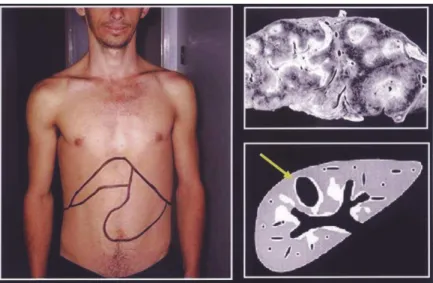
Note in Figure 1 the patient with hepatosplenomegaly, the aspect of a liver with advanced periportal fibrosis and the Ec pattern of WHO for ultrasound in schistosomiasis mansoni ( advanced central e peripheral fibrosis + thickening of the gallbladder wall) . US of the abdomen with a SIEMENS Sonoline Prima devic e showed enlarged periportal bands with increased echogenicity suggesting inte nse pe r ipo r tal fib r o sis ( Figur e 2 ) . Evide nc e o f po r tal
Figure 2 - Ultra so und o f the pa tient sho wing peripo rta l thick ening ( yello w a rro ws o n the le ft side ) , thick e ning o f the ga llb la dde r wa ll ( G = ga llb la dde r; ye llo w a rro w = thick e ne d wa ll) a nd the e nla rge d sple e n ( S) .
Figure 4 - MRI T2-weighted im age of liver and spleen – transversal section. There are four im ages showing different aspects of peripo rta l fibro sis in the liver ( the vein in the center a ppea rs da rk – red a rro w) . Peripo rta l thick ening ( yello w a rro ws) . White a rro ws = sto m a ch; S = spleen.
hypertensio n, with po rtal vein and spleen enlargement and collateral veins, and thickening of the gallbladder wall were also noticed. MRI of the abdomen was performed using a Giroscan Intera superc onduc ting 1 .5 Tesla magnetic system ( Philips – Netherlands) ( Figures 3 , 4 and 5 ) . The sequences demonstrated the broad periportal bands seen on liver ultrasound. On T1
sequences, these bands were hypointense to the liver, while they had increased signal on T2 -weighted images. Thickening of the gallbladder wall, enlargement of spleen, splenic and portal veins and collateral vessels were detected. After contrast administration, T1 -weighted images revealed enhanc ement of the gallbladder wall and periportal space ( Figure 6 ) .
DISCUSSION
In the patient reported herein, the US and MRI were in accordance. However, MRI scans gave a more objective and familiar view of the intra-abdominal organs. It is easier to pinpoint the alterations caused by the disease in the walls of the gallbladder, the periportal thickening, and to identify the abdominal venous system.
Until very recently the diagnosis of hepatosplenic schistosomiasis was based on the evaluation of hepatosplenomegaly by abdominal palpation7. In the last two decades, however, studies comparing spleen
palpation to more accurate methods for the diagnosis of spleen enlargement have shown the limitations of abdominal palpation3 6 1 0.
Intense periportal thickening and Symmers’ fibrosis have been unveiled in patients without spleen enlargement both on autopsy Fi gu re 5 - MRI T2 - w e i gh te d i m a ge o f li ve r a n d sp le e n – tra n sve rsa l se c ti o n . Th e ve sse ls a re e a si ly
i d e n ti f i e d . Bla c k a rro w = sp le n i c ve i n ; ye llo w a rro w = th i c k e n i n g a ro u n d th e ga llb la d d e r w a ll. G = ga llb la d d e r; S = sp le e n .
and by US examination 9 ,1 7. In addition, enlarged spleens were
revealed by US examination in people with ac tive sc histosomiasis, living in e nde mic ar e as, c o nc ur r e ntly with a no r mal live r appearanc e 9. So, the definition of hepatosplenic sc histosomiasis
based on the finding of S. m a nso ni eggs in the stools of a person with liver and spleen enlargement is no longer appropriate.
Th e s e a r e e xa m ple s o f h o w n e w a n d m o r e a c c ur a te tec hniques allow a more c omprehensive view of sc histosomal morbidity2 1 1 1 9. MRI has been shown to be a very accurate imaging
method in the evaluation of a variety of diseases. The most frequent findings reported in hepatosplenic schistosomiasis using MRI were accentuation of periportal signal in T2 -weighted sequences, and hypointense signal in relation to the normal liver parenchyma in T1 -weighted sequences with fat suppression. T1 --weighted sequences s ho we d a c c e ntua tio n o f pe r ipo r ta l s igna l a fte r c o ntr a s t administration. It has been suggested that the hyperintense signal observed in T2 -weighted sequences may differentiate periportal inflammation from fibrosis, what c an not be ac hieved by US examination. It is also of great interest to know whether MRI will be able to recognize patients with less advanced lesions of the liver in schistosomiasis mansoni and help to detect patients in the earlier phases of the disease - a well known limitation of ultrasonography. The data presented abo ve suggest that MRI findings are characteristic and diagnostic of hepatosplenic schistosomiasis and that it can be a more accurate method for the identification of morbidity, progression of the disease and possibly of involution of fibrosis after treatment. MRI may come to be the gold standard procedure for the evaluation of periportal fibrosis and inflammation in schistosomiasis mansoni. Study of a large series of patients is under way.
REFERENCES
1 . Am a r a l R S , P o r to MAS . E vo l u ç ã o e s i tu a ç ã o a tu a l d o c o n tr o l e d a e sq uisto sso mo se no B r asil. Re vista da So c ie dade B r asile ir a de Me dic ina Tr o pic al 2 7 ( supl III) : 7 3 - 9 0 , 1 9 9 4 .
2 . B ar b o sa MM, Lamo unie r J A, Olive ir a EC, So uza MV, Mar q ue s DS, Silva AA, La m b e r tuc c i J R. Pulm o na r y hype r te nsio n in sc histo so m ia sis m a nso ni. Tr a n s a c tio n s o f th e R o ya l So c ie ty o f Tr o pic a l Me dic in e a n d Hygie n e 9 0 : 6 6 3 - 6 6 5 , 1 9 9 6 .
3 . B ar k un NA, Camus M, Gr e e n L, Me aghe r T, Co upal L, De Ste mpe l J , Gr o ve r AS. The b e dside asse ssme nt o f sple nic e nlar ge me nt. Ame r ic an J o ur nal o f Me dic ine 9 1 : 5 1 2 - 5 1 8 , 1 9 9 1 .
4 . B ar ata CH, Pinto - Silva RA, Lamb e r tuc c i J R. Ab do minal ultr aso und in ac ute sc histo so miasis manso ni. B r itish Jo ur nal o f Radio lo gy 7 2 : 9 4 9 -9 5 2 , 1 9 9 9 .
5 . Fataar S, B assio ny H, Satyanath S, Rudwan MA, Khaffaj i S, e l Magdy W, Al-Ansar i AG, Hanna R. CT o f he patic sc histo so miasis manso ni. Ame r ic an J o ur nal o f Radio lo gy 1 4 5 : 6 3 - 6 6 , 1 9 8 5 .
6 . Gerspac her-Lara R, Pinto-Silva RA, Serufo JC, Rayes AAM, Drummond SC,
Lamb er tuc c i JR. Spleen palpatio n fo r the evaluatio n o f mo r b idity due to sc histosomiasis mansoni. Memórias do Instituto Oswaldo Cruz 9 3 : 6 7 -7 1 , 1 9 9 8 .
7 . Klo e tze l K. Sple no me galy in sc histo so miasis manso ni. Ame r ic an J o ur nal o f Tr o pic al Me dic ine 1 1 : 4 7 2 - 4 7 6 , 1 9 6 2 .
8 . Lamb e r tuc c i J R, Andr ade LM, Pinto - Silva RA. Magne tic r e so nanc e imaging o f the live r in he pato sple nic sc histo so miasis manso ni. Re vista da So c ie dade B r asile ir a de Me dic ina Tr o pic al 3 5 : 6 7 9 - 6 8 0 , 2 0 0 2 .
9 . Lamb e r tuc c i JR, Co ta GF, Pinto -Silva RA, Se r ufo JC, Ge r spac he r-Lar a R,
Drummond SC, Antunes CM, Nobre V, Rayes AA. Hepatosplenic sc histosomiasis mansoni in field-based studies: a c ombined c linic al and sonographic definition. Memórias do Instituto Oswaldo Cruz 9 6 : 1 4 7 -1 5 0 , 2 0 0 1 .
1 0 . La m b e r tu c c i J R , Ge r s p a c h e r- La r a R , P i n to - S i l va R A, B a r b o s a MM, Te ix e ir a R , B a r b o s a HF, Se r ufo J C, R e ze n de DF, Dr um m o n d SC, R a ye s
AAM . O p r o j e t o Q u e i x a d i n h a : a m o r b i d a d e e o c o n t r o l e d a e s q u i s to s s o m o s e e m á r e a e n dê m i c a n o n o r de s te de Mi n a s Ge r a i s , B r a s i l . R e vi s ta d a S o c i e d a d e B r a s i l e i r a d e Me d i c i n a Tr o p i c a l 2 9 : 1 2 7 - 1 3 5 , 1 9 9 6 .
1 1 . Lamb e r tuc c i JR, Se r ufo JC, Ge r spac he r-Lar a R, Raye s AA, Te ixe ir a R, No b r e V, Antune s CM. Sc h i sto so m a m a n so n i: asse ssme nt o f mo r b idity b e fo r e and afte r c o ntr o l. Ac ta Tr o pic a 7 7 : 1 0 1 - 1 0 9 , 2 0 0 0 .
1 2 . Me r go PJ , Ro s PR. Imaging o f diffuse live r dise ase . Radio lo gic al Clinic s o f No r th Ame r ic a 3 6 : 3 6 5 - 4 7 5 , 1 9 9 8 .
1 3 . Niame y Wo r k ing Gr o up. Ultr aso und in sc histo so miasis. A pr ac tic al guide to th e s ta n d a r d i ze d u s e o f u l tr a s o n o gr a p h y fo r th e a s s e s s m e n t o f sc histo so miasis-r e late d mo r b idity. Wo r ld He alth Or ganizatio n/TDR/SCH/ ULTRASON/do c ume nt. Ge ne va, Switze r land, 2 0 0 0 .
1 4 . Patel AS, Castillo DF, Hibbeln JF, Watkins JL. Magnetic resonanc e imaging appe ar anc e o f he patic sc histo so m iasis, with ultr aso und and c o m pute d tomography c orrelation. Americ an Journal of Gastroenterology 8 8 : 1 1 3 -1 1 6 ,
1 9 9 3 .
1 5 . Petroianu A. Tratamento c irúrgic o da hipertensão porta na esquistossomose mansoni. Revista da Soc iedade Brasileira de Medic ina Tropic al 3 6 : 2 5 3 -2 6 5 ,
2 0 0 3 .
1 6 . Pinto - Silva RA, Ab r ante s WL, Antune s CM, Lamb e r tuc c i J R. So no gr aphic
fe atur e s o f po r tal hype r te nsio n in sc histo so miasis manso ni. Re vista do Instituto de Me dic ina Tr o pic al de São Paulo 3 6 : 3 5 5 - 3 6 1 , 1 9 9 4 .
1 7 . Pr ata A, Andr ade ZA. Fib r o se he pátic a de Symme r s se m e sple no me galia. O Ho spital 6 3 : 6 1 7 - 6 2 3 , 1 9 6 3 .
1 8 . Ric hte r J , Do mingue s ALC, B ar ata CH, Pr ata AR, Lamb e r tuc c i J R. Re po r t
o n th e s e c o n d s a te llite s ym po s ium o n ultr a s o un d in s c h is to s o m ia s is . Me mó r ias do Instituto Oswaldo Cr uz 9 6 : 1 5 1 - 1 5 6 , 2 0 0 1 .
1 9 . Se r ufo J C, Antune s CM, Pinto - Silva RA, Ge r s pa c he r- La r a R, Ra ye s AA,
Dr ummo nd SC, Re is CM, Mar tins MJ , Mingo ti AS, Lamb e r tuc c i J R. Chr o nic c a r r i e r s o f h e p a t i t i s B s u r fa c e a n t i g e n i n a n e n d e m i c a r e a fo r sc histo so miasis manso ni in B r azil. Me mó r ias do Instituto Oswaldo Cr uz
9 3 : 2 4 9 - 2 5 3 , 1 9 9 8 .
2 0 . Str auss E. He patitis C. Re vista da So c ie dade B r asile ir a de Me dic ina Tr o pic al
3 4 : 6 9 - 8 2 , 2 0 0 1 .
2 1 . Wille mse n UF, Pfluge r TH, Zo lle r WG, Kue ffe r G, Hahn K. MRI o f he patic