21 2121 2121 Mem Inst Oswaldo Cruz, Rio de Janeiro, Vol. 99(Suppl. I): 21-26, 2004
Cytokine Profile Associated with H uman Chronic Schistosomiasis
M ansoni
Andréa M agalhães, D elfin Gonzalez M iranda* , Roberval Gonzalez M iranda* , M aria Ilma Araújo, Adriana Almeida de Jesus, Angela Silva* * ,Luciana B Santana* * ,
Edward Pearce* * * ,Edgar M Carvalho/+,Amélia Ribeiro de Jesus
Serviço de Imunologia, Hospital Universitário Professor Edgard Santos, Universidade Federal da Bahia, Rua João das Botas s/no, 40110-160 Salvador, BA, Brasil *Clínica Gonzalez Miranda Ltda, Salvador, BA, Brasil **Departamento de Doenças
Infecciosas, Universidade Federal de Sergipe, Aracaju, SE, Brasil ***Department of Pathobiology, School of Veterinary Medi-cine, University of Pennsylvania, Philadelphia, US
This study objective was to evaluate the cytokines associated with early events of hepatic fibrosis in schistoso-miasis mansoni. Hepatic fibrosis was classified by ultrasonography in 94 patients. Immunological evaluation was performed by measurement of secreted cytokines (interleukin IL-5, IL-10, IL-13, interferon-γ, tumor necrosis
factor-α and transforming growth factors-β) in peripherl blood mononuclear cells stimulated by Schistosoma mansoni antigens. Significantly, higher levels of IL-5, IL-10 and IL-13 were found in supernatants of SEA-stimulated PBMC from subjects with degree III hepatic fibrosis as compared to patients with degree I or II fibrosis,Significant in-creases in IL-5 and IL-13 levels were also observed in some of the subjects who remained untreated for one year following initial assessment and developed more serious fibrosis during this period. The data suggests a role for type 2 cytokines in early stages of hepatic fibrosis in human schistosomiasis mansoni.
Key words: schistosomiasis - hepatic fibrosis - interleukin-5 - interleukin-13
The pathology resulting from infection with the helm-inth parasite Schistosoma mansoni is predominantly caused by the host immune response to parasite eggs that are laid in the portal venous system, and which be-come trapped in hepatic sinusoids and sequestered within granulomatous lesions (Warren 1968, Pessoa & Martins 1978, Cheever et al. 1994, Bica et al. 2000, Chiaramonte et al. 2001, Monica et al. 2003). The fibrosis associated with granuloma formation can lead to portal hypertension, which causes much of the morbidity and mortality associ-ated with schistosomiasis (Doumenge 1987).
The formation of granulomas around schistosome eggs is mediated by CD4+ T cells (Warren et al. 1967). In the murine model of schistosomiasis, type 2 associated cytokines, including interlukin (IL-4, IL-5, IL-13) contrib-ute to granuloma formation and the presence of eosino-phils in these lesions (Chiaromonte et al. 2001). However, in human schistosomiasis, studies have shown that high levels of tumor necrosis factor-α and low levels of inter-feron-γ produced by in SEA stimulated peripheral blood mononuclear cells (PBMC) are significantly associated with the presence of severe liver fibrosis (Mwatha et al. 1998, Henri et al. 2002, Booth et al. 2004). As hepatoesplenic
Financial support: Pronex, NIH grant AI-30639
+Corresponding author. Fax: +55-71-245.7110. E-mail:
++Investigators of CNPq +++Fellowship from Fapesb
Received 28 May 2004 Aceepted 26 July 2004
disease is a long-term complication of this schistosomia-sis mansoni, it is conceivable that the immune mecha-nisms responsible for this lesion occur much earlier dur-ing infection and precedes the downstream development of hepatosplenomegaly. Consequently, it is important to evaluate the immune response in the early events of he-patic fibrosis. In the present study the cytokine profiles in supernatants of soluble egg Ag-stimulated PBMC from schistosomiasis patients with different stages of hepatic fibrosis were measured, and an association between type 2 cytokine production and progression to fibrosis was detected.
MATERIALS AND METHODS
Endemic area, subjects selected and ultrasonogra-phy - This study included 94 patients with
2 2 2 2 2 2 2 2
2 2 Cytokine in Chronic Shistosomiasis • Andréa M agalhães et al.
ribs) and no past digestive bleeding or other signs of severe portal hypertension. The inclusion criteria were to have positive Kato-Katz parasitological examinations for schistosomiasis, no history of therapy for schistosomia-sis in the last 3 years, and hepatic fibroschistosomia-sis as assessed by ultrasonography. Exclusion criteria included being under 4 or above 60 years or having positive serology for HIV (human immunodeficiency virus), HTLV-1 (human T cell leukemia virus type I) or hepatitis virus type B and C, as well as other conditions that could interfere in the immu-nological evaluation or ultrasonographycal classification. Clinical and parasitological stool exams are performed in these populations every 3 years, followed by specific treat-ment for S. mansoni with oxaminiquine (20 mg/kg of weight). All patients or the guardians of minors signed an informed consent and the study was approved by the Ethical Committee of the Hospital Universitário Prof. Edgard Santos.
Ultrasonographical examination - Were performed using the WHO criteria established in 1993 for classifica-tion of hepatic fibrosis, (Abdel-Wahab et al. 1992, de Jesus et al. 2000). This classification considers degree 0, if peri-portal tract is < 3 mm; degree I, if it is from 3 to 5 mm; degree II, if it is from > 5 to 7 mm; and degree III, if it is > 7 mm. These exams were performed by two independent persons, and we have shown a correlation between peri-portal thickness and peri-portal vein diameter and spleen size (de Jesus et al. 2000). Twenty-one of these subjects who failed to be treated, even after several attempts to localize them for schistosomicidal drugs, were re-evaluated by ultrasonographical exam after one year of the first evalu-ation. Nineteen of these patients had shown an increase in the periportal tract thickness, but in only 12 had this increase induced a change in their degrees of hepatic fi-brosis. For 3 patients, this had changed from degree 0 to I and for 9 patients, this had changed from degree I to II. One year after treatment with oxaminiquine, all patients had improved and returned to degree 0 or I of fibrosis (de Jesus et al. 2000).
Immunological methods - PBMC were isolated from
heparinized blood by density gradient centrifugation us-ing histopaque 1077 (Sigma Diagnostic, St. Louis, MO) as previously described (Ribeiro de Jesus et al. 2000) and were left without stimulus or were stimulated with S. mansoni specific antigens: soluble extract of whole adult
S. mansoni SWAP (10 µg/ml) and soluble extract egg Ag, SEA (10 µg/ml), prepared as previously described (Pearce et al. 1986, Ribeiro de Jesus et al. 2000). The optimal con-centration was determined previously by testing unin-fected controls from Salvador. Levels of the cytokines (IL-5, IL-10, IFN-γ, TGF-β, IL-13, and TNF-α) were mea-sured in supernatants using sandwich ELISAs, and the results were expressed as pg/ml, based on comparisons with standard curves, according to a previously described technique (Ribeiro de Jesus et al. 2000, 2002). Commercial kits from R&D (R&D systems Inc. Minneapolis, MN, US) were used for IFN-γ, IL-10, TNF-α, IL-13, and TGF-β. Duo set antibodies (PharMingen, San Diego, CA, US) were used to test IL-5. Immulon 2 ELISA plates were from Dynatech (Dynatech Laboratories, Inc., US). The sensi-tivity cut-offs for the ELISAs for each of the cytokines were: 30 pg/ml for IFN-γ, IL-5, IL-10, and TNF-α; 14 pg/ml for IL-13 and 7 pg/ml for transforming growth factor-β. The results considered for analysis were the differences of cytokine concentrations from the supernatants of stimu-lated culture versus unstimustimu-lated PBMC cultures.
Statistical methods - The differences in all variables analyzed between the groups of subjects with degrees I, II, and III of hepatic fibrosis were analyzed by the Mann-Whitney test. A non-parametric test was chosen because the data did not follow a Gaussian distribution. The dif-ferences in cytokine levels in the group of patients who increased the degree of fibrosis in 2 consecutive evalua-tions were compared by Wilcoxon signed rank test for matched groups. These statistical analyses were per-formed using the program Instat for Machintosh. An α value of 5% was considered for significance.
RESULTS
Description of the study groups - The 94 schistoso-miasis patients selected for study had an age range of 5 to 55 with mean and standard deviation of 16 ± 8, with 46 male and 48 females. Their infection levels ranged from 24 to 1784 e.p.g of feces. According to ultrasonographycal examination, 8 subjects were classified as degree 0, 50 as degree I, 22 as degree II, and 14 as degree III of hepatic fibrosis. The age, sex, and infection levels in the sub-groups of patients with different degrees of hepatic fibro-sis are described in the Table. Patients with degree 0 and I of hepatic fibrosis were included in the same subgroup.
TABLE
Demographic data and infection intensity for schistosomiasis subjects with different degrees of hepatic fibrosis classified by ultrasonography
Degrees of hepatic fibrosis
0 - I II III
Number of patients 58 22 14
Age range 5-55 9-45 7-50
Mean age ± standard deviation (SD) 16 ± 11.5 a 21 ± 10.5a 21 ± 13.3
Sex (M:F) 31:27 11:11 8:6
Range of number of eggs/g stool 24-1784 40-1616 24-216
Mean ± SD number of eggs/g stool 379 ± 426b 412 ± 399b 117 ± 98b/c
2 3 2 32 3 2 3 2 3 Mem Inst Oswaldo Cruz, Rio de Janeiro, Vol. 99(Suppl. I), 2004
Differences in age were observed between patients with degrees 0-I (mean ± SD of 16 ± 11.5) and II (21 ± 10.5) of hepatic fibrosis (p = 0.01, Mann-Whitney test). Differ-ences in infection levels were also observed between the patients with degrees I and III (379 ± 426 and 117 ± 98, respectively) and between degrees II and III (412 ± 399 and 117 ± 98, respectively) of hepatic fibrosis (p = 0.01, Mann Whitney test).
The differences in the numbers of patients evaluated for each cytokine reflects the limited availability of cul-ture supernatants, which itself reflects the limited num-bers of PBMC obtained. Based on previous reports, we prioritized the evaluation of IFN-γ (SWAP n = 94, SEA n = 62) TNF-α (SWAP n = 74, SEA n = 56), and IL-5 (SWAP n = 82, SEA n = 73), leaving less supernatants for the mea-surement of IL-10 (SWAP n = 74, SEA n = 54), IL-13 (SEA n = 39), and TGF-β (SEA n = 41). Because patients were treated after donating PBMC samples, we were unable to perform repeat analyses. We were unable to assess IL-4 production due to the difficultly of measuring thus cytokine in PBMC supernatants (Sabin et al. 1996).
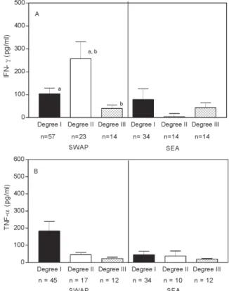
Cytokine response to S. mansoni antigens - A great variability in cytokine levels was observed in general, showing the heterogeneity of the response to S. mansoni antigens. A significantly (p = 0.03, Mann Whitney test) higher level of IFN-γ was observed in supernatants of SWAP-stimulated PBMC from patients with degree II (n = 23) versus degree I (n = 57) versus degree III (n = 14) hepatic fibrosis (257 ± 354 versus 104 ± 189 vs 39 ± 58 pg/ ml, respectively). No significant differences in IFN-γ lev-els were observed in supernatants from SEA stimulated PBMC from the three groups of patients (79 ± 295.8, 3 ± 14.2 and 44 ± 79 pg/ml), (Fig. 1A). The levels of TNF-α in SWAP and SEA-stimulated PBMC supernatants also did not show any differences between the groups (Fig. 1B).
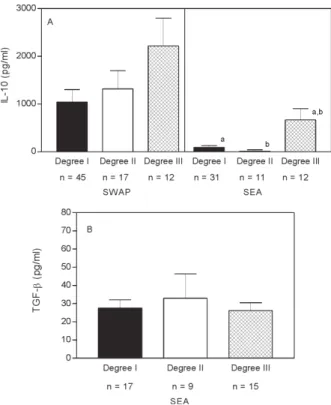
High levels of IL-5 were produced by SWAP-stimu-lated PBMC from all groups of patients (degree I 1043 ± 1844.5, degree II 1319 ± 1706.7, and degree III 2219 ± 2011), with significantly higher levels in patients with degree III versus degree I fibrosis (p = 0.03, Mann Whitney test). Levels of IL-5 were significantly lower in supernatants from SEA-stimulated PBMC of subjects with degrees I (93 ± 248.4 pg/ml) and II (11 ± 29.8 pg/ml) of hepatic fibro-sis, than in supernatants from patients with degree III hepatic fibrosis (669 ± 815.6pg/ml), (p = 0.01 and p = 0.009, respectively, Mann Whitney test) (Fig. 2A). IL-13 levels in supernatants of SEA stimulated PBMC were also higher in subjects with degree III fibrosis (116 ± 120.9 pg/ml), as compared to those with degrees I (24 ± 11.4 pg/ml) or II (86 ± 166.5 pg/ml) of hepatic fibrosis, (p = 0.009 and p = 0.05, respectively, Mann Whitney test) (Fig. 2B). Levels of IL-10 in SWAP- stimulated PBMC was not statistically significant. Higher levels of IL-10 were observed in su-pernatants of SEA-stimulated PBMC from patients with degree III hepatic fibrosis (187 ± 110 pg/ml) compared to those with degree I (63 ± 62.9 pg/ml) or II (66 ± 53 pg/ml) of hepatic fibrosis (p = 0.01 and p = 0.03, respectively, Mann Whitney test) (Fig. 3A). No differences were detected in TGF-β levels in SEA-stimulated PBMC supernatants be-tween the groups of patients with degrees I, II, and III hepatic fibrosis. (Fig. 3B).
Fig.1: interferon-γ (A) and tumor necrosisfactor-α (B) levels in SWAP and SEA-stimulated peripheral blood mononuclear cells su-pernatants in schistosomiasis patients with different degrees of hepatic fibrosis. Data represents the mean± Standard Error (SEM). a:p = 0.03; b:p = 0.02, Mann Whitney test
2 4 2 4 2 4 2 4
2 4 Cytokine in Chronic Shistosomiasis • Andréa M agalhães et al.
Responses of patients who remained untreated fol-lowing initial assessment. Even after an active search, 21
patients missed treatment that should be performed at the end of the study, and remained untreated after one year from their first ultrasonographyexamination. Ultrasonog-raphy was repeated on these individuals and 19 (90%) of them were found to have increased periportal thickness and 2 of them remained with similar measurements. Within this group, 12/19 (57%) exhibited more severe fibrosis compared to the previous exam. In 3 patients the fibrosis index had changed from degree 0 to degree I, and in 9 patients the fibrosis index had increased from degree I to degree II. Immunological evaluation was repeated in these patients and compared with the first evaluation. An in-crease in the levels of some cytokines was observed in SEA-stimulated PBMC supernatants. Levels of IFN-γ were evaluated in 18 patients and 2 patient presented a de-crease but the others did not show any significant change in the second versus first evaluations (Fig. 4A). TNF-α levels increased in 5 patients and decreased in 2 patients and remained at similar levels in 8 of the 15 patients tested (Fig. 4B). IL-5 levels increased in 9 patients, decreased in 1 patient and remained at similar levels in 8 of the 18 pa-tients tested (Fig. 5A), and IL-13 levels showed a major increased in 5 patients, a minor increase in 2 patients and decreased orremained at similar levels in 2 of the 9 pa-tients tested (Fig. 5B). The levels of IL-5 and IL-13 were significantly higher in the second evaluation (206 ± 434
and 89.7 ± 56 pg/ml, respectively), in comparison to the first evaluation (19 ± 62 pg/ml and 9.6 ± 11pg/ml, respec-tively), (p = 0.03 and p = 0.04 respectively, Wilcoxon signed rank test). IL-10 levels also increased in 7 of 13 patients, decreased in 3 and remained at similar levels in other 3 patients (Fig. 6A), and TGF-β levels increased in 6 of 14 patients, decreased in 2 and remained at similar levels in other2 patients (Fig. 6B). No statistically significant dif-ferences were observed in the levels of IFN-γ, TNF-α, IL-10 and TGF-β in SEA-stimulated PBMC supernatants from the second to the first evaluations (p = 0.4, p = 0.4, p = 0.3 and p = 0.4, respectively, Wilcoxon signed rank test). No significant differences were observed between the levels of cytokines in SWAP-stimulated PBMC supernatants from the second to the first evaluations.
Fig. 3: interleukin-10 (A) and transforming growth factor-β (B) levels in SWAP and SEA-stimulated peripheral blood mononuclear cells supernatants in schistosomiasis patients with different degrees of hepatic fibrosis. Data represents the mean± Standard Error (SEM). a: p = 0.01; b: p = 0.03, Mann Whitney test
Fig. 4: interferon-γ (A) and tumor necrosis factor-α (B) levels in SEA-stimulated peripheral blood mononuclear cells supernatants from subjects who increased the periportal tract thickness after one-year follow-up without treatment. a: Wilcoxon signed rank test
Fig. 5: interleukin-5 (A) and IL-13 (B) levels in SEA-stimulated peripheral blood mononuclear cells supernatants from subjects who increased the periportal tract thickness after one-year follow-up without treatment. IL-5 and IL-13 levels increased in (50%) of the patients. a: Wilcoxon signed rank test
2 5 2 52 5 2 5 2 5 Mem Inst Oswaldo Cruz, Rio de Janeiro, Vol. 99(Suppl. I), 2004
DISCUSSION
A type-2 cytokine pattern dominates the immune re-sponse in mice and humans chronically infected by S. mansoni. In mice, IL-4 and IL-13 play a major role in egg granuloma formation, and IL-13 was shown to play a sig-nificant role in the development of hepatic fibrosis (Chiaramonte et al. 1999, 2001, Monica et al. 2003). In hu-mans, although low IFN-γ and high TNF-α are associated to severe hepatic fibrosis (Henri et al. 2002, Booth et al. 2004), it is still not clear which cytokines are involved in the progression of schistosomiasis mansoni pathology. The present study evaluated the cytokine profile in schis-tosomiasis patients developing hepatic fibrosis in pre-hepatosplenic and early pre-hepatosplenic stages of the dis-ease, and showed an association between a type 2 cytokine profile (the production of IL-5, IL-10, and IL-13) and degree III hepatic fibrosis. IL-5 and IL-13 showed the strongest association with severe hepatic fibrosis, and also increased significantly in patients who exhibited in-creased hepatic fibrosis after 1 year without treatment. Besides these Th2 cytokines, TGF-β also increased in about 50% of thepatients who exhibit increased hepatic fibrosis after 1 year without treatment. remained at similar. However TGF-β levels did not differ in the groups with different degrees of hepatic fibrosis.
Many variables may influence the magnitude of the immune response in human schistosomiasis, including age, intensity of infection, and the type of Ag used to assess immune responsiveness. Moreover, increasing age is associated with an enhanced type 1 immune response (Ribeiro de Jesus et al. 1993) and heavy infections (more than 200 eggs/g stool) are associated with enhanced type 2 response (Araujo et al. 1996). These variables did not explain the results of the present study, since patients with degree III hepatic fibrosis made more IL-5 and IL-13 despite the fact that their age were higher than the other groups, and that they had the lowest intensity of infec-tion.
Recent studies have pointed out an important role of IL-13 in the development of liver fibrosis (Chiaramonte et al. 2001, Monica et al. 2003).Although IL-4 and IL-13 share the same receptor and many biological activities, there are functional differences between the two cytokines. While granuloma formation was partially reduced in IL-4-deficient mice, blocking IL-13 and the IL-4 receptor in these animals almost completely abrogated granuloma devel-opment and tissue fibrosis (Chiaramonte et al. 2001). Our data, by showing a correlation between the production of high levels of IL-13 and the development of more severe fibrosis, suggests that this cytokine might also play a significant role in human schistosomiasis fibrosis. A re-cent study has found a protective effect of IFN-γ in liver fibrosis in Sudanese S. mansoni-infected subjects (Henri et al. 2002). Another recent study has also associated low IFN-γ production with liver fibrosis(Booth et al. 2004).In fact, earlier studies on schistosomiasis pathology in mice demonstrated that IFN-γ reduces the cellularity of granu-lomas, and downmodulates granuloma size (Boros & Lukacs 1992). These data are in concordance with the data from the present study, because IFN-γ is the major
type 1 cytokine involved in down modulate T helper type 2 cells.
In contrast to our data that showed increased IL-5 and IL-13 associated with liver fibrosis and that showed any significant associations between TNF-α levels and liver fibrosis, studies in schistosomiasis patients from Africa showed that patients with more severe hepatic fibrosis had higher TNF-α in PBMC supernatants (Mwatha et al. 1998, Henri et al. 2002, Booth et al. 2004). The contradic-tory results might be explained by the differences in the stages of the disease evaluated in these 2 studies: while the present study evaluated patients in the pre-hepatosplenic and early pre-hepatosplenic stages of schisto-somiasis mansoni, these studies evaluated hepatosplenic patients. Since the degree III hepatic fibrosis patients are in the early hepatosplenic phase of disease evolution, it is possible that the TNF-α is produced in later stages of the disease. Another possible explanation for the oppos-ing results of these 2 studies is that the African popula-tion has a high prevalence of malaria, a protozoan infec-tion that is known to cause hepatosplenomegaly and also to induce high levels of TNF-α (Mwathaet al. 1998, Clark 2003, Naus 2003).Further studies are necessary to better clarify the cytokines involved in advanced stages of hepatosplenic schistosomiasis, and the role of co-infec-tions with malaria and other pathogens.
The documentation that IL-10 was found to be higher in subjects with degree III compared to degrees I and II hepatic fibrosis was unexpected. IL-10 is a modulatory cytokine which down regulates macrophage activation, MHC class I and II expression and reduces activation of both Th1 and Th2 cells (De Wall-Malefyt et al. 1993). Pvious studies have shown that IL-10 is important to re-duce pathology of acute schistosomiasis and it is de-creased in patients with hepatosplenomegaly (Falcão et al. 1998). Since the patients in the present study were in the pre-hepatosplenic and early stages of hepatosplenom-egaly it is possible that the documentation herein of in-creased levels of IL-10 in patients developing liver fibro-sis may represent an attempt of this cytokine to down regulate the high levels of IL-5 and IL-13. The increment of TGF-β levels during evolution of hepatic lesions and lack of differences in subjects with degrees I, II, and III hepatic fibrosis could be due to an intermittent induction of fibrosis.
The present study presents evidence for a role of type 2 immune response in the development of liver fibrosis in human schistosomiasis. Since IL-5, IL-10, and IL-13 were associated with degree III hepatic fibrosis, and IL-5, IL-13 increased significantly in patients who developed more serious hepatic fibrosis over the course of the study.
ACKNOWLEDGMENTS
To Antônio E de Souza and Sonia B de Souza for their major help in the endemic area. To Elbe M, Silva, Lúcia dos Reis, and Jackson Lemos for secretarial and computer assistance.
REFERENCES
2 6 2 6 2 6 2 6
2 6 Cytokine in Chronic Shistosomiasis • Andréa M agalhães et al.
Abdel-Wahab MF, Ramzy I, Esmat G, el Kafass H, Strickland GT 1992. Ultrasound for detecting Schistosoma haema-tobium urinary tract complications: comparison with ra-diographic procedures. J Urol 148: 346-50.
Bica I, Hamer DH, Stadecker MJ 2000. Hepatic schistosomia-sis. Infect Dis Clin North Am 14: 583-604.
Booth M, Mwatha JK, Joseph S, Jones FM, Kadzo H, Ireri E, Kazibwe F, Kemijumbi J, Kariuki C, Kimani G, Ouma JH, Kabatereine NB, Vennervald BJ, Dunne DW 2004, Peri-portal fibrosis in human Schistosoma mansoni infection is associated with low IL-10, low IFN-gamma, high TNF-alpha, or low RANTES, depending on age and gender. J Immunol 172: 1295-303.
Boros DL, Lukacs NW 1992. The role of egg antigens, cytokines in granuloma formation in murine schistosomiasis mansoni. Mem Inst Oswaldo Cruz 87: 75-79.
Cheever AW, Williams ME, Wynn TA, Finkelman FD, Seder RA, Cox TM, Hieny S, Caspar P, Sher A 1994. Anti-IL-4 treatment of Schistosoma mansoni-infected mice inhibits development of T cells and non-B, non-T cells expressing Th2 cytokines while decreasing egg-induced hepatic fibro-sis. J Immunol 153: 753-759.
Chiaramonte MG, Cheever AW, Malley JD, Donaldson DD, Wynn TA 2001. Studies of murine schistosomiasis reveal interleukin-13 blockade as a treatment for established and progressive liver fibrosis. Hepatology 34: 273-282. Chiaramonte MG, Donaldson DD, Cheever AW, Wynn TA 1999.
An IL-13 inhibitor blocks the development of hepatic fi-brosis during a T-helper type 2-dominated inflammatory response. J Clin Invest 104: 777-785.
Clark IA 2003. The pathophysiology of falciparum malaria. Pharmacol Ther 99: 221-260.
de Jesus AR, Miranda DG, Miranda RG, Araujo I, Magalhaes A, Bacellar M, Carvalho EM 2000. Morbidity associated with Schistosoma mansoni infection determined by ultra-sound in an endemic area of Brazil, Caatinga do Moura. Am J Trop Med Hyg 63: 1-4.
De Wall-Malefyt HR, Spits JH, Rancarolo M, De Vries J 1993. L-10 and viral IL-10 strongly reduce antigen-specific hu-man T cell proliferation by diminishing the antigen-pre-senting capacity of monocytes via down regulation of class II MHC expression. JExp Med 174: 915-924.
Doumenge JP 1987. Atlas of Global Distribution of Schistoso-miasis, Université de Bordeaux Press, Bordeaux, France. Falcão PL, Malaquias LC, Martins-Filho OA, Silveira AM,
Passos VM, Prata A, Gazzinelli G, Coffman RL, Correa-Oliveira R 1998. Human schistosomiasis mansoni: IL-10 modulates the in vitro granuloma formation. Parasite Immunol 20: 447-454.
Henri S, Chevillard C, Mergani A, Paris P, Gaudart J, Camilla C, Dessein H, Montero F, Elwali NE, Saeed OK, Magzoub M, Dessein AJ 2002. Cytokine regulation of periportal fibro-sis in humans infected with Schistosoma: IFN-gamma is
associated with protection against fibrosis and TNF-alpha with aggravation of disease. J Immunol 169: 929-936. Monica G, Chiaramonte MM-K, Jacobson BA, Cheever AW,
Whitters MJ, Goad MEP, Wong V, Collins M, Donaldson DD, Grusby MJ, Wynn TA 2003. Regulation and function of the interleukin 13 receptor alpha 2 during a T helper cell type 2-dominant immune response. J Exp Med 197: 687-701.
Mwatha JK, Kimani G, Kamau T, Mbugua GG, Ouma JH, Mumo J, Fulford AJ, Jones FM, Butterworth AE, Roberts MB, Dunne DW 1998. High levels of TNF, soluble TNF receptors, soluble ICAM-1, and IFN-gamma, but low lev-els of IL-5, are associated with hepatosplenic disease in human schistosomiasis mansoni. J Immunol 160: 1992-1999.
Naus CW, Jones FM, Satti MZ, Joseph S, Riley EM, Kimani G, Mwatha JK, Kariuki CH, Ouma JH, Kabatereine NB, Vennervald BJ, Dunne DW 2003. Serological responses among individuals in areas where both schistosomiasis and malaria are endemic: cross-reactivity between Schistosoma mansoni and Plasmodium falciparum. J Infect Dis 187: 1272-1282.
Pearce EJ, James SL, Dalton J, Barrall A, Ramos C, Strand M, Sher A 1986. Immunochemical characterization and purification of Sm-97, a Schistosoma mansoni antigen monospecifically recognized by antibodies from mice pro-tectively immunized with a nonliving vaccine. J Imunol 137: 3593-3600.
Pessoa SB, Martins AV 1978. Parasitologia Medica, 10th ed., Guanabara Koogan, Rio de Janeiro.
Ribeiro de Jesus A, Araujo I, Bacellar O, Magalhães A, Pearce E, Harn D, Strand M, Carvalho EM 2000. Human immune responses to Schistosoma mansoni vaccine candidate anti-gens. Infect Immun 68: 2797-2803.
Ribeiro de Jesus AR, Silva A, Santana LB, Magalhães A, de Jesus AA, de Almeida RP, Rego MA, Burattini MN, Pearce EJ, Carvalho EM 2002. Clinical and immunologic evalua-tion of 31 patients with acute schistosomiasis mansoni. J Infect Dis 185: 98-105.
Ribeiro de Jesus A, Almeida RP, Bacellar O, Araújo MI, De-meure C, Bina JC, Dessein AJ, Carvalho EM 1993. Corre-lation between cell-mediated immunity and degree of infec-tion in subjects living in na endemic area of schistosomiasis. Eur J Immunol 23: 152-158.
Sabin EA, Araújo MI, Carvalho EM, Pearce EJ 1996. Impair-ment of tetanus toxoid-specific Th1-like immune responses in humans infected with Schistosoma mansoni. J Infect Dis 173: 269-272.
Warren KS 1968. Pathophysiology and pathogenesis of hepatosplenic schistosomiasis mansoni. Bull NY Acad Med 44: 280-294.