SCYPHOZOA, SEMAEOSTOMEAE AND RHIZOSTOMEAE) IN THE
SOUTHEASTERN BRAZILIAN COAST
1 Centro de Biologia Marinha, Universidade de São Paulo, Caixa Postal 83, 11600-970, São Sebastião, SP, Brasil. Tel.:+55-12-38627149 Fax: +55-12-4626646
2 Departamento de Zoologia, Instituto de Biociências, Universidade de São Paulo, Caixa Postal 11461, 05422-970, São
Paulo, SP, Brasil.
Tel.:+55-11-30917619 Fax: +55-11-30917513
e-mails: valbadtr@usp.br, acmorand@usp.br, aemigott@usp.br
Resumo – A ocorrência de éfiras de cifozoários das ordens Semaeostomeae e Rhizostomeae é registrada pela primeira vez para a costa brasileira. Os espécimes, coletados com arrastos de plâncton no Canal de São Sebastião e no Sistema estuarino-lagunar de Cananéia, são: Chrysaora lactea (Semaeostomeae), Phyllorhiza punctata (Rhizostomeae), e uma espécie não identificada de Pelagia (Semaeostomeae). Uma tabela, com todas as espécies de cifozoários com ciclo de vida conhecido, é apresentada.
Palavras-chave : Scyphozoa, Semaeostomeae, Rhizostomeae, éfira, ciclo de vida, Brasil.
Abstract – The occurrence of ephyrae of the scyphozoan orders Semaeostomeae and Rhizostomeae is reported for the first time for the Brazilian coast. The specimens, caught in plankton tows in the São Sebastião Channel and the Cananéia lagoon estuarine system, are: Chrysaora lactea (Semaeostomeae), Phyllorhiza punctata (Rhizostomeae), and an unidentified species of Pelagia (Semaeostomeae). A table with all species of scyphozoan with the known life cycle is provided.
Palavras-chave : Scyphozoa, Semaeostomeae, Rhizostomeae, ephyra, life cycle, Brazil.
Biota Neotropica v2 (n2) – http://www.biotaneotropica.org.br/v2n2/pt/abstract?article+BN02102022002
Date Received 07/04/2002 Revised 08/03/2002
Accepted 10/02/2002
Tronolone,V. B.; Morandini, A. C.; Migotto, A. E. - Biota Neotropica, v2 (n2) - BN02102022002 2
Introduction
The class Scyphozoa is defined, besides other fea-tures, by the process of strobilation (Schuchert, 1993), i.e. successive vegetative (asexual reproduction) cutting-off of a sequence of new individuals (ephyrae) from one end of a parent benthic polyp (scyphistomae) (Mianzan & Cornelius, 1999: 525, the text in parenthesis was added). These disks (ephyrae) are the young (immature) free-swimming stages of the scyphozoans (Stachowitsch, 1992). Strobilation can be divided into monodisc (one disc produced at a certain time) or polydisc (more than one disc produced at a certain time) (Arai, 1997).
In the life cycle of Stylocoronella riedli and S. variabilis (members of the order Stauromedusae), the inter-stitial polyp metamorphoses into a medusa (see Kikinger & Salvini-Plawen, 1995), thus not presenting an ephyra stage. Collins (2002) proposed that the Stauromedusae be sepa-rated from the other Scyphozoa, due to the absence of the strobilation process and, consequently, not producing ephyrae. The absence of strobilation and ephyrae, plus the presence of a claustrum led several authors to the conclu-sion that the Stauromedusae are closely related to the class Cubozoa (cf. Haeckel, 1880; Uchida, 1929; one topology found by Collins, 2002).
Some scyphomedusae present a holopelagic life cycle (absence of the polyp stage, and thus, of strobilation), e.g., the semaeostome Pelagia noctiluca (see Rottini Sandrini & Avian, 1983) and the coronate Periphylla periphylla (see Jarms et al., 1999).
Besides the general morphological similarity of the ephyra stage of most species, which makes their specific identification difficult, there are few life cycle studies of Scyphozoa, that include detailed accounts of this plank-tonic stage, to rely on. Out of ca. 200 species of scyphozo-ans (Mianzan & Cornelius, 1999), only about 42 species (almost a quarter of those known) had their life cycles de-scribed (see Table 1 in Results and Discussion). Russell (1970) provided a comparative plate of the ephyra stages of the British species. Even among the works devoted to the life cycle of scyphozoan species, only a few [e.g., Silveira & Morandini (1997) and Jarms (2001), for Coronatae; Avian (1986) and Pitt (2000), for Rhizostomeae; Calder (1972) and Gershwin & Collins (2002), for Semaeostomeae] included descriptions of ephyrae. For some species, information con-cerning life cycle stages is scattered throughout the litera-ture, included in systematic or faunistc reports among de-scriptions of other species, or referring to one or another stage only.
None of the 23 scyphozoans recorded for Brazil (see Migotto et al., 2002) refer to ephyrae collected in nature. Despite their efforts, Silveira & Morandini (1998) could not find the ephyra and medusa of the coronate Linuche
unguiculata, a species associated with a calcareous sub-strate and abundant in the region of São Sebastião. Goy (1979) mentioned the presence of young specimens of Chrysaora lactea (as C. hysoscella) on the coast of Uru-guay, and young Aurelia aurita on the coast of Bahia State (Brazil).
This work reports and describes three different ephyrae found on the southeastern coast of Brazil. A table covering the knowledge from literature and our own experi-ences in life cycles of Scyphozoa (except Stauromedusae) is provided.
Material and Methods
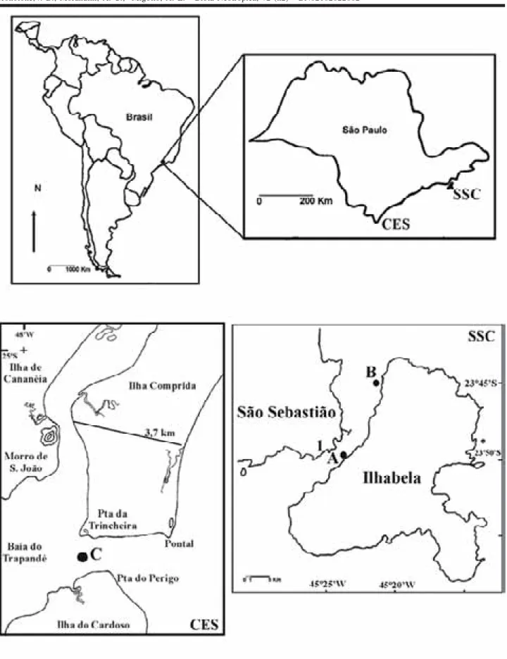
Specimens came from two localities on the coast of São Paulo State, southeastern Brazil: the São Sebastião Channel (23ºS – 45ºW) (SSC) and the Cananéia lagoon es-tuarine system (25ºS – 48ºW) (CES) (see Figure 1).
In the SSC, two ephyrae were collected with vertical plankton tows (300 and 500 µm mesh sizes; maximum depth 40 m) in August and October 1999, and reared in the labora-tory until the first signs of gonadal tissue were detected. The ephyrae were isolated in glass containers filled with filtered seawater (changed daily) and kept in a constant temperature chamber at 20-21ºC. The medusae were fed daily with Artemia sp. nauplii, besides other planktonic organ-isms (especially copepods) and small pieces of muscle and gonads of mussels (Perna perna).
In the CES, ephyrae were collected with horizontal (0.5 m below surface) and vertical (maximum depth 12 m) plankton tows (500 and 200 µm mesh sizes) in April 2001, and January and February 2002. The ephyrae were trans-ferred to culture dishes, kept in a constant temperature cham-ber at 22ºC and reared as described by Jarms et al. (in press). Mature medusae of Chrysaora lactea collected at CES and reared in “planktonkreisel” (according to Greve, 1968) pro-duced planulae that settled on the “planktonkreisel” walls and originated scyphistomae; these were transferred to cul-ture dishes, there producing ephyrae.
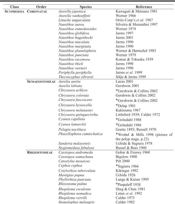
Table 1. Scyphozoan species with known life cycles (except Stauromedusae), adapted from several sources. The most recent references or those with the most complete description regarding their life cycle were included. A reference which mentions one or another stage was included and marked with an “*”.
Class Order Species Reference
SCYPHOZOA CORONATAE Atorella japonica Kawaguti & Matsuno 1981
Atorella vanhoeffeni Werner 1966
Linuche unguiculata Ortiz-Corp’set al. 1987 Nausithoe aurea Silveira & Morandini 1997 Nausithoe eumedusoides Werner 1974
Nausithoe globifera Jarms 1997 Nausithoe hagenbecki Jarms 2001 Nausithoe maculata Jarms 1990 Nausithoe marginata Jarms 1990
Nausithoe planulophora Werner & Hentschel 1983 Nausithoe punctata Werner 1979
Nausithoe racemosa Komai & Tokuoka 1939 Nausithoe thieli Jarms 1990
Nausithoe werneri Jarms 1990 Periphylla periphylla Jarmset al. 1999 Thecoscyphus zibrowii Sötje & Jarms 1999
SEMAEOSTOMEAE Aurelia aurita Lucas 2001
Aurelia labiata Gershwin 2001
Chrysaora achlyos
*
Gershwin & Collins 2002 Chrysaora colorata Gershwin & Collins 2002 Chrysaora fuscescens*
Gershwin & Collins 2002 Chrysaora hysoscella*
Delap 1901Chrysaora melanaster Kakinuma 1967
Chrysaora quinquecirrha Littleford 1939; Calder 1972 Cyanea capillata
*
Gröndahl 1988Cyanea lamarckii
*
Gröndahl 1988Pelagia noctiluca Goette 1893; Russell 1970
Phacellophora camtschatica
*
Wrobel & Mills 1998 (picture of the polyp stage, p.22)Sanderia malayensis Uchida & Sugiura 1978 Stygiomedusa fabulosa Russel & Rees 1960
RHIZOSTOMEAE Cassiopea andromeda Gohar & Eisawy 1960
Cassiopea xamachana Bigelow 1900 Catostylus mosaicus Pitt 2000 Cephea cephea
*
Sugiura 1966 Cotylorhiza tuberculata Kikinger 1992 Mastigias papua Uchida 1926Tronolone,V. B.; Morandini, A. C.; Migotto, A. E. - Biota Neotropica, v2 (n2) - BN02102022002 4
Results and Discussion Known life cycle
A review of scyphozoan literature was performed to build up a list with all scyphozoan species (except for the order Stauromedusae) in which the life cycle is known. This list is presented here as Table 1. We tried to provide the most complete or the most recent reference for each spe-cies. But, for some of them there is little information on life cycle stages, and we decided to include references which mentioned one or another stage (these are marked with a “*” in the table). As mentioned in the Introduction, life cycles have been described of only about 42 species, this means almost a quarter of the known scyphozoan species. The order with more species with known life cycles is Coronatae (16 spp.), followed by Semaeostomeae (14 spp.), the last being Rhizostomeae (12 spp.).
Order Rhizostomeae
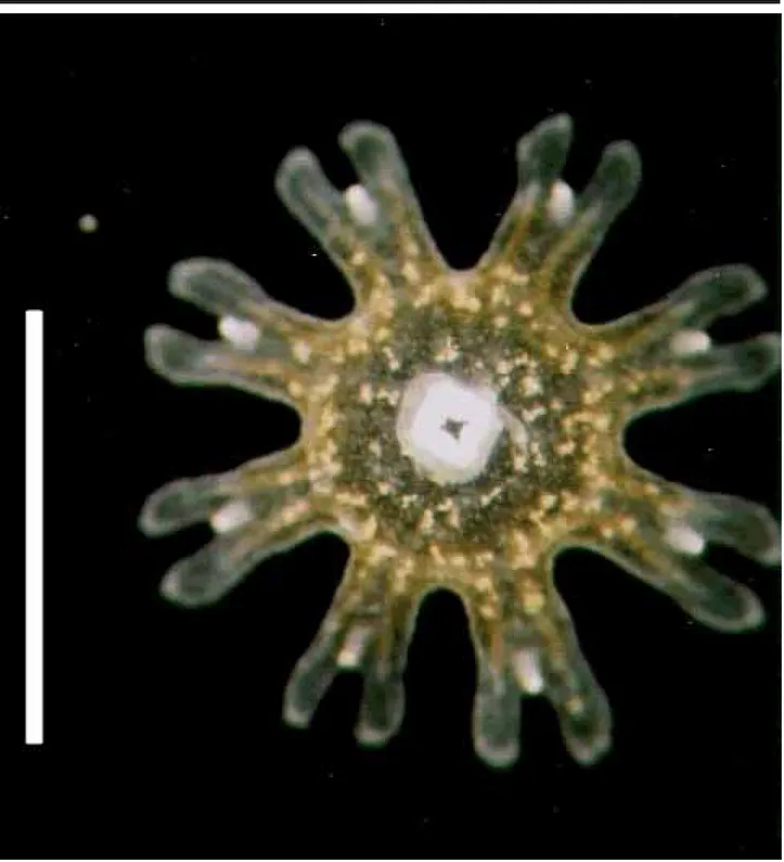
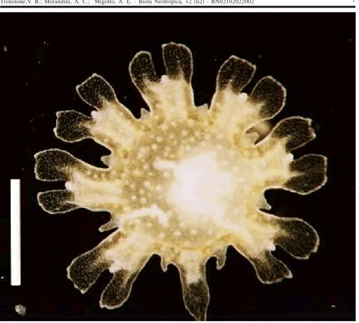
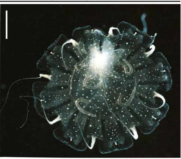
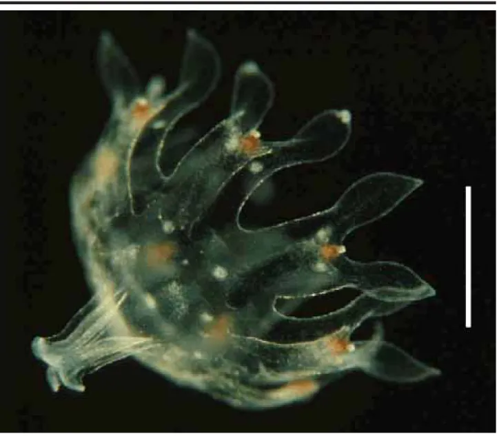
Phyllorhiza punctata von Lendenfeld, 1884 Seven specimens (Figure 2), found in the waters of CES (salinity 20-24‰; temperature 28ºC) measuring 1.5-2.5 mm in diameter when collected, were reared for 54 days. Their tissues were filled with zooxanthellae, and they pre-sented 8 lobes with rounded lappets and 4 gastric filaments; some specimens had small warts on the exumbrella. By the 7th day (Figure 3), the marginal lappets started to grow
be-tween the lobes, and the manubrium became more elon-gated. By the 10th day the oral arms showed the first signs of
oral tentacles and began to bifurcate. On the 40th day the
mouth closed and the exumbrellar warts were more con-spicuous.
These ephyrae were identified as Phyllorhiza punctata by the presence of zooxanthellae and the white warts on the exumbrella. In the same period that the studied specimens were found, large medusae were also present in the area. Ephyrae obtained from laboratory cultures mea-sured 1.5 mm when detached from the scyphistomae, sug-gesting that the specimens collected in the plankton tows had been newly released. Although recent articles (Garcia, 1990; Rippingale & Kelly, 1995) mention the occurrence of the ephyrae stage of P. punctata, until now there is no mor-phological description of it in the literature. D’Ambra et al. (2001) commented on the flow and prey capture of young P. punctata (1.4-7.4 cm in diameter).
Order Semaeostomeae
Chrysaora lactea Eschscholtz, 1829
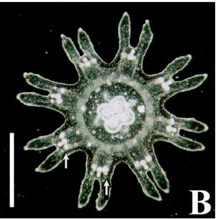
One ephyra (Figure 4a) was found in SSC (salinity: 34‰; temperature 20.8ºC), and reared for 38 days. When collected it had a transparent body, was 1.2 mm in diameter, had a manubrium with red pigment spots, pointed lappets (3
lappets were probably damaged during collecting, regener-ated during the first week), and nematocyst clusters at the base of each marginal lappet (i.e., in the typical Chrysaora-pattern, cf. Russell, 1970; Gershwin & Collins, 2002). From days 10-19 the beginning of the 8 marginal tentacles were noted, and from days 20-29 the pigmentation of the manu-brium became pale and smaller (Figure 5). From days 30-37, 5-9 gastric filaments were observed in each quadrant. At the 38th day the young medusa reached a diameter of 2 cm
and the manubrium a length of 4 cm (Figure 6). At this stage, secondary tentacle buds were observed growing from be-low the lappets (Figure 7), totaling 24 tentacles.
The number of tentacles and the nematocyst clus-ters arranged in a pattern are diagnostic characclus-ters of Chrysaora; as the only species of the genus that occurs on the Brazilian coast is Chrysaora lactea, the specimen is identified as that. With this, secondary tentacles arising below the marginal lappets are diagnostic characters of C. lactea. The ephyrae obtained in the laboratory from mature medusae of C. lactea collected at CES were identical to the nature-collected specimen, except for the lack of pigmenta-tion (Figure 4b). Calder (1972) noted that ephyra of C. quinquecirrha obtained from scyphistomae raised in the laboratory had no pigmentation. Little information on the biology of the species exists. Mianzan (1989a, 1989b) men-tioned that some ephyrae of C. lactea were collected in plankton tows along the Argentinean coast.
Pelagia Péron & Lesueur, 1810
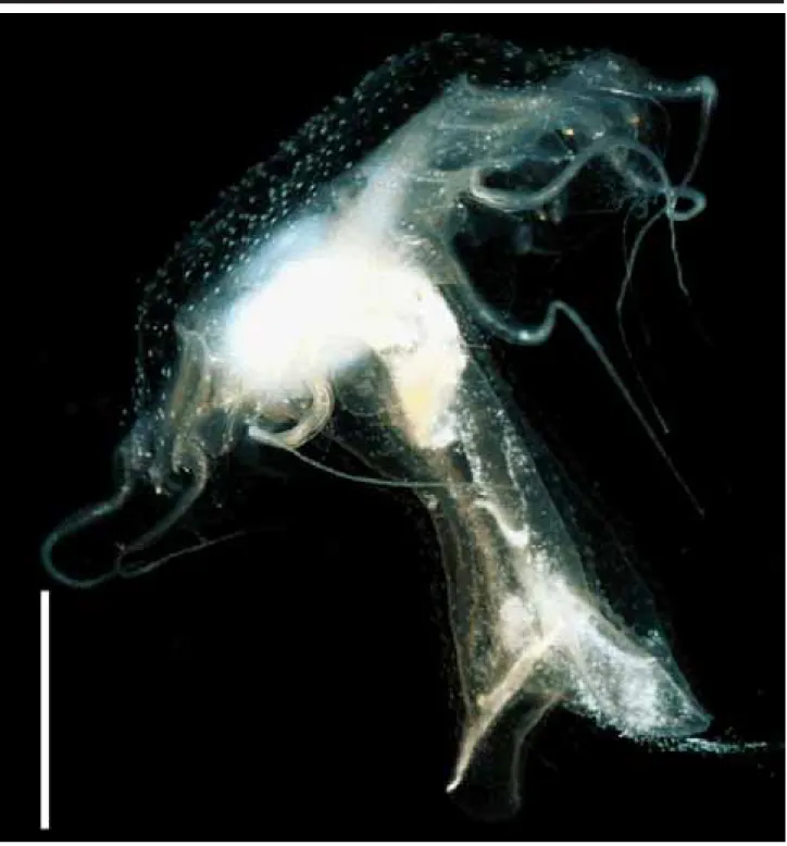
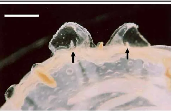
One ephyra (Figure 8), found in the waters of SSC (salinity: 34-36‰; temperature: 20.5ºC), had a diameter of 2.4 mm when collected. It was reared for 76 days. In the laboratory, from days 1-19, it acquired 2 gastric filaments, 4 milky spots on the stomach, red pigment on the manubrium and nematocyst clusters around the central disk. From days 20-39, 2 marginal tentacles and 4-5 gastric filaments on each quadrant developed, the manubrium attained a length of 4.3 mm, and warts on the umbrella and manubrium (Figure 9) were visible; at the same time the milky spots on the stom-ach disappeared. From days 40-70 many gastric filaments (up to 10 in each quadrant) were observed in each quadrant, the number of warts increasing. On the 76th day, the young
medusa was 3 cm in diameter, the manubrium was 5.3 cm long, there were 8 tentacles and the beginning of the go-nads was noted.
Tronolone,V. B.; Morandini, A. C.; Migotto, A. E. - Biota Neotropica, v2 (n2) - BN02102022002 6
Tronolone,V. B.; Morandini, A. C.; Migotto, A. E. - Biota Neotropica, v2 (n2) - BN02102022002 8
Tronolone,V. B.; Morandini, A. C.; Migotto, A. E. - Biota Neotropica, v2 (n2) - BN02102022002 10
Figure 5. Oral view of ephyra of Chrysaora lactea Eschscholtz, 1829, collected in the São Sebastião Channel in August
Tronolone,V. B.; Morandini, A. C.; Migotto, A. E. - Biota Neotropica, v2 (n2) - BN02102022002 12
Tronolone,V. B.; Morandini, A. C.; Migotto, A. E. - Biota Neotropica, v2 (n2) - BN02102022002 14
Tronolone,V. B.; Morandini, A. C.; Migotto, A. E. - Biota Neotropica, v2 (n2) - BN02102022002 16
Acknowledgements
We are grateful to Dr Fábio L. da Silveira (IB-USP, Brazil) for assistance and the critical reading of the manu-script. We also thank the Departamento de Zoologia (IB-USP), Instituto Oceanográfico da Universidade de São Paulo (IO-USP), and Centro de Biologia Marinha (CEBIMar-USP) for providing collecting and logistic support. Dr. H.W. Mianzan (INIDEP, Argentina) and MSc. A. Lindner (Duke University, USA) provided important literature. VBT and AEM had financial support from the Conselho Nacional de Desenvolvimento Científico e Tecnológico (CNPq) and from CAPES/DS/PROAP. ACM received financial support from the Fundação de Amparo à Pesquisa do Estado de São Paulo (FAPESP 99/05374-7).
References
ARAI, M.N., 1997. A Functional Biology of Scyphozoa. Chapman & Hall, London, 316 p.
AVIAN, M., 1986. Biological cycle of Cotylorhiza tubercolata (Macri 1778): morphological aspects of the development from ephyra to young medusa. Nova Thalassia, 8(suppl.2): 47-58.
BIGELOW, R.P., 1900. The anatomy and development of Cassiopea xamachana. Mem. Boston Soc. Nat. Hist., 5: 193-237.
CALDER, D.R., 1972. Development of the sea nettle Chrysaora quinquecirrha (Scyphozoa, Semaeostomeae). Ches. Sci., 13: 40-44.
CALDER, D.R., 1973. Laboratory observations on the life history of Rhopilema verrilli (Scyphozoa: Rhizostomeae). Mar. Biol., 21: 109-114.
CALDER, D.R., 1982. Life history of the cannonball jelly-fish, Stomolophus meleagris L. Agassiz, 1860 (Scyphozoa, Rhizostomida). Biol. Bull., 162: 149-162. COLLINS, A.G., 2002. Phylogeny of Medusozoa and the
evolution of cnidarian life cycles. J. Evol. Biol., 15: 418-432.
D’AMBRA, I.; J.H. COSTELLO & F. BENTIVEGNA, 2001. Flow and prey capture by the scyphomedusa Phyllorhiza punctata von Lendenfeld, 1884. Hydrobiologia, 451 (Dev. Hydrobiol. 155): 223-227.
DELAP, M.J., 1901. Notes on the rearing of Chrysaora isos-celes in an aquarium. Ir. Nat., 10: 25-28.
DING, G. & J. CHEN, 1981. The life history of Rhopilema esculenta Kishinouye. J. Fish. China, 5: 93-104. [In Chi-nese]
GARCÍA, J.R., 1990. Population dynamics and production of Phyllorhiza punctata (Cnidaria: Scyphozoa) in La-guna Joyuda, Puerto Rico. Mar. Ecol. Prog. Ser., 64: 243-251.
GERSHWIN, L., 2001. Systematics and biogeography of the jellyfish Aurelia labiata (Cnidaria: Scyphozoa). Biol. Bull., 201: 104-119.
GERSHWIN, L. & A.G. COLLINS, 2002. A preliminary phy-logeny of Pelagiidae (Cnidaria, Scyphozoa), with new observations of Chrysaora colorata comb. nov. J. Nat. Hist., 36(2): 127-148.
GOETTE, A., 1893. Vergleichende Entwicklungsgeschichte von Pelagia noctiluca Pér. Z. wiss. Zool., 55: 645-695. GOHAR, H.A.F. & A.M. EISAWY, 1960. The development
of Cassiopea andromeda. Publ. Mar. Biol. Sta., Al-Ghardaqa, 11: 148-190.
GOY, J. 1979. Campagne de la Calypso au large des côtes atlantiques de l’Amérique du Sud (1961-1962) - 35. Méduses. Rés. scient. Camp. Calypso, 11: 263-296. GREVE, W., 1968. The “planktonkreisel”, a new device for
culturing zooplankton. Mar. Biol., 1: 201-203.
GRÖNDAHL, F., 1988. A comparative ecological study on the scyphozoans Aurelia aurita, Cyanea capillata and C. lamarckii in the Gullmar Fjord, western Sweden, 1982 to 1986. Mar. Biol., 97: 541-550.
HAECKEL, E. 1880. Das system der medusen. I, 2: System der Acraspeden. Zweite Hälfte des Systems der Medusen. Gustav Fischer, Jena, 361-672.
JARMS, G., 1990. Neubeschreibung dreier Arten der Gattung Nausithoe (Coronata, Scyphozoa) sowie Wiederbeschreibung der Art Nausithoe marginata Kölliker, 1853. Mitt. hamb. zool. Mus. Inst., 87: 7-39. JARMS, G., 1997. The polyps of Coronatae (Scyphozoa), a
review and some new results. In: den HARTOG, J.C. (ed.), Proceedings of the 6th International Conference on
Co-elenterate Biology, 1995. Nationaal Natuurhistorisch Museum, Leiden, p. 271-278.
JARMS, G., 2001. The life cycle of Nausithoe hagenbecki sp. nov. (Scyphozoa, Coronatae). Mitt. hamb. zool. Mus. Inst., 98: 13-22.
JARMS, G.; U. BÅMSTEDT; H. TIEMANN; M.B. MARTINUSSEN & J.H. FOSSÅ, 1999. The holopelagic life cycle of the deep-sea medusa Periphylla periphylla (Scyphozoa, Coronatae). Sarsia, 84: 55-65.
KAKINUMA, Y., 1967. Development of a scyphozoan, Dactylometra pacifica Goette. Bull. Mar. Biol. Sta. Asamushi, 13: 29-32.
KAWAGUTI, S. & A. MATSUNO, 1981. A new species of the Coronatae, Scyphozoa, from the Japan Sea; Atorella japonica n.sp. Bull. Kawasaki Paramed. coll., 1: 15-21. KIKINGER, R., 1992. Cotylorhiza tuberculata (Cnidaria:
Scyphozoa) - life history of a stationary population. P.S.Z.N. I: Mar. Ecol., 13(4): 333-362.
KIKINGER, R. & L. von SALVINI-PLAWEN, 1995. Develop-ment from polyp to stauromedusa in Stylocoronella (Cnidaria: Scyphozoa). J. mar. biol. Ass. U. K., 75: 899-912.
KOMAI, T. & Y. TOKUOKA, 1939. Further observations on the strobilation of Stephanoscyphus. Mem. Coll. Sci., Kyoto Imp. Univ., ser. B, 15(2): 127-133.
LANGE, J. & R. KAISER, 1995. The maintenance of pelagic jellyfish in the Zoo-Aquarium Berlin. Int. Zoo Yb., 34: 59-64.
LITTLEFORD, R.A., 1939. The life cycle of Dactylometra quinquecirrha. L. Agassiz in the Chesapeake Bay. Biol. Bull., 77: 368-381.
LOTAN, A.; R. BEN-HILLEL & Y. LOYA, 1992. Life cycle of Rhopilema nomadica: a new immigrant scyphomedusan in the Mediterranean. Mar. Biol., 112: 237-242.
LUCAS, C.H., 2001. Reproduction and life history strate-gies of the common jellyfish, Aurelia aurita, in relation to its ambient environment. Hydrobiologia, 451 (Dev. Hydrob. 155): 229-246.
MIANZAN, H.W., 1989a. Las medusas Scyphozoa de la Bahía Blanca, Argentina. Bolm Inst. oceanogr., 37(1): 29-32.
MIANZAN, H.W., 1989b. Sistematica y zoogeografía de Scyphomedusae en aguas neriticas argentinas. Invest. Mar. CICIMAR, 4(1): 15-34.
MIANZAN, H.W. & P.F.S. CORNELIUS, 1999. Cubomedusae and Scyphomedusae. In: D. BOLTOVSKOY (ed.), South Atlantic Zooplankton, Vol. I. Blackhuys Publishers, Leiden, 513-559 pp.
MIGOTTO, A.E.; A.C. MARQUES; A.C. MORANDINI & F.L. da SILVEIRA, 2002. Checklist of the Cnidaria Medusozoa of Brazil. Biota Neotropica, 2(1), 30p. ORTIZ-CORP’S, E.; C.E. CUTRESS & B.M. CUTRESS, 1987.
Life history of the coronate scyphozoan Linuche unguiculata (Swartz, 1788). Carib. J. Sci., 23: 432-443. PASPALEFF, B.W., 1938. Über die Entwicklung von
Rhizostoma pulmo Agass. Arb. biol. Meeresst Varna, 7: 1-17.
PITT, K.A., 2000. Life history and settlement preferences of the edible jellyfish Catostylus mosaicus (Scyphozoa: Rhizostomeae). Mar. Biol., 136: 269-279.
RIPPINGALE, R.J. & S.J. KELLY, 1995. Reproduction and survival of Phyllorhiza punctata (Cnidaria: Rhizostomeae) in a seasonally fluctuating salinity re-gime in western Australia. Mar. Freshw. Res., 46: 1145-1151.
ROTTINI SANDRINI, L. & M. AVIAN, 1983. Biological cycle of Pelagia noctiluca: morphological aspects of the de-velopment from planula to ephyra. Mar. Biol., 74: 169-174.
RUSSELL, F.S., 1970. The medusae of the British Isles II. Pelagic Scyphozoa with a supplement to the first vol-ume on hydromedusae. Cambridge University Press, Cambridge, 284 pp.
RUSSELL, F.S. & W.J. REES, 1960. The viviparous scyphomedusa Stygiomedusa fabulosa Russell. J. mar. biol. Ass. U. K., 39: 303-322.
SCHUCHERT, P., 1993. Phylogenetic analysis of the Cnidaria. Z. zool. syst. evolutionsforsch., 31: 161-173.
SILVEIRA, F.L. da & A.C. MORANDINI, 1997. Nausithoe aurea n. sp. (Scyphozoa, Coronatae, Nausithoidae), a species with two pathways of reproduction after strobi-lation: sexual and asexual. Contr. Zool., 66(4): 235-246. SILVEIRA, F.L. da & A.C. MORANDINI, 1998. Asexual
re-production in Linuche unguiculata (Swartz, 1788) (Scyphozoa: Coronatae) by planuloid formation through strobilation and segmentation. Proc. biol. Soc. Wash., 111(4): 781-794.
SÖTJE, I. & G. JARMS, 1999. Detailed description of Thecoscyphus zibrowii Werner, 1984 (Scyphozoa, Coronatae) with remarks on the life cycle. Mitt. hamb. zool. Mus. Inst., 96: 5-13.
STACHOWITSCH, M., 1992. The invertebrates. An illus-trated glossary. New York, Wiley. [Scyphozoa: 19-24 + one unnumbered plate.]
SUGIURA, Y., 1966. On the life history of rhizostome medu-sae. IV. Cephea cephea. Embryologia, 9: 105-122. UCHIDA, T., 1926. The anatomy and development of a
rhizostome medusa, Mastigias papua L.Agassiz, with observations on the phylogeny of Rhizostomae. J. Fac. Sci., Imp. Univ. Tokyo, Sec. VI – Zool., 1(1): 45-95. UCHIDA, T., 1929. Studies on the Stauromedusae and
Cubomedusae, with special reference to their metamor-phosis. Jap. J. Zool., 2(2): 103-193.
UCHIDA, T. & Y. SUGIURA, 1978. On the polyp of the scyphomedusa, Sanderia malayensis and its reproduc-tion. J. Fac. Sci., Hokkaido Univ. (Zool.), 21, 279–286. WERNER, B., 1966. Stephanoscyphus (Scyphozoa,
Tronolone,V. B.; Morandini, A. C.; Migotto, A. E. - Biota Neotropica, v2 (n2) - BN02102022002 18
WERNER, B., 1974. Stephanoscyphus eumedusoides n. spec. (Scyphozoa, Coronatae) ein Höhlenpolyp mit einem neuen Entwicklungsmodus. Helgoländer wiss. Meeresunters., 26: 434-463.
WERNER, B., 1979. Coloniality in the Scyphozoa: Cnidaria. In: D. LARWOOD & B.R. ROSEN (eds.), Biology and systematics of colonial organisms, Academic Press, Lon-don, 81-103 pp.
WERNER, B. & J. HENTSCHEL, 1983. Apogamous life cycle of Stephanoscyphus planulophorus. Mar. Biol., 74: 301-304.
WROBEL, D. & C.E. MILLS, 1998. Pacific coast pelagic in-vertebrates. A guide to the common gelatinous animals. Sea Challengers & Monterrey Bay Aquarium, Monterrey, 108 p.
Title: On the occurrence of scyphozoan ephyrae
(Cnidaria, Scyphozoa, Semaeostomeae and Rhizostomeae) in the southeastern Brazilian coast
Authors: Valquiria Baddini Tronolone, André Carrara Morandini,, Alvaro Esteves Migotto
Biota Neotropica, Vol. 2 (number 2): 2002
h t t p : / / w w w. b i o t a n e o t r o p i c a . o rg . b r / v 2 n 2 / p t / a b s t r a c t ? a r t i c l e + B N 0 2 1 0 2 0 2 2 0 0 2
Date Received 07/04/2002 Revised 08/03/2002 Accepted 10/02/2002