Ropinirole alters gene expression pro
fi
les in SH-SY5Y
cells: a whole genome microarray study
M.Z. Zhu
1, W.D. Le
2,3and G. Jin
2,4,51School of Public Health, Shanghai University of Traditional Chinese Medicine, Shanghai, China 2Institute of Health Sciences, Shanghai Institutes for Biological Sciences, Chinese Academy of Science/Shanghai Jiao Tong
University School of Medicine, Shanghai, China 3Department of Neurology, Baylor College of Medicine, Houston, TX., USA 4ShanghaiBio Corporation, North Brunswick, NJ, USA 5Shanghai Biochip Co., Ltd and National Engineering Center for Biochip at Shanghai, Shanghai, China
Abstract
Ropinirole (ROP) is a dopamine agonist that has been used as therapy for Parkinson’s disease. In the present study, we aimed to detect whether gene expression was modulated by ROP in SH-SY5Y cells. SH-SY5Y cell lines were treated with 10mM ROP for 2 h, after which total RNA was extracted for whole genome analysis. Gene expression profiling revealed that 113 genes were differentially expressed after ROP treatment compared with control cells. Further pathway analysis revealed modulation of the phosphatidylinositol 3-kinase (PI3K) signaling pathway, with prominent upregulation of PIK3C2B. Moreover, batches of regulated genes, includingPIK3C2B, were found to be located on chromosome 1. Thesefindings were validated by quantitative RT-PCR and Western blot analysis. Our study, therefore, revealed that ROP altered gene expression in SH-SY5Y cells, and future investigation ofPIK3C2Band other loci on chromosome 1 may provide long-term implications for identifying novel target genes of Parkinson’s disease.
Key words: Parkinson’s disease; Gene expression; Ropinirole; SH-SY5Y
Introduction
Parkinson’s disease (PD) is a progressive neurologi-cal disorder with primary symptoms of bradykinesia, tremor, and rigidity, and patients with advanced disease also show postural instability. It is the second most common neurodegenerative disorder after Alzheimer’s disease, and is responsible for significant morbidity as well as shortened life expectancy. It also places a substantial economic burden on the patient, their family, and the society (1). Therefore, any therapy proven to modify the course of PD would be extremely valuable.
To date, treatments for PD have been confined to symptomatic therapies, which have focused on motor deficits and the loss of dopaminergic neurons in the substantia nigra. However, the past few years have seen important advances in the development of new drugs for PD, and importantly how existing drugs are used as part of a long-term strategy for disease management (2).
Ropinirole (ROP) is a novel dopamine receptor agonist with a high affinity for all dopamine D2 subfamily receptors, but the highest affinity for the D3 receptor subtype (3). It has been demonstrated to have neuroprotective effects and has been used for clinical PD therapy (4). Researchers
have made many attempts to clarify the potential mechan-ism of ROP action over recent years. For instance, it has been thought to increase the concentration of glutathione, catalase, and superoxide dismutase (5). Moreover, both bromocriptine and ROP were shown to reduce hydroxyl radical generation in the rodent striatum after infusion of the neurotoxin 1-methyl-4-phenylpyridinium (6). However, the exact mechanisms appear complicated and controversial and require further investigation. The advent of microarray chips provides an entirely new approach to the molecular characterization of neurodegenerative diseases and their models (7).
In the present study, therefore, we used genome-wide Affymetrix microarrays to identify genes that were regulated following ROP treatment of SH-SY5Y cells to illustrate the potential mechanisms of its effects.
Material and Methods
Cell culture
The human neuroblastoma cell line SH-SY5Y, sub-cloned from the SK-N-SH cell line, is often used as a
Correspondence: G. Jin:<[email protected]>| W. Le:<[email protected]>
model of human dopaminergic neurons, and thus was used in the current study. Cells were routinely cultured in Dulbecco’s modified Eagle’s medium supplemented with 10% heat-inactivated fetal bovine serum (FBS, Gibco, USA) and maintained at 37°C under a humidified 5% CO2 atmos-phere. ROP stock was freshly made in water prior to each experiment, and cells were divided into two groups (control and treated groups). Cells of treated groups were exposed for 2 h to 10mM ROP, which is thought to be a clinically relevant dose (8). Control cells received no ROP treatment.
Total RNA extraction and microarray experiments Total RNA fractions were isolated from cultured cells after specific treatment using the SV total RNA isolation system (Promega, USA). Total RNA samples were spectro-photometrically scanned from 220 to 320 nm; A260/A280 was typically41.9. Formaldehyde agarose gel electrophor-esis was used as a quality control for total RNA. RNA samples were then used to generate biotinylated cRNA targets for the Affymetrix GeneChip Human genome U133 Set. Six microarray chips were prepared, including three biological replicates for control and treated groups. All experiments were performed according to manufacturer protocols (Affymetrix Inc., USA).
After hybridization, arrays were stained in the Gene-Chip Fluidics Station 450 and scanned on the Affymetrix Scanner 3000. Fluorescent signal intensities were ana-lyzed using the Gene Chip Operating System (Affymetrix). Ratios comparing treated and control groups were calculated to represent fold-changes in gene expression. Regulated genes were shown to be consistent across all biological replicate sets. Annotation and categorization of the regulated genes were based on gene ontology performed by the Database for Annotation, Visualization and Integrated Discovery (DAVID; https://david.ncifcrf. gov/summary.jsp). KEGG and BIOCARTA functional path-ways were evaluated for regulated genes, and DAVID 2007 was used to detect chromosomal loci containing ROP-modulated genes.
TaqMansreal-time PCR assay
Total RNA was isolated as described above and reverse transcription was performed using the High Capacity cDNA Reverse Transcription kit (Applied Biosystems, USA). PCR amplification was performed using TaqMan Gene Expres-sion Assays and the TaqMan Universal PCR Master Mix according to the manufacturer’s instructions (Applied Bio-systems). Assay IDs of genes CALM3, EPS15, RIPK5, and PIK3C2B were Hs00270914_m1, Hs00179978_m1, Hs00418647_m1, and Hs00153248_m1, respectively. Ampli-fication was conducted on duplicate samples using the ABI 7900 Detection System according to the manufacturer’s instructions.GAPDHwas used as an endogenous control to normalize all assays. Relative quantification of gene expression levels was determined using the Comparative Ct method.
Western blot analysis
Proteins were extracted using RIPA Lysis Buffer (Beyotime Institute of Biotechnology, China) with Phospha-tase Inhibitor Cocktail Tablets (Roche, Switzerland) accord-ing to the manufacturer’s instructions. Briefly, 100mg of protein was run on a 12% denaturing polyacrylamide gel and transferred onto a polyvinylidene fluoride membrane. After incubation with an anti-PIK3C2B primary antibody (Abcam, USA), the membrane was washed and incubated with a corresponding horseradish peroxidase-labeled sec-ondary antibody (BD Pharmingen, USA). Detection was performed using an ECL kit (Amersham Pharmacia Biotech, Japan) according to the manufacturer’s instructions. Absor-bance was analyzed using Image-Pro Plus software (Media Cybernetics, USA). PIK3C2B protein levels were was nor-malized to those ofb-actin and compared among groups.
Statistical analysis
Data are reported as means±SE. Statistical analysis was performed using analysis of variance and the Student’s t-test. Genes were deemed significantly different between groups if Po0.05, and if fold-changes were greater than 1.5 or less than 0.67. P values were corrected by the Benjamini-Hochberg false discovery rate method using R software.
Results
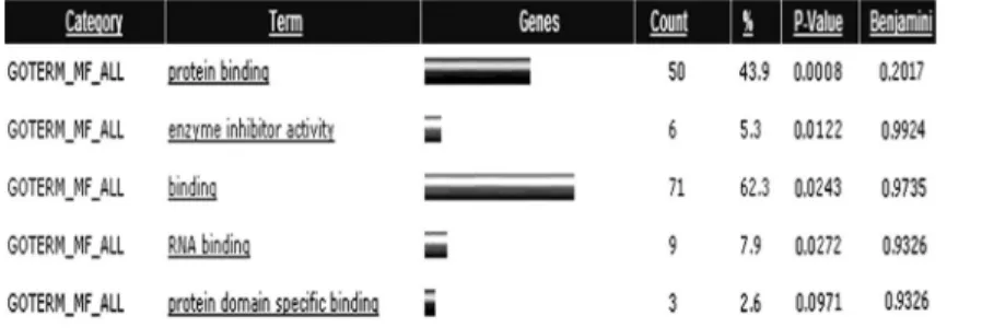
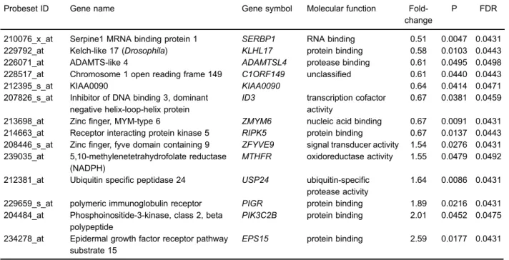
ROP modulated gene expression in SH-SY5Y cells We identified a total of 113 genes as differentially regulated by ROP treatment, of which 48 were upregulated and 65 were downregulated. Among the 113 genes, 101 were known genes and 12 were expressed sequence tags. As shown in Figure 1, GOTERM_Molecular Function_ALL revealed that most of these genes had functions in protein and RNA binding, and enzyme inhibitor activity. Table 1 lists 20 genes representative of the complete list. Further pathway analysis (KEGG and BIOCARTA functional pathways) revealed that only the phosphatidylinositol 3-kinase (PI3K) signaling pathway was over-represented, including genes CALM3,INPP4A, andPIK3C2B. Notably, PIK3C2B expres-sion was strongly promoted by ROP treatment in this pathway. We also identified a number of modulated genes that are located nearPIK3C2Bon chromosome 1, including KLHL17, USP24, C1ORF149, ID3, MTHFR, KIAA0090, ADAMTSL4, SERBP1, RIPK5, EPS15, PIGR, ZFYVE9, and ZMYM6. Of these, EPS15 expression was clearly induced by ROP (Table 2).
TaqMan real-time PCR and Western blot
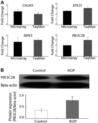
as representative loci on chromosome 1 and their putative relationship with PD. As shown in Figure 2, a strong increase in the mRNA levels ofPIK3C2BandEPS15was detected following ROP treatment, which supported micro-array data. A change in expression ofCALM3andRIPK5 was also confirmed. Western blotting showed that PIK 3C2B protein levels were 2.5 times higher in the ROP-treated group than the control group (Figure 3).
We validated our microarray data in HeLa cells by performing cell culture, TaqMan real-time PCR, and Western
blotting as described previously. As shown in Figure 4, similar expression patterns were observed in HeLa cells to those seen in SH-SY5Y cells.
Discussion
PD is a neuropathological disorder involving the degen-eration of dopaminergic neurons in the substantia nigra, and subsequent loss of their terminals in the striatum. The ensuing loss of dopamine causes most of the debilitating
Figure 1.Molecular function categories of genes regulated by ropinirole (ROP). GOTERM_Molecular Function_ALL revealed that most of these genes functioned in protein and RNA binding, and in enzyme inhibitor activity.
Table 1.Representative 20 genes of all 113 regulated by ropinirole.
Probeset ID Gene name Gene symbol Molecular function
Fold-change
P FDR
234278_at Epidermal growth factor receptor pathway substrate 15
EPS15 protein binding 2.59 0.0177 0.0431
242560_at Fanconi anemia, complementation group D2 FANCD2 protein binding 2.57 0.0191 0.0431 204484_at Phosphoinositide-3-kinase, class 2, beta
polypeptide
PIK3C2B protein binding 2.01 0.0452 0.0475
231830_x_at RAB11 family Interacting protein 1 (class I) RAB11FIP1 protein binding 1.94 0.0229 0.0431 235395_at SEC63-like (S. cerevisiae) SEC63 heat shock
protein binding
1.94 0.0471 0.0431
222297_x_at Ribosomal protein L18 RPL18 RNA binding 1.91 0.0118 0.0431 234082_at Chromosome 21 open reading frame 116 C21ORF116 unclassified 1.91 0.0074 0.0431 229659_s_at Polymeric immunoglobulin receptor PIGR protein binding 1.89 0.0216 0.0431 232808_at Hypothetical protein FLJ10601 ANTXR1 protein binding 1.78 0.0029 0.0431 233059_at Potassium inwardly-rectifying channel,
subfamily j, member 3
KCNJ3 ion channel
activity
1.77 0.0187 0.0431
210076_x_at Serpine1 MRNA binding protein 1 SERBP1 RNA binding 0.51 0.0047 0.0431 227721_at C3 and PZP-like, alpha-2-macroglobulin
domain containing 8
CPAMD8 enzyme inhibitor
activity
0.54 0.0151 0.0431
227404_s_at Early growth response 1 EGR1 transcription activator activity
0.57 0.0269 0.0431
239289_x_at KIAA1018 MTMR15 unclassified 0.58 0.0299 0.0471 244360_at F-box and leucine-rich repeat protein 17 FBXL17 unclassified 0.58 0.0437 0.0435 239419_at Protein tyrosine phosphatase, receptor type A PTPRA catalytic activity 0.58 0.0039 0.0431 229792_at Kelch-Like 17 (DrosoIphila) KLHL17 protein binding 0.58 0.0103 0.0443 219480_at Snail homolog 1 (Drosophila) SNAI1 protein binding 0.58 0.0080 0.0431 226864_at Protein kinase (camp- dependent, catalytic)
inhibitor alpha
PKIA enzyme inhibitor activity
0.58 0.0004 0.0431
201694_s_at Early growth response 1 EGR1 transcription regulator activity
0.59 0.0285 0.0150
motor disturbances associated with PD. Current PD medications treat the symptoms of the disease, focusing on halting or retarding the degeneration of dopaminergic neurons. Recently, there has been considerable interest in neuroprotection as a therapeutic strategy for PD, and several drugs such as ROP have been proposed as candidate agents (9). However, the molecular mechanism of neuroprotection is elusive.
In the present study, we treated SH-SY5Y cells with ROP and applied whole-genome microarray to screen changes in gene expression with the aim of uncovering the underlying molecular mechanism. Using bioinformatics, we identified genes that were differentially regulated after
ROP treatment, which are known to function in protein and RNA binding, and enzyme inhibitor activity. We also observed that the PI3K signaling pathway was over-represented and thatPIK3C2Bexpression was distinctly increased in this pathway.
The PI3K family is evolutionarily conserved and is implicated in many biological processes including cell survival, proliferation, inflammation, adhesion, glucose metabolism, chemotaxis, and cancer. It can be classified into three distinct sub-groups (I, II, and III) based on substrate specificity and sequence homology. PIK3C2B is a family member of class II proteins, which contain a C2 domain and PX domain (10,11). Although diverse biological
Table 2.Ropinirole-regulated genes located on chromosome 1.
Probeset ID Gene name Gene symbol Molecular function Fold-change
P FDR
210076_x_at Serpine1 MRNA binding protein 1 SERBP1 RNA binding 0.51 0.0047 0.0431 229792_at Kelch-like 17 (Drosophila) KLHL17 protein binding 0.58 0.0103 0.0443 226071_at ADAMTS-like 4 ADAMTSL4 protease binding 0.61 0.0495 0.0498 228517_at Chromosome 1 open reading frame 149 C1ORF149 unclassified 0.61 0.0440 0.0443
212395_s_at KIAA0090 KIAA0090 0.64 0.0414 0.0471
207826_s_at Inhibitor of DNA binding 3, dominant negative helix-loop-helix protein
ID3 transcription cofactor activity
0.67 0.0381 0.0459
213698_at Zincfinger, MYM-type 6 ZMYM6 nucleic acid binding 0.67 0.0091 0.0431 214663_at Receptor interacting protein kinase 5 RIPK5 protein binding 0.67 0.0137 0.0443 208446_s_at Zincfinger, fyve domain containing 9 ZFYVE9 signal transducer activity 1.54 0.0276 0.0431 239035_at 5,10-methylenetetrahydrofolate reductase
(NADPH)
MTHFR oxidoreductase activity 1.55 0.0479 0.0492
212381_at Ubiquitin specific peptidase 24 USP24 ubiquitin-specific protease activity
1.64 0.0086 0.0431
229659_s_at polymeric immunoglobulin receptor PIGR protein binding 1.89 0.0216 0.0431 204484_at Phosphoinositide-3-kinase, class 2, beta
polypeptide
PIK3C2B protein binding 2.01 0.0452 0.0475
234278_at Epidermal growth factor receptor pathway substrate 15
EPS15 protein binding 2.59 0.0177 0.0431
FDR: False discovery rate. The Student’st-test was used for statistical analysis.
Figure 2.TaqMansreal-time PCR con
roles have been assigned to class I and class III PI3Ks, the functions of class II PI3Ks are still unknown. However, PIK3C2B has recently been implicated in cell growth, cell
migration, and differentiation (12–14). Moreover, activation of a major neuroprotective signaling pathway, the PI3K/Akt pathway, can prevent cell death in a PD model of SH-SY5Y cells (9).
Intriguingly, we observed the distinct promotion of PIK3C2B transcript and protein expression levels in SH-SY5Y cells following ROP treatment. This indicated that ROP might exert neuroprotective effects through the PI3K pathway, and that PIK3C2B might play a role in this process. Additionally, we previously observed that the PI3K/Akt pathway modulates the expression of Nurr1, which is a transcription factor essential for the differentia-tion and maturadifferentia-tion of central dopaminergic cells (15). This suggested that ROP might induce PIK3C2B and modulate Nurr1 to exert neuroprotection. However, the present study found no direct evidence of Nurr1 modula-tion by either ROP or PIK3C2B. Further investigamodula-tions may shed new light on the mechanism of ROP neuropro-tection and the role of PIK3C2B in PD.
Nine loci in the human genome have previously been linked to PD. Mutations in alfa-synuclein, parkin,DJ-1, and, arguablyUCH-L1genes have been associated with familial PD (16). Recently a locus on chromosome 1 was linked to common late-onset PD in the Icelandic population (16). Meanwhile, linkage studies have also defined susceptibility regions for late-onset PD on chromosomes 1 and 2 (17). We observed that ROP regulated several genes located on chromosome 1, suggesting that this might be its main way of exerting neuroprotective effects. Only three of these genes, USP24, MTHFR, and EPS15, have been asso-ciated with PD in earlier studies (17–19). For instance,in vitro experiments showed that EPS15 enhanced the ubiquitin ligase activity of PARKIN, and PARKIN-mediated EPS15 ubiquitination is crucial in promoting the PI3K/Akt signaling pathway (19,20).
In the present study, we observed that ROP distinctly increased the expression ofEPS15. Considering the role of EPS15 and PI3K/Akt in neuronal survival, our observation is likely to further our understanding of the role of ROP in PD therapy. Despite other chromosomal 1 genes having no known link with PD, their underlying biological functions may nevertheless provide new implications for disease. RIPK5, a member of the RIP serine/threonine kinase family, was previously reported to induce both caspase-dependent apoptosis and caspase-independent cell death (21). Con-sidering the important role of cell death pathways in PD (22), future work may identify a novel role for RIPK5 in the pathogenesis of PD.
In conclusion, we used genome-wide microarray analysis to identify genes that were regulated after ROP treatment. Pathway analysis suggested that ROP mainly modulated the PI3K signaling pathway in SH-SY5Y cells. Further extensive investigation of PIK3C2B and other loci on chromosome 1 may open up a new avenue to understand the pathology of PD and provide novel pharmaceutical targets to improve patient care.
Figure 3.Elevation of PIK3C2B protein expression by ropinirole (ROP) treatment in SH-SY5Y cells. b-actin was used as the loading control. PIC3C2B protein levels were significantly increased by treatment with 10mM ROP. *Po0.05, 10mM ROP
compared to the control group (Student’st-test).
Figure 4.Validation of microarray data in HeLa cells.A,CALM3,
EPS15, RIPK5, and PIK3C2B underwent TaqMans real-time
PCR in HeLa cells. The y-axis represents the fold-change in expression after ROP treatment, and microarray and TaqMan data are plotted on the x-axis. *Po0.05, TaqMan data compared
to microarray data (Student’st-test).B, Western blot analysis in HeLa cells.b-actin was used as the loading control. PIC3C2B protein levels were elevated by treatment with 10mM. *Po0.05,
References
1. Pan T, Xie W, Jankovic J, Le W. Biological effects of pramipexole on dopaminergic neuron-associated genes: relevance to neuroprotection. Neurosci Lett 2005; 377: 106–109, doi: 10.1016/j.neulet.2004.11.080.
2. Schapira AH. Dopamine agonists and neuroprotection in Parkinson’s disease.Eur J Neurol2002; 9 (Suppl 3): 7–14. 3. Singh A, Althoff R, Martineau RJ, Jacobson J. Pramipexole, ropinirole, and mania in Parkinson’s disease.Am J Psychiatry 2005; 162: 814–815.
4. Schapira AH, Olanow CW. Rationale for the use of dopamine agonists as neuroprotective agents in Parkinson’s disease. Ann Neurol 2003; 53 (Suppl 3): S149–S157, doi: 10.1002/ ana.10514.
5. Iida M, Miyazaki I, Tanaka K, Kabuto H, Iwata-Ichikawa E, Ogawa N. Dopamine D2 receptor-mediated antioxidant and neuroprotective effects of ropinirole, a dopamine agonist.Brain Res1999; 838: 51–59, doi: 10.1016/S0006-8993(99)01688-1. 6. Olanow CW, Jenner P, Brooks D. Dopamine agonists and neuroprotection in Parkinson’s disease.Ann Neurol1998; 44: S167–S174, doi: 10.1002/ana.410440725.
7. Mandel S, Weinreb O, Youdim MB. Using cDNA microarray to assess Parkinson’s disease models and the effects of neuroprotective drugs. Trends Pharmacol Sci 2003; 24: 184–191, doi: 10.1016/S0165-6147(03)00067-1.
8. Alonso CA, Luquin PR, G. Ruiz-Espiga P, Burguera JA, Campos AV, Castro A, et al. Dopaminergic agonists in Parkinson’s disease.Neurologia 2015; 29: 230–241, doi: 10.1016/j.nrl.2011.04.012.
9. Nakaso K, Ito S, Nakashima K. Caffeine activates the PI3K/ Akt pathway and prevents apoptotic cell death in a Parkinson’s disease model of SH-SY5Y cells.Neurosci Lett 2008; 432: 146–150, doi: 10.1016/j.neulet.2007.12.034. 10. Fougerat A, Gayral S, Malet N, Briand-Mesange F,
Breton-Douillon M, Laffargue M. Phosphoinositide 3-kinases and their role in inflammation: potential clinical targets in atherosclero-sis?Clin Sci2009; 116: 791–804, doi: 10.1042/CS20080549. 11. Harada K, Truong AB, Cai T, Khavari PA. The class II phosphoinositide 3-kinase C2beta is not essential for epidermal differentiation.Mol Cell Biol2005; 25: 11122–11130, doi: 10.1128/MCB.25.24.11122-11130.2005.
12. Sindic A, Crljen V, Matkovic K, Lukinovic-Skudar V, Visnjic D, Banfic H. Activation of phosphoinositide 3-kinase C2 beta in the nuclear matrix during compensatory liver growth. Adv Enzyme Regul2006; 46: 280–287, doi: 10.1016/j.advenzreg. 2006.01.008.
13. Maffucci T, Cooke FT, Foster FM, Traer CJ, Fry MJ, Falasca M. Class II phosphoinositide 3-kinase defines a novel signaling pathway in cell migration.J Cell Biol 2005; 169: 789–799, doi: 10.1083/jcb.200408005.
14. Visnjic D, Crljen V, Curic J, Batinic D, Volinia S, Banfic H. The activation of nuclear phosphoinositide 3-kinase C2beta in all-trans-retinoic acid-differentiated HL-60 cells.FEBS Lett 2002; 529: 268–274, doi: 10.1016/S0014-5793(02)03357-4. 15. Fan X, Luo G, Ming M, Pu P, Li L, Yang D, et al. Nurr1 expression and its modulation in microglia. Neuroimmuno-modulation2009; 16: 162–170, doi: 10.1159/000204229. 16. Toft M, Aasly J. [The genetics of Parkinson disease].Tidsskr
Nor Laegeforen2004; 124: 922–924.
17. Li Y, Schrodi S, Rowland C, Tacey K, Catanese J, Grupe A. Genetic evidence for ubiquitin-specific proteases USP24 and USP40 as candidate genes for late-onset Parkinson disease. Hum Mutat2006; 27: 1017–1023, doi: 10.1002/humu.20382. 18. Caccamo D, Gorgone G, Curro M, Parisi G, Di IW, Menichetti C, et al. Effect of MTHFR polymorphisms on hyperhomocysteine-mia in levodopa-treated Parkinsonian patients.Neuromolecular Med2007; 9: 249–254, doi: 10.1007/s12017-007-8006-x. 19. Fallon L, Belanger CM, Corera AT, Kontogiannea M,
Regan-Klapisz E, Moreau F, et al. A regulated interaction with the UIM protein Eps15 implicates parkin in EGF receptor trafficking and PI(3)K-Akt signalling.Nat Cell Biol2006; 8: 834–842, doi: 10.1038/ncb1441.
20. Husnjak K, Dikic I. EGFR trafficking: parkin’in a jam.Nat Cell Biol2006; 8: 787–788, doi: 10.1038/ncb0806-787. 21. Zha J, Zhou Q, Xu LG, Chen D, Li L, Zhai Z, et al. RIP5 is a