Hemodynamic monitoring in the intensive care
unit: a Brazilian perspective
INTRODUCTION
Hemodynamic monitoring (HM) is an important component of critically ill patient care. Knowledge of the cardiovascular function, monitoring of therapeutic interventions, and the need for diferential diagnosis make HM techniques an essential component for the outcomes of these patients.
Since the early 1970s, with the introduction of the pulmonary artery catheter (PAC),(1) the cardiovascular variables began to be routinely monitored Fernando Suparregui Dias1, Ederlon Alves de
Carvalho Rezende2, Ciro Leite Mendes3, João Manoel Silva Jr3, Joel Lyra Sanches2
1. Intensive Care Department, Hospital Pompéia - Caxias do Sul (RS), Brazil.
2. Intensive Care Unit, Hospital do Servidor Público Estadual “Francisco Morato de Oliveira” - São Paulo (SP), Brazil.
3. Adult Intensive Care Unit, Hospital Universitário, Universidade Federal da Paraíba - Campus I - João Pessoa (PB), Brazil.
Objective: In Brazil, there are no data on the preferences of intensivists regarding hemodynamic monitoring methods. he present study aimed to identify the methods used by national intensivists, the hemodynamic variables they consider important, the regional diferences, the reasons for choosing a particular method, and the use of protocols and continued training.
Methods: National intensivists were invited to answer an electronic questionnaire during three intensive care events and later, through the Associação de Medicina Intensiva Brasileira portal, between March and October 2009. Demographic data and aspects related to the respondent preferences regarding hemodynamic monitoring were researched.
Results: In total, 211 professionals answered the questionnaire. Private hospitals showed higher availability of resources for hemodynamic monitoring than did public institutions. he pulmonary artery catheter was considered the most trusted by 56.9% Conflicts of interest: None.
Submitted on August 28, 2014 Accepted on September 22, 2014
Corresponding author: Fernando Suparregui Dias
Linha de Cuidados Intensivos do Hospital Pompéia Avenida Júlio de Castilhos, 2163 - Centro Zip code: 95010-001 - Caxias do Sul (RS), Brazil E-mail: [email protected]
Responsible editor: Luciano César Pontes de Azevedo
Monitorização hemodinâmica em unidade de terapia intensiva:
uma perspectiva do Brasil
ABSTRACT
Keywords: Monitoring/physiology; Monitoring, physiologic; Catheterization, Swan-Ganz; Echocardiography; Cardiac output; Questionnaires; Brazil
of the respondents, followed by echocardiograms, at 22.3%. Cardiac output was considered the most important variable. Other variables also considered relevant were mixed/central venous oxygen saturation, pulmonary artery occlusion pressure, and right ventricular end-diastolic volume. Echocardiography was the most used method (64.5%), followed by pulmonary artery catheter (49.3%). Only half of respondents used treatment protocols, and 25% worked in continuing education programs in hemodynamic monitoring.
Conclusion: Hemodynamic
monitoring has a greater availability in intensive care units of private institutions in Brazil. Echocardiography was the most used monitoring method, but the pulmonary artery catheter remains the most reliable. he implementation of treatment protocols and continuing education programs in hemodynamic monitoring in Brazil is still insuicient.
in intensive care units (ICU). PAC allows for assessing the hemodynamic status in detail, which is not possible only by clinical means.(2-5) Because PAC is a monitoring technique, it has no therapeutic properties, and as an invasive technique, it has caused a heated debate regarding its eiciency and safety in the decades since its introduction.(6,7) his debate is a product of the results of some observational(8) and subgroup(9) studies, which have led to reduced use of this tool.(10) he decreased use of PAC coincided with the emergence and dissemination of other less invasive HM techniques, such as determination of the cardiac output (CO) by arterial pulse wave contour analysis, transpulmonary thermodilution,
echocardiography, Doppler, carbon dioxide (CO2)
rebreathing, and bioimpedance and bioreactance.(11) However, although many forms of HM are available, most physicians still need to better understand how to use them and how to interpret the provided information(12-14) because there is a large variability in decisions based on the HM-derived parameters.(15,16)
his study aimed to investigate the Brazilian status regarding the use of HM devices, considering the preferences of physicians, the variables used in HM, how much HM is used in the ICU, reasons for choosing a particular method, the existence or non-existence of hemodynamic management and continuing training protocols, and potential regional diferences.
METHODS
During three scientiic meetings, (II Congresso
Luso-Brasileiro de Medicina Intensiva, held in 2010 in Pernambuco; XIV Congresso Brasileiro de Medicina Intensiva, held in São Paulo in 2009; and IV Simpósio Internacional de Monitorização em UTI, also held in São Paulo in 2010), an electronic form was made available for completion. Over a six-month period (March - October 2009), the form was also made available for completion on the website of the Associação de Medicina Intensiva Brasileira (AMIB). Physicians present at these events, intensivists or non-specialists in intensive care medicine, whether intensivists or not, were invited to participate in the study. he project was approved by the Ethics and Research Committee of the Instituto de Assistência Médica ao Servidor Público Estadual (IAMSPE) under number 760.253/14, and there was exemption from obtaining the informed consent form because the present study does not involve patients or interventions.
he following demographic data were collected: age, gender, time since graduation, title in intensive care, region of the country (North, Northeast, Southeast, Midwest, and South), hospital type (public or private), and number of ICU beds where the respondent worked. he responses were stored in a database created exclusively for this purpose. he identity of respondents was kept conidential.
Respondents were asked whether the ICU where they worked was able to assess central venous pressure (CVP) and mean arterial pressure (MAP) via an invasive arterial
line and whether PAC, esophageal Doppler LiDCO®
,
PiCCO®
, FloTrac/Vigileo®
, and echocardiography were available in the service. Furthermore, respondents were asked how many patients had CVP and invasive blood pressure measured per month.
Aiming to evaluate the importance of each method, respondents were asked which of the methods they considered the most reliable. To determine which variables were most valuable in a patient with hemodynamic instability, respondents were asked to grade the variables in order of importance from 0 and 10. To identify which were the most used monitoring methods, respondents were asked which methods were used in the ICU they worked at. he need for HM and the way it was performed was assessed using the following questions: (1) to how many patients per month was HM recommended; (2) how many patients underwent HM; (3) whether a HM protocol was used; and (4) whether there was a training and retraining HM program.
he statistical analysis involved describing the demographic characteristics of the individuals included in the study. he health centers were divided into two groups (private and public hospitals) and compared. To describe categorical variables, the frequencies and percentages were calculated. he quantitative variables were described using central tendency and dispersion measures (mean and standard deviation).
RESULTS
Table 1 shows the demographic characteristics of the 211 respondents. Respondents were divided into two groups according to the characteristics of their institution: private (n=113) or public (n=98). hese two groups did not difer regarding age, gender, time since graduation, geographic region, or title in intensive care. here was a predominance of units with 10-20 beds, compared to units with less than 10 or more than 20 beds.
Table 1 - Demographic variables
Variables Public hospitals N=98
Private hospitals N=113
All N=211
p value
Age 38.3±8.6 37.3±7.7 37.8±8.1 0.353
Male 71.4 72.6 72.0 0.88
Time since graduation 0.50
Less than 5 years 16.3 15.9 16.1
5-10 years 24.5 31.0 28.0
10-20 years 43.9 40.7 42.2
More than 20 years 15.3 12.4 13.7
Region 0.50
Southeast 52.0 53.1 52.6
Northeast 14.3 21.2 18.0
South 15.3 9.7 12.3
Mid-West 11.2 11.5 11.4
North 7.1 4.4 5.7
Title 52.0 59.3 55.9 0.30
No title 48.0 40.7 44.1
ICU beds 0.02
<10 26.5 15.9 20.9
10-20 54.1 54.0 54.0
>20 19.4 30.1 25.1
ICU - intensive care unit. Results are expressed as means±standard deviations and percentages.
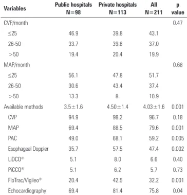
Table 2 presents the most available HM methods. here was greater availability of HM resources in private hospitals than in public hospitals, with a signiicant diference in MAP (p=0.001), PAC (p=0.005), esophageal Doppler (p=0.002), FloTrac/Vigileo®
(p=0.001), and echocardiogram (p=0.004).
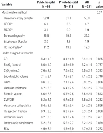
Table 3 shows the degree of conidence of respondents in each monitoring type and the importance given to certain variables. PAC was considered the most reliable method by 56.9% of the respondents; echocardiography, by 22.3%; FloTrac/Vigileo®
, by 12.3%; and LiDCO®
, by
4.7%. PiCCO®
and esophageal Doppler were the most
Table 2 - Available methods and variables used in hemodynamic monitoring
Variables Public hospitals N=98
Private hospitals N=113
All N=211
p value
CVP/month 0.47
≤25 46.9 39.8 43.1
26-50 33.7 39.8 37.0
>50 19.4 20.4 19.9
MAP/month 0.68
≤25 56.1 47.8 51.7
26-50 30.6 43.4 37.4
>50 13.3 8. 10.9
Available methods 3.5±1.6 4.50±1.4 4.03±1.6 0.001
CVP 94.9 98.2 96.7 0.18
MAP 69.4 88.5 79.6 0.001
PAC 49.0 68.1 59.2 0.005
Esophageal Doppler 35.7 57.5 47.4 0.002
LiDCO® 5.1 8.0 6.6 0.40
PiCCO® 5.1 6.2 5.7 0.73
FloTrac/Vigileo® 20.4 42.5 32.2 0.001
Echocardiography 69.4 81.4 75.8 0.04
CVP - central venous pressure; MAP - mean arterial pressure; PAC - pulmonary artery catheter. Results are expressed as means±standard deviations and percentages.
reliable methods for only 1.9% of respondents for each of the methods. Regarding the variables used for HM, the ive that were considered the most important were CO, central venous oxygen saturation (SvcO2), mixed venous oxygen saturation (SvO2), right ventricular end-diastolic volume (RVEDV), and pulmonary artery wedge pressure (PAWP).
Respondents indicated echocardiography as the most used monitoring method, followed by PAC, FloTrac/Vigileo®
and esophageal Doppler, with 64.5%,
49.3%, 31.3%, and 25.6%, respectively. LiDCO®
and
PiCCO®
had usage percentages of 5.2% and 4.7%, respectively (Table 4).
Table 5 lists the number of patients and HM procedures per month and the percentage of services that used protocols with therapeutic goals and that had training and retraining programs.
DISCUSSION
Table 3 - Methods considered more reliable and importance of the assessed variables
Variable Public hospital
N=98
Private hospital N=113
All N=211
p value
Most reliable method 0.57
Pulmonary artery catheter 52.0 61.1 56.9
LiDCO® 6.1 3.5 4.7
PiCCO® 3.1 0.9 1.9
Echocardiography 25.5 19.5 22.3
Esophageal Doppler 2.0 1.8 1.9
FloTrac/Vigileo® 11.2 13.3 12.3
Grades assigned to variables
CO 8.3±1.9 8.4±1.9 8.4±1.9 0.855
SvcO2 (central) 8.0±1.9 8.3±1.9 8.2±1.9 0.757
SvO2 (mixed) 7.6±2.6 7.5±2.3 7.5±2.4 0.313
End-diastolic volume 7.1±2.4 7.2±2.1 7.1±2.2 0.740
PAWP 6.6±2.6 7.1±2.4 6.9±2.5 0.086
Vascular resistance 6.7±2.6 6.4±2.5 6.5±2.5 0.733
Systolic volume 6.6±2.6 6.4±2.5 6.5±2.6 0.543
CVP/DBP 6.2±2.7 6.7±2.5 6.5±2.6 0.232
Vena cava collapsibility 6.4±2.7 6.5±2.4 6.4±2.5 0.866
RV ejection fraction 6.3±2.3 6.3±2.4 6.3±2.4 0.868
Ventricular work 6.2±2.5 6.1±2.6 6.1±2.6 0.401
Intrathoracic blood volume 5.2±2.4 5.2±2.7 5.2±2.6 0.879
ELW 4.9±2.4 4.5±3.0 4.7±2.8 0.273
CO - cardiac output; ScvO2 - central venous oxygen saturation; SvO2 - mixed venous oxygen saturation; PAWP - pulmonary artery wedge pressure; CVP - central venous pressure; DBP - diastolic blood pressure; DV - diastolic volume; ELW - extrapulmonary lung water. Results are expressed as means±standard deviations and percentages.
Table 4 - Methods used for hemodynamic monitoring
Method used Public hospitals N=98
Private hospitals N=113
All N=211
p value
PAC 42.9 54.9 49.3 0.08
LiDCO® 3.1 7.1 5.2 0.19
PiCCO® 5.1 4.4 4.7 0.81
FloTrac/Vigileo® 22.4 38.9 31.3 0.01
Echocardiography 61.2 67.3 64.5 0.36
Esophageal Doppler 21.4 29.2 25.6 0.20
PAC - pulmonary artery catheter.
Table 5 - Number of procedures performed, existence of therapeutic goals, and training programs
Variables Public hospitals N=98 (%)
Private hospitals N=113 (%)
All N=211 (%)
p value
Procedures/month 0.48
Less than 5 26.5 20.4 23.2
6-10 27.6 31.9 29.9
11-15 22.4 20.4 21.3
16-20 7.1 9.7 8.5
>20 16.3 17.7 17.1
Protocol with defined
therapeutic goals 41.8 61.1 52.1 0.005*
Training/retraining
program 25.5 23.9 24.6 0.78
* Private hospitals compared with public hospitals.
greater among physicians who were more experienced, had a title in intensive medicine, and were from more economically developed regions.
Although CVP was the method most mentioned as available, the arterial line was the hemodynamic variable most used to determine MAP. In the approach for patients with circulatory shock, it is recommended that MAP be measured by an arterial line whenever the patient is using vasoactive drugs,(17) and CVP, although it is contested,(18) is used to guide therapy in patients with
severe sepsis/septic shock.(19) Echocardiography was the most cited non-invasive HM method; however, it is worth noting that the way the question was presented might have led some interviewees to interpret the diagnostic use of echocardiography as an HM method. he fact that PAC was mentioned by 59% of respondents suggests that, even with the emergence of new methods, this resource is still used frequently in patients who undergo intensive treatment. Less invasive technologies such as PiCCO®
,
LiDCCO®
, and FloTrac/Vigileo®
were used by 44.5% of respondents. hese numbers suggest that less invasive methods are becoming an alternative for HM in critically ill patients.
Despite the importance of less invasive methods, PAC, which is a technique known to be invasive, was considered the most reliable. From the 1990s onward, the usefulness and safety of PAC began to be questioned.(8) Despite its reduced use in recent years(10) and its being an invasive technology associated with potentially serious complications,(20) the fact that, in this series, it was considered by a population of experienced physicians to be the most reliable technique is noteworthy. his inding is most likely because PAC is the older HM technology at the bedside and because of the multiple studies using this method in many diferent clinical scenarios. Echocardiography, despite being a non-invasive technology that has been increasingly used,(8,9) was considered less reliable than PAC. Echocardiography is highly operator-dependent, requires speciic training,(7,21) and is a technique still little dominated by intensivists, which may be factors related to this belief.
he most valued hemodynamic variables were CO,
ScvO2/SvO2, RVEDV, and PAWP. he importance of
correctly predicted by clinical evaluation in only 50% of critically ill patients, which gives it a signiicant value in the critically ill this situation.(2,3) Another aspect to be considered is that CO can be determined using various technologies in addition to thermodilution with PAC, such as transpulmonary thermodilution, arterial pulse wave contour analysis, echocardiography and esophageal Doppler.(11) he importance of measuring blood low in critically ill patients is crucial because CO is an important determinant of oxygen transport. Determining CO is useful for establishing the diagnosis, guiding therapy, and determining the prognosis.(22)
ScvO2/SvO2 are variables used to determine the balance between global oxygen availability and consumption by the body. he occurrence of a drop in oxygen saturation suggests imbalance between availability and tissue requirements.(23) he present study revealed that in the evaluation of respondents, venous oxygen saturation is considered an important marker of the cardiocirculatory condition. A greater appreciation of ScvO2 in relation to SvO2 may be a result of the popularization of ScvO2 with early goal-directed therapy,(19) the advantage of not requiring the insertion of a PAC, or even the lack
of knowledge that mixed SvO2 relects the oxygen
consumption in circulatory shock conditions.(23)
RVEDV and PAWP are two measures that estimate the preload. he irst is a variable that measures the volumetric preload of the right ventricle, and the second evaluates the preload of the left ventricle. Because PAWP is a static measure of responsiveness to luid, it has a low ability to correctly estimate the preload in healthy individuals(24) and to predict the response to luid infusion in critically ill patients.(25) However, it must be considered that global variables, such as inlation pressures, are useful until hemodynamic stabilization occurs, after which the regional variables should guide the therapy.(26) In this context, a study of patients with acute myocardial infarction treated in the pre-thrombolytic era showed that an initial PAWP above 18mmHg was a signiicant predictor of 30-day mortality,(27) and in the last decade, the use of CVP as a therapeutic target was shown to be important in the initial management of patients with severe sepsis and septic shock.(19) hus, the controversy remains on how useful blood pressure variables may be to estimate the preload and the response to luid infusion.
Most physicians performed up to 15 HM procedures per month, which indicates a high incidence of patients with circulatory disorders in the ICU. Despite this frequency, just over half of respondents worked with protocols of deined therapeutic goals, which was a more
frequent characteristic in private hospitals than in public hospitals. It is established that delayed hemodynamic stabilization and lack of treatment protocols are two of the leading causes of failure in HM studies. Once organ dysfunctions are present, hemodynamic optimization has no positive efect.(28) A post hoc analysis of a randomized study(29) showed that when PAC was used after 16 hours of the indication or when the degree of organ dysfunction corresponded to a Sequential Organ Failure Assessment score (SOFA) score >6, the use of PAC was not beneicial.(30) Because HM with PAC is a technology subjected to great variability between observers when obtaining pressure curves(31) and interpreting data,(16) minimum speciic training is required. A standard from the American College of Physicians, American College of Cardiology, and American Heart Association discusses the need for technical and cognitive knowledge to perform HM. he conclusion is that these requirements vary according to individual ability and dexterity and that a minimum of 25 procedures would be necessary to make the physician proicient.(32) Regarding the use of echocardiography as an HM tool, in recent years, there has been much excitement regarding this method.(33) Recently, a French study stipulated that a 12-hour training merging clinical discussions with use of the technique would be suicient to make resident physicians proicient.(34) Conversely, Cholley et al.(33) divide the technical proiciency into three levels. he basal level corresponds to an operator with minimal training, the medium level corresponds to an operator in training, and the higher level corresponds to a well-trained operator - this individual being able to fully perform an echocardiographic examination. However, these authors do not discuss how long it would take for the operator to be fully proicient in this technology.(33) Regarding CO determination using esophageal Doppler, a study showed that after 12 procedures, the operator was able to obtain a clear and audible signal with a well-deined wavelength.(35)
Among the participants of the present study, only 25% worked with a program of continuing education in HM, which was more frequently available in private institutions. his is an important factor demonstrating that despite hemodynamic instability being a major cause of ICU admission and mortality, there is still a signiicant shortage of intensivist training.
low number of responses may relect the lack of interest of the average intensivist in diferent HM techniques. It is also possible that there was a selection bias, as physicians interested in hemodynamics may have greater tendency to being willing to answer the questionnaire. Physicians who attend the meetings mentioned may also belong to a portion of the medical community more interested in improving quality. In addition, the predominance of respondents from the Southeast Region may not relect how HM is being performed in Brazil. his may be a result of various aspects such as availability of resources and training in diferent HM methods in other regions. Finally, the questionnaire was not previously validated, which may in fact be a limiting factor for the interpretation of respondents.
CONCLUSION
In conclusion, the present study indicates that physicians working >5 years in intensive care units use hemodynamic monitoring more. Resources are available more frequently in private hospitals than in public hospitals. Blood pressure variables are the most utilized; however, cardiac output was considered the most important. Echocardiography and pulmonary artery catheter are the most used monitoring methods, but pulmonary artery catheter is still regarded the most reliable method. Treatment protocols are still seldom used, and most intensive care units do not have continuing education programs for hemodynamic monitoring.
Objetivo: No Brasil, não há dados sobre as preferências do intensivista em relação aos métodos de monitorização hemodinâmica. Este estudo procurou identiicar os métodos utilizados por intensivistas nacionais, as variáveis hemodinâmicas por eles consideradas importantes, as diferenças regionais, as razões para escolha de um determinado método, o emprego de protocolos e treinamento continuado.
Métodos: Intensivistas nacionais foram convidados a responder um questionário em formato eletrônico durante três eventos de medicina intensiva e, posteriormente, por meio do portal da Associação de Medicina Intensiva Brasileira, entre março e outubro de 2009. Foram pesquisados dados demográicos e aspectos relacionados às preferências do entrevistado em relação à monitorização hemodinâmica.
Resultados: Responderam ao questionário 211 proissionais. Nos hospitais privados, foi evidenciada maior disponibilidade de recursos de monitorização hemodinâmica do que nas instituições públicas. O cateter de artéria pulmonar foi considerado o mais idedigno por 56,9%, seguido do ecocardiograma, com 22,3%. O
débito cardíaco foi considerado a variável mais importante. Outras variáveis também julgadas relevantes foram débito cardíaco, saturação de oxigênio venoso misto/saturação de oxigênio venoso central, pressão de oclusão da artéria pulmonar e volume diastólico inal do ventrículo direito. O ecocardiograma foi apontado como o método mais utilizado (64,5%), seguido pelo cateter de artéria pulmonar (49,3%). Apenas metade dos entrevistados utilizava protocolos de tratamento e 25% trabalhava com programas de educação continuada em monitorização hemodinâmica.
Conclusão: A monitorização hemodinâmica é mais
disponível nas unidades de terapia intensiva de instituições privadas do Brasil. O ecocardiograma foi apontado como método de monitorização mais utilizado, porém o cateter de artéria pulmonar permanece o mais coniável. A implantação de protocolos de tratamento e de programas de educação continuada em monitorização hemodinâmica no Brasil ainda é insuiciente.
RESUMO
Descritores: Monitoramento/isiologia; Monitorização isiológica; Cateterismo de Swan-Ganz; Ecocardiograia; Débito cardíaco; Questionários; Brasil
REFERENCES
1. Swan HJ, Ganz W, Forrester J, Marcus H, Diamond G, Chonette D. Catheterization of the heart in man with use of a flow-directed balloon-tipped catheter. N Engl J Med. 1970;283(9):447-51.
2. Connors AF Jr, McCaffree DR, Gray BA. Evaluation of right-heart catheterization in the critically ill patient without acute myocardial infarction. N Engl J Med. 1983;308(5);263-7.
3. Steingrub JS, Celoria G, Vickers-Lahti M, Teres D, Bria W. Therapeutic impact of pulmonary artery catheterization in a medical/surgical ICU. Chest. 1991;99(6):1451-5.
4. Coles NA, Hibberd M, Russell M, Love T, Ory D, Field TS, et al. Potential impact of pulmonary artery catheter placement on short-term management decisions in the medical intensive care unit. Am Heart J. 1993;126(4):815-9. 5. Mimoz O, Rauss A, Rekik N, Brun-Buisson C, Lemaire F, Brochard L.
Pulmonary artery catheterization in critically ill patients: a prospective analysis of outcome changes associated with catheter-prompted changes in therapy. Crit Care Med. 1994;22(4):573-9.
6. Dalen JE, Bone RC. Is it time to pull the pulmonary artery catheter? JAMA. 1996;276(11):916-8.
8. Connors AF Jr, Speroff T, Dawson NV, Thomas C, Harrell FE Jr, Wagner D, et al. The effectiveness of right heart catheterization in the initial care of critically ill patients. SUPPORT Investigators. JAMA. 1996;276(11):889-97. 9. Cohen MG, Kelly RV, Kong DF, Menon V, Shah M, Ferreira J, et al.
Pulmonary artery catheterization in acute coronary syndromes: insights from the GUSTO IIb and GUSTO III trials. Am J Med. 2005;118(5):482-8. 10. Wiener RS, Welch HG. Trends in the use of the pulmonary artery catheter
in the United States, 1993-2004. JAMA. 2007;298(4):423-9.
11. Vincent JL, Rhodes A, Perel A, Martin GS, Della Rocca G, Vallet B, et al. Clinical review: Update on hemodynamic monitoring-a consensus of 16. Crit Care. 2011;15(4):229.
12. Iberti TJ, Fischer EP, Leibowitz AB, Panacek EA, Silverstein JH, Albertson TE. A multicenter study of physicians’ knowledge of the pulmonary artery catheter. Pulmonary Artery Catheter Study Group. JAMA. 1990;264(22):2928-32.
13. Gnaegi A, Feihl F, Perret C. Intensive care physicians’ insufficient knowledge of right-heart catheterization at the bedside: time to act? Crit Care Med. 1997;25(2):213-20.
14. Johnston IG, Fraser JF, Sabapathy S, Kruger PS. The pulmonary artery catheter in Autralasia: a survey investigating intensive care physicians’ knowledge and perception of future trends in use. Anaesth Intensive Care. 2008;36(1):84-9.
15. Jain M, Canham M, Upadhyay D, Corbridge T. Variability in interventions with pulmonary artery catheter data. Intensive Care Med. 2003;29(11):2059-62. 16. Mendes CL, Rezende E, Dias FS, Réa-Neto A. Avaliação da variabilidade
de intervenções baseadas no cateter de artéria pulmonar: experiência brasileira. Rev Bras Ter Intensiva. 2006;18(2):137-42.
17. Rhodes A, Grounds RM, Bennett ED. Hemodynamic monitoring. In: Vincent JL, Abraham E, Moore FA, Kochanek PM, Fink MP. Textbook of critical care. 6th ed. Philadelphia: Elsevier Saunders; 2011.
18. Osman D, Ridel C, Ray P, Monnet X, Anguel N, Richard C, et al. Cardiac filling pressures are not appropriate to predict hemodynamic response to volume challenge. Crit Care Med. 2007;35(1):64-8.
19. Rivers E, Nguyen B, Havstad S, Ressler J, Muzzin A, Knoblich B, Peterson E, Tomlanovich M; Early Goal-Directed Therapy Collaborative Group. Early goal-directed therapy in the treatment of severe sepsis and septic shock. N Engl J Med. 2001;345(19):1368-77.
20. Peters SG, Afessa B, Decker PA, Schroeder DR, Offord KP, Scott JP. Increased risk associated with pulmonary artery catheterization in the medical intensive care unit. J Crit Care. 2003;18(3):166-71.
21. Brown JM. Use of echocardiography for hemodynamic monitoring. Crit Care Med. 2002;30(6):1361-4.
22. Pinsky MR. Why measure cardiac output? Crit Care. 2003;7(2):114-6. 23. Reinhart K, Bloos F. The value of venous oxymetry. Curr Opin Crit Care.
2005;11(3):259-63.
24. Kumar A, Anel R, Bunnell E, Habet K, Zanotti S, Marshall S, et al. Pulmonary artery occlusion pressure and central venous pressure fail to predict ventricular filling volume, cardiac performance, or the response to volume infusion in normal subjects. Crit Care Med. 2004;32(3):691-9. 25. Nahouraii RA, Rowell SE. Static measures of preload assessment. Crit
Care Clin. 2010;26(2):295-305, table of contents.
26. Poeze M, Solberg BC, Greve JW, Ramsay G. Monitoring global volume-related hemodynamic or regional variables after initial resuscitation: What is a better predictor of outcome in critically ill septic patients? Crit Care Med. 2005;33(11):2494-500.
27. Shell WE, DeWood MA, Peter T, Mickle D, Prause JA, Forrester JS, et al. Comparison of clinical signs and hemodynamic state in the early hours of transmural myocardial infarction. Am Heart J. 1982;104(3):521-8. 28. Kern JW, Shoemaker WC. Meta-analysis of hemodynamic optimization in
high-risk patients. Crit Care Med. 2002;30(8):1686-92.
29. Harvey SE, Welch CA, Harrison DA, Rowan KM, Singer M. Post hoc insights from the PAC-Man-the U.K. pulmonary artery catheter trial. Crit Care Med. 2008;36(6):1714-21.
30. Vincent JL, de Mendonça A, Cantraine F, Moreno R, Takala J, Suter PM, et al. Use of the SOFA score to assess the incidence of organ dysfunction/failure in the intensive care units: results of a multicenter, prospective study. Working group on “sepsis-related problems” of the European Society of Intensive Care Medicine. Crit Care Med. 1998;26(11):1793-800.
31. Komadina KH, Schenk DA, LaVeau P, Duncan CA, Chambers SL. Interobserver variability in the interpretation of pulmonary artery catheter pressure tracings. Chest. 1991;100(6):1647-54.
32. Clinical competence in hemodynamic monitoring. A statement for physicians from the ACP/ACC/AHA Task Force on Clinical Privileges in Cardiology. Circulation. 1990;81(6):2036-40.
33. Cholley BP, Vieillard-Baron A, Mebazaa A. Echocardiography in the ICU: time for widespread use! Intensive Care Med. 2006;32(1):9-10. Erratum in Intensive Care Med. 2006;32(4):634.
34. Vignon P, Mücke F, Bellec F, Marin B, Croce J, Brouqui T, et al. Basic critical care echocardiography: validation of a curriculum dedicated to noncardiologist residents. Crit Care Med. 2011;39(4):636-42.