Inefectiveness of using the pressure relief valve
technique during cuf inlation
INTRODUCTION
Tracheal prostheses are important for pulmonary ventilation of patients in intensive care units (ICU), but they can also damage the histological structure of the trachea when the pressure exerted by the cuf is greater than the tracheal perfusion.(1,2) he severity of the injury depends on both the contact time and
the pressure exerted between the cufand the tracheal wall,(3,4) which may result
in the loss of cilia, epithelial erosion, rupture of blood capillaries with tracheal ulceration, stenosis, and tracheoesophageal istulas.(5,6)
Conversely, cuf pressures below 20cmH2O may lead to the aspiration
of contaminated oropharyngeal contents into the lower respiratory tract. Raquel Annoni1, Ruy Camargo Pires-Neto1
1. Department of Pathology, School of Medicine, Universidade de São Paulo - São Paulo (SP), Brazil.
Objective: To test the efectiveness of using a cuf pressure relief valve technique to maintain cuf pressure levels within the normal in vitro range (Phase 1) in patients admitted to the intensive care unit (Phase 2) and to test the reproducibility of the technique using diferent syringes.
Methods: In Phase 1, a tracheal tube was inserted into a trachea model. Ten- and 20mL syringes were used to inlate the cuf through the tracheal tube. he cuf was slowly and steadily inlated until the syringe plunger would move in the opposite direction of the application. After the plunger stopped, the cuf pressures were recorded. In Phase 2, the same maneuvers for inlating the cuf were performed on 20 patients using 5, 10, and 20mL syringes and were compared with manometer measurements. he intraclass correlation coeicient and Bland-Altman analysis were employed to determine the reproducibility and agreement between
Conflicts of interest: None.
Submitted on May 25, 2014 Accepted on October 5, 2014
Corresponding author: Raquel Annoni
Faculdade de Medicina da Universidade de São Paulo
Av. Dr. Arnaldo, 455, sala 1155
Zip code: 01246-903 - São Paulo (SP), Brazil E-mail: [email protected]
Responsible editor: Flávia Ribeiro Machado
Ineficácia da técnica de alívio de pressão por meio de válvula em
insuflar o cuff
ABSTRACT
Keywords: Transducers, pressure; Respiration, artiicial; Airway management; Intubation, intratracheal; Respiratory therapy; Intensive care units syringes. Data were expressed as medians (interquartile range).
Results: here was no reproducibility between syringes with an intraclass correlation coeicient ranging between -0.33 and 0.8 (p>0.05). he pressures generated with the syringes were higher than the pressures generated using a standard manometer: the
5mL syringe pressure was 105cmH2O
(82.5-120cmH2O), the 10mL
syringe pressure was 69cmH2O
(47.5-111.3cmH2O), and the 20mL
syringe pressure was 45cmH2O
(35-59.5cmH2O). he Bland-Altman
analysis conirmed the large bias and variability between the syringes used, compared with the manometer.
Conclusion: he use of syringes is not an efective technique for determining the cuf pressure in patients admitted to the intensive care unit.
According to the American horacic Society and the Infectious Diseases Society of America, the primary route of bacterial entry into the lower respiratory tract is the aspiration of oropharyngeal pathogens through the cuf, predisposing patients to mechanical ventilator-associated pneumonia,(1,7) the prevalence of which varies between
10% and 27% in critically ill patients.(8-10)
To avoid injuries caused by the hyperinlation or underinlation of the cuf, many authors recommend that the intra-cuf pressures be maintained at between 20 and 30cmH2O.(11-13) However, maintaining these pressures
within these levels has been a challenge in clinical practice because many factors inluence the variation in cuf pressures, such as changes in the tracheal muscle tone, hypothermia, and the position of both the patient and cuf,(14-16) reinforcing the necessity of frequently
monitoring and adjusting the cuf pressures.
Although numerous techniques have been described to measure cuf pressure, it is believed that the most efective methods for achieving the recommended pressures are those that use an aneroid manometer (usually portable) speciically designed for this function.(17) However, in Brazil, many
hospitals do not have such a device because of its high cost; instead, pressures are checked using indirect measurements.
he cuf pressure relief valve technique was described by Somri et al. as an economic alternative for measuring and maintaining the cuf pressure in patients undergoing anesthesia. his technique uses a 20mL (P20) syringe to inlate the pilot balloon with 15mL of air. Tracheal distension caused by air insulation generates an opposing force of resistance, which acts on the cuf and is then transferred to the syringe plunger, moving it in the opposite direction of the application. When the syringe plunger stops moving, the tracheal wall pressure is considered equal to the cuf pressure, thus maintaining it at safe levels.(18)
In a surgical environment where the gases used in inhalation anesthetics directly inluence cuf pressure, it has been demonstrated that the cuf pressure relief valve technique can maintain pressures within the recommended limits.(18) Because this technique is an
economical alternative to a manometer, it has spread beyond the surgical center. However, its efectiveness has not yet been established in patients admitted to ICU, where cuf pressure is not afected by anesthetic gases. herefore, the aim of this study was to test the efectiveness of the cuf pressure relief valve technique in maintaining adequate cuf pressure levels in ICU patients using 20mL (P20), 10mL (P10), and 5mL (P5) syringes. Furthermore, we analyzed the reproducibility of this technique using diferent P20 and P10 syringes in a trachea model.
METHODS
his study was a prospective cross-sectional investigation that was conducted in two phases: (1) in vitro and (2) in vivo. he study was approved by the Research Ethics Committee of the Hospital das Clínicas at the Universidade de São Paulo (1070/09). Family members of all the patients agreed to participate in the study and signed an informed consent form.
he irst phase assessed the reproducibility of ive P10 and P20 syringes (BD, Becton, Dickinson and Company, USA) using a trachea model. For this purpose, a PVC tube was used that had an inner diameter of approximately 4cm, into which a tracheal tube with an inner diameter of 8mm was inserted until the cuf portion of the tracheal tube reached the middle third of the tube. A three-way connector was attached to the pilot balloon, to a manometer speciically designed to measure cuf pressures (VBM Medizintechnik GmbH, Germany) and to the syringe being used in the experiment.
he syringes were preilled with air, and the cuf was slowly and steadily inlated until the syringe plunger moved in the opposite direction of the application. Small pauses were taken while inlating the cuf to allow passive recoil of the syringe plunger until it reached pressure equilibrium. he cuf pressure was measured with the manometer as soon as the syringe plunger stopped moving. For this phase, ive P10 and ive P20 syringes from the same manufacturer were used, and ive measurements were performed for each syringe.
he second phase consisted of an in vivo experiment, in which the maneuver described in Phase 1 was conducted on a convenience sample of 20 patients intubated less than 48 hours who were ≥18 years of age. Patients who had a prior history of intubation and/or tracheostomy, head and neck surgery (previous or current), or mechanical ventilation at high peak airway pressures (positive end-expiratory pressure (PEEP) >10cmH2O) were excluded. In this phase, a three-way connector was connected to the pilot balloon of the endotracheal tube of each patient, to the manometer, and to a syringe. After a bronchial clearance session and suctioning the upper airways and supra-cuf region, the initial cuf pressure (P-initial) was evaluated. hen, the pilot balloon was completely emptied, and the same maneuver performed in Phase 1 was sequentially and randomly performed with P5, P10, and P20 syringes. When the syringe plunger stopped moving (as in Phase 1), the cuf pressure was measured.
single cuf pressure measurement was performed with each syringe on each patient. After collecting the data, the initial cuf pressure was reestablished. he calculation of the sample power (1-β) with a conidence interval (CI) of 99% (two-sided) indicated that our sample had a power of 100%.
New syringes with less than one year since the manufacturing date and stored at room temperature in a dry place were used in all phases of the study. Prior to the experiments, the syringes were “tested”; in other words, we moved the plunger within the syringe barrel two or three times to unstick the rubber plunger from the barrel (due to general storage of the syringe).
Statistical analysis
In Phase 1, we used a simple analysis of variance
(one-wayANOVA) to determine the variability between
the ive P10 syringes and between the ive P20 syringes used. At this stage, the pressures between the P10 and P20 syringes were not compared. he intraclass correlation coeicient (ICC) with a 95% CI was used to determine the reproducibility of each P10 and P20 syringe. he ICC can range from -1 to +1, where values >0.75 indicate excellent reproducibility, values between 0.4 and 0.75 indicate reasonable reproducibility, and values <0.4 indicate poor reproducibility. Negative values indicate cases with an extreme lack of consistency between the data analyzed.(19)
In Phase 2 of the study, the Wilcoxon test was used to compare the initial cuf pressures to the pressures generated using maneuvers involving the P5, P10, and P20 syringes. his test was selected because the data were not normally distributed. A Bland-Altman plot was used to analyze the degree of agreement between the P5, P10, and P20 syringes compared with the P-initial. his method assesses the level of agreement between two diferent instruments. To apply this method, the mean diference between the two measurement methods and the 95% limits of agreement were calculated, which generates a scatterplot that can be used to visualize the bias (how the diferences deviate from zero), the error (dispersion of the individual diferences around the mean diference), and the outliers and trends.(20)
he level of signiicance was set at p<0.05, and the results are expressed as the median (interquartile range), mean±standard deviation (SD), or as speciied. Statistical analyses were performed using Statistical Package for the Social Sciences (SPSS, Chicago, USA) software, version 15.0 and GraphPad Prism 5 (GraphPad, San Diego, California, USA).
RESULTS
In the tracheal model used in Phase 1, the cuf pressures for all of the syringes during maneuvers with the P10 and P20 syringes exhibited high levels. Upon comparing the diferent syringes (the ive P10 and ive P20 syringes), we observed a statistically signiicant diference between P10 syringes and between P20 syringes (Table 1).
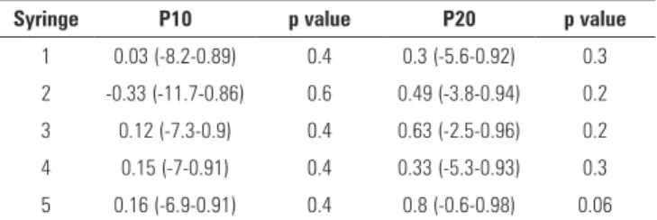
Table 1 - Cuff pressures observed after inflating the pilot balloon in Phase 1 of the study
Syringe 1 Syringe 2 Syringe 3 Syringe 4 Syringe 5 p value
P10 45.2±7.5 54.8±10.1 48.2±4.3 47±6.1 59.6±9.2 p=0.043*
P20 84±16.5 53.2±5 48.8±4.6 51.6±14.5 35±2.4 p<0.001**
Values are expressed as the mean±standard deviation of the five measurements performed with each syringe. P10 - maneuver performed with a 10mL syringe; P20 - maneuver performed with a 20mL syringe. * comparisons between 10mL syringes; ** comparisons between 20mL syringes.
he ICC between the ive tests performed with the syringes in Phase 1 was not signiicant, and the 95% CI ranged from a low negative value to a high positive value, which implies that the reproducibility of each syringe was zero (Table 2).
Table 2 - Intraclass correlation coefficient between the five tests performed with each syringe in Phase 1.
Syringe P10 p value P20 p value
1 0.03 (-8.2-0.89) 0.4 0.3 (-5.6-0.92) 0.3
2 -0.33 (-11.7-0.86) 0.6 0.49 (-3.8-0.94) 0.2
3 0.12 (-7.3-0.9) 0.4 0.63 (-2.5-0.96) 0.2
4 0.15 (-7-0.91) 0.4 0.33 (-5.3-0.93) 0.3
5 0.16 (-6.9-0.91) 0.4 0.8 (-0.6-0.98) 0.06
P10 - maneuver performed with a 10mL syringe; P20 - maneuver performed with a 20mL syringe. Values for the intraclass correlation coefficients and the 95% confidence intervals (95%CI).
Of the 20 patients included in Phase 2 of this study, 11 (55%) were male and had a median height of 163cm (153-170cm) and a median age of 57 years (27-74 years). he patients were intubated with endotracheal tubes of various sizes: 7 (n=3), 7.5 (n=3), 8 (n=12), and 8.5 (n=2). Most patients (n=15) were not sedated at the time the data were collected.
he cuf pressures generated with the P5 [105 (82.5-120)
cmH2O], P10 [69 (47.5-111.3) cmH2O], and P20 [45
(35-59.5) cmH2O] syringes were higher than the P-initial values [20 (20-25) cmH2O] (p<0.001) (Figure 1).
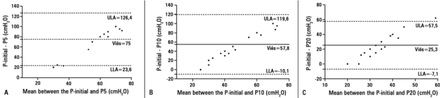
he Bland-Altman plots revealed large biases
between the P-initial and P5 (75±26.2cmH2O), P10
and bias in the Bland-Altman analysis, compared with the initial pressure.
he use of a syringe to measure cuf pressure (cuf pressure relief valve technique) is an alternative, inexpensive, and rapid method to ensure that the pressure on the trachea is not too high (thereby injuring the trachea) or too low (to prevent microaspiration) in hospitals that do not have a manometer speciically designed to perform such measurements. However, our indings demonstrate that this technique is not efective at maintaining the cuf pressure within the recommended limits and is therefore not safe to use. he use of this technique may cause the loss of cilia or even the formation of tracheoesophageal istulas due to high pressures exerted on the trachea.(5,6)
he use of syringes with diferent volumes and from diferent manufacturers for determining the cuf pressure using the pressure relief valve technique was previously tested by Mac et al. hese authors used a trachea model to analyze three diferent brands of P20 syringes and found that only one brand was able to maintain the cuf pressure within safe limits. Upon comparing syringes with diferent volumes (P10 and 60mL), the authors found cuf pressures of 57 and 23cmH2O, respectively, demonstrating not only that the syringe brand is important for determining cuf pressure using this technique but also that the syringe volume is paramount.(21) Our study is consistent with and
complements this previous study, as we observed large variations between the same brand of syringe with the same volume in sequential tests.
Testing the cuf pressure relief valve technique in patients who were not anesthetized, we found that as the size of the syringe decreased, cuf pressure increased. his observation can be explained by the physical principle in which the pressure is determined by the ratio of the force applied and the surface area on which the force is applied. he increased surface area of the larger syringe plungers Figure 1 - Comparisons between the initial cuff pressures (cmH2O) (P-initial) and
the cuff pressures obtained during maneuvers with 5mL (P5), 10mL (P10), and 20mL (P20) syringes in Phase 2 of the study; in vivo experiments (individual data for all patients). * p<0.001 compared with the P-initial.
he variability in the values, as determined using the 95% upper and lower limits of agreement (bias±1.96 SD), ranged from 23.6 to 126.4cmH2O for P5, -10.1 to 119.6cmH2O for P10, and -7.1 to 57.5cmH2O for P20, compared to the P-initial (Figure 2).
DISCUSSION
his study examined the efectiveness of the cuf pressure relief valve technique in maintaining the cuf pressure within the recommended limits in ICU patients and the reproducibility of P20 and P10 syringes in an experimental trachea model. he reproducibility was tested using ive tests for each of the syringes. he ICC, which indicates the degree of agreement between measurements, indicated a lack of reproducibility between tests for each syringe. he cuf pressure values observed in the maneuver with the P5, P10, and P20 syringes exceeded the limit considered safe (20-30cmH2O) and exhibited signiicant variability
implies that less pressure is required to overcome the force resulting from the dynamic friction.(21)
Substituting one instrument or evaluation technique for another is only possible if the new device is equivalent to the previous one and has been tested prior to its clinical use.(20)
It is very unlikely that a clinical measurement evaluated using two diferent devices will yield identical results. Bland et al. therefore proposed that the diferences between instruments be as small as possible to provide equivalent, accurate, and reliable clinical measurements.(20) However,
the acceptable degree of divergence between instruments depends on the clinical measurement being examined.(22)
In the case of cuf pressures in tracheal tubes for which the normal range is extremely narrow (20 to 30cmH2O), the variability between measurements should also be as small as possible. Using the Bland-Altman method, we observed biases of 75cmH2O, 57.8cmH2O, and 25.3cmH2O using P5, P10, and P20 syringes, respectively. Furthermore, the 95% lower and upper limits of agreement were excessively high (between -10.1cmH2O and 126.4cmH2O), showing clinically impractical values.
Our study does have some limitations. In Phase 1 of the study, we used a PVC tube to simulate the trachea. Due to its rigidity, this material does not accurately simulate the resistance and elasticity of the trachea. However, in Phase 1, only the reproducibility of the technique was tested, without making comparisons between the diferent volumes of syringes and the standard manometer. In both phases of the study, the pauses taken while inlating the pilot balloon with the syringes were not standardized, and the perceived
recoil of the plunger was always “researcher-dependent.” Furthermore, the design of our study did not allow us to “blind” the researcher for the data collection. However, the same researcher performed all of the measurements, and the order in which the syringes were used during the data collection was random, which might have partially minimized these efects. Our study employed a convenience sample that consisted of only 20 patients. Although it was calculated after the data were collected, the power of our sample was 100%; therefore, we do not believe that increasing the sample size would change the results. Finally, diferent brands of syringes can have diferent plunger sizes and thicknesses, although they have the same overall volume. hus, we cannot conclude whether the results would be diferent if other brands of syringes were used. Moreover, regardless of the dimensional characteristics (thickness and size of the plunger), the concepts of static and dynamic resistance (where the static resistance is always greater than the dynamic resistance) would have a predominant efect; consequently, we believe that our results would be similar for any brand of syringe.
CONCLUSION
he present study demonstrates that the cuf pressure relief valve technique is not reproducible in a trachea model and is not efective for using 5mL, 10mL, or 20mL syringes to determine the pressure of cufs secured in intubated intensive care unit patients. herefore, the use of a manometer speciically designed to measure the cuf pressure of endotracheal tubes is recommended for this population.
Objetivo: Testar a eicácia da técnica de alívio de pressão de
cuff por meio de uma válvula em manter níveis de pressão de cuff
dentro da normalidade in vitro (Fase 1) e em pacientes interna-dos em unidade de terapia intensiva (Fase 2), bem como testar a reprodutibilidade da técnica utilizando diferentes seringas.
Métodos: Na Fase 1, uma cânula orotraqueal foi inserida em um modelo de traqueia. Seringas de 10 e 20mL foram uti-lizadas para insular o cuff da cânula. O cuff foi insulado lenta e progressivamente até que o êmbolo da seringa se deslocasse em direção contrária da aplicação. Após a pausa do êmbolo, as pressões do balonete foram registradas. Na Fase 2, a mesma manobra de insulação do cuff foi realizada em 20 pacientes, utilizando-se seringas de 5, 10 e 20mL, e foi comparada com as medidas de um manômetro. O índice de correlação intraclasse e a análise de Bland-Altman foram realizados para veriicar a
reprodutibilidade e a concordância entre as seringas. Os dados foram expressos como mediana (intervalo interquartil).
Resultados: A reprodutibilidade entre as seringas foi nula, com índice de correlação intraclasse variando entre -0,33 e 0,8 (p>0,05). As pressões geradas com as seringas foram supe-riores à pressão obtida com o manômetro padrão: seringa de
5mL teve 105cmH2O (82,5-120cmH2O); seringa de 10mL
teve 69cmH2O (47,5-111,3cmH2O) e seringa de 20mL teve
45cmH2O (35-59,5cmH2O). O teste de Bland-Altman verii-cou grandes vieses e variabilidade entre as seringas utilizadas, quando estas foram comparadas ao manômetro.
Conclusão: O uso de seringas não é eicaz em determinar valores de pressão de cuff seguros em pacientes internados em unidade de terapia intensiva.
RESUMO
REFERENCES
1. Rello J, Soñora R, Jubert P, Artigas A, Rué M, Vallés J. Pneumonia in intubated patients: role of respiratory airway care. Am J Respir Crit Care Med. 1996;154(1):111-5.
2. Seegobin RD, van Hasselt GL. Endotracheal cuff pressure and tracheal mucosal blood flow: endoscopic study of effects of four large volume cuffs. Br Med J (Clin Res Ed). 1984;288(6422):965-8.
3. Barbosa PM, Santos BM. Alterações morfológicas em traqueias de pacientes intubados em função do tempo de intubação. Rev Latinoam Enferm. 2003;11(6):727-33.
4. Curiel García JA, Guerrero-Romero F, Rodríguez-Morán M. [Cuff pressure in endotracheal intubation: should it be routinely measured?]. Gac Med Mex. 2001;137(2):179-82. Spanish.
5. Castilho EC, Braz JR, Catâneo AJ, Martins RH, Gregório EA, Monteiro ER. Efeitos da pressão limite (25cmH2O) e mínima de “selo” do balonete de tubos traqueais sobre a mucosa traqueal do cão. Rev Bras Anestesiol. 2003;53(6):743-55.
6. Nseir S, Duguet A, Copin MC, De Jonckheere J, Zhang M, Similowski T, et al. Continuous control of endotracheal cuff pressure and tracheal wall damage: a randomized controlled animal study. Crit Care. 2007;11(5):R109. 7. American Thoracic Society; Infectious Diseases Society of America. Guidelines for the management of adults with hospital-acquired, ventilator-associated, and healthcare-associated pneumonia. Am J Respir Crit Care Med. 2005;171(4):388-416.
8. Resende MM, Monteiro SG, Callegari B, Figueiredo PM, Monteiro CR, Monteiro-Neto V. Epidemiology and outcomes of ventilator-associated pneumonia in northern Brazil: an analytical descriptive prospective cohort study. BMC Infect Dis. 2013;13:119.
9. Safdar N, Dezfulian C, Collard HR, Saint S. Clinical and economic consequences of ventilator-associated pneumonia: a systematic review. Crit Care Med. 2005;33(10):2184-93. Review.
10. Alberti C, Brun-Buisson C, Burchardi H, Martin C, Goodman S, Artigas A, et al. Epidemiology of sepsis and infection in ICU patients from an international multicentre cohort study. Intensive Care Med. 2002;28(2):108-21. Erratum in: Intensive Care Med. 2002;28(4):525-6.
11. Bernhard WN, Yost L, Joynes D, Cothalis S, Turndorf H. Intracuff pressures in endotracheal and tracheostomy tubes. Related cuff physical characteristics. Chest. 1985;87(6):720-5.
12. Nseir S, Zerimech F, De Jonckheere J, Alves I, Balduyck M, Durocher A. Impact of polyurethane on variations in tracheal cuff pressure in critically ill patients: a prospective observational study. Intensive Care Med. 2010;36(7):1156-63.
13. Ramirez P, Bassi GL, Torres A. Measures to prevent nosocomial infections during mechanical ventilation. Curr Opin Crit Care. 2012;18(1):86-92. Review.
14. Godoy AC, Vieira RJ, Capitani EM. Endotracheal tube cuff pressure alteration after changes in position in patients under mechanical ventilation. J Bras Pneumol. 2008;34(5):294-7.
15. Souza Neto EP, Piriou V, Durand PG, George M, Evans R, Obadia JF, et al. Influence of temperature on tracheal tube cuff pressure during cardiac surgery. Acta Anaesthesiol Scand. 1999;43(3):333-7.
16. Sultan P, Carvalho B, Rose BO, Cregg R. Endotracheal tube cuff pressure monitoring: a review of the evidence. J Perioper Pract. 2011;21(11):379-86. 17. Galinski M, Tréoux V, Garrigue B, Lapostolle F, Borron SW, Adnet F.
Intracuff pressures of endotracheal tubes in the management of airway emergencies: the need for pressure monitoring. Ann Emerg Med. 2006;47(6):545-7.
18. Somri M, Fradis M, Malatskey S, Vaida S, Gaitini L. Simple on-line endotracheal cuff pressure relief valve. Ann Otol Rhinol Laryngol. 2002;111(2):190-2. 19. Deyo RA, Diehr P, Patrick DL. Reproducibility and responsiveness of health
status measures. Statistics and strategies for evaluation. Control Clin Trials. 1991;12(4 Suppl):142S-58S.
20. Bland JM, Altman DG. Statistical methods for assessing agreement between two methods of clinical measurement. Lancet. 1986;1(8476):307-10. 21. Mac Murdo SD, Buffington CW. Brand and size matter when choosing
a syringe to relieve pressure in a tracheal tube cuff. Anesth Analg. 2004;99(5):1445-9; table of contents.