Humeral internal rotation osteotomy for the
treat-ment of Erb-Duchenne-type obstetric palsy: clinical
and radiographic results
Jorge Henrique Assunc¸a˜o, Arnaldo Amado Ferreira Neto, Eduardo Benegas, Raul Bolliger Neto,
Fla´via Santis Prada, Eduardo Angeli Malavolta, Mauro Emilio Conforto Gracitelli, Gilberto Luis Camanho Faculdade de Medicina da Universidade de Sa˜o Paulo, Department of Orthopedics and Traumatology, Sa˜o Paulo/SP, Brazil.
OBJECTIVE:To evaluate the functional and radiographic results in patients undergoing shoulder anterior soft tissue stretching in association with open reduction and internal rotation osteotomy to centralize the humeral head as a treatment for Erb-Duchenne obstetric palsy sequelae.
METHOD:A total of 35 patients underwent this surgical treatment, and the mean follow-up was 4.6 years. The Mallet scale was applied before and after the surgical procedure. A total of 20 patients underwent computed tomography to assess the glenoid version and humeral head subluxation.
RESULTS: Functional improvement was achieved, as evidenced by an increase in the Mallet scale score from 12.14 to 16.46 (p,0.001). The correction of retroversion was achieved once the glenoid version ranged from -21.4 to -12 degrees (p,0.001). The humeral head subluxation improved from 6.5 to 35.2% (p,0.001). Patients older than 6 years of age did not achieve glenohumeral joint improvement with respect to dysplastic abnormalities.
CONCLUSION: Internal rotation osteotomy in association with the stretching of anterior soft tissues of the shoulder in patients under the age of 7 years provided improvements in the function, retroversion, and subluxation of the glenohumeral joint.
KEYWORDS: Obstetric Palsy; Glenohumeral Deformity; Shoulder Dislocation; Osteotomy.
Assunc¸a˜o JH, Neto AAF, Benegas E, Neto RB, Prada FS, Malavolta EA, et al. Humeral internal rotation osteotomy for the treatment of Erb-Duchenne-type obstetric palsy: clinical and radiographic results. Clinics. 2013;68(7):928-933.
Received for publication onJanuary 16, 2013;First review completed onJanuary 28, 2013;Accepted for publication onMarch 8, 2013 E-mail: [email protected]
Tel.: 55 11 2661-7812
& INTRODUCTION
Obstetric palsy results from traction of the brachial plexus in above-average-weight newborns in cases of difficult delivery (such as breech presentation) and in cases of shoulder dystocia (1). The incidence of obstetric palsy ranges from 1 to 4.6 cases per 1,000 live births (2).
Although complete motor recovery may occur in many patients, some patients develop growth deformities due to nerve injury resulting from the secondary muscle imbalance. One of the more common deformities is a contracture in adduction and internal rotation of the shoulder because of the predominance of the adductor and internal rotator muscles with respect to the elevators and external rotators. This deformity is mainly caused by
injury to the suprascapular nerve (3,4). This imbalance leads to misalignment of the humeral head in relation to the glenoid, which promotes both increased retroversion and hypoplasia of the humeral head and retroversion of the glenoid. The persistence of this abnormal positioning evolves to posterior glenohumeral subluxation or even dislocation within a short time (5).
Microsurgical nerve reconstruction, soft-tissue releases, tendon transfers, open reduction, and humeral osteotomies have been proposed as methods to improve upper-extremity function in appropriately selected patients (6-15). Waters et al. (8) demonstrated that glenoid abnormal-ities showed little improvement with muscle-tendon stretch-ing, even when performed in association with muscle transfers, if the presence of bony deformities was neglected. There have been descriptions of an internal rotation osteotomy of the proximal humerus in addition to the reduction of the humeral head in the glenoid cavity associated with tendon stretching performed during the same operation. The objective is to reduce the humeral head retroversion, yielding better positioning of the head in relation to the glenoid cavity. This operation has benefits related to osteoarticular modeling, this modeling Copyrightß2013CLINICS– This is an Open Access article distributed under
the terms of the Creative Commons Attribution Non-Commercial License (http:// creativecommons.org/licenses/by-nc/3.0/) which permits unrestricted non-commercial use, distribution, and reproduction in any medium, provided the original work is properly cited.
No potential conflict of interest was reported.
is potentially large-scale in very young children (14,15). There have been no studies assessing the effect of this technique on glenohumeral joint development as it applies to the remodeling of a retroverted glenoid.
The objective of the present study was to present functional and radiographic results from patients who underwent internal rotator stretching and open reduction of the humeral head in the glenoid cavity in association with an internal-rotation osteotomy to center the humeral head.
& MATERIALS AND METHODS
Between 1996 and 2008, a total of 42 children who sustained obstetric palsy sequelae in adduction and internal rotation shoulder contracture underwent stretching of the anterior soft tissues (Sever’s procedure), open reduction of the humeral head in the glenoid cavity, and internal-rotation osteotomy of the proximal humerus.
This surgical treatment was indicated for patients with adduction and internal rotation contracture (Erb-Duchenne palsy) with posterior subluxation or dislocation of the shoulder. To obtain and maintain joint congruity of the glenohumeral joint, we indicated this procedure for children beginning at 18 months of age in consideration of the greater potential for bone remodeling in younger children. Patients were excluded from the study if they did not complete a minimum postoperative follow-up period of 2 years.
The patients were clinically assessed pre- and post-operatively according to the modified Mallet scale (16). They were also assessed radiographically before and after the operation. The radiographic exams were performed during the preoperative and postoperative evaluations, which were performed at 1, 2, 4, 6, 12, and 24 weeks after the surgery and then annually. The radiographic assessment of the shoulder included the anteroposterior view, axillary view, and true lateral view. The humeral head was described as centered, subluxated, or dislocated with respect to the glenoid cavity.
A total of 20 patients underwent computed tomography (CT) scans of both shoulders before and after the operation. The post-operative CT scans were performed at least 2 years after the surgical procedure. On the CT scan, the glenoid version was measured on the axial slices (17) in the following manner: a line was drawn parallel to the scapula body through the midpoint of the glenoid (scapular line), and another line was drawn parallel to the glenoid cavity and joined the anterior and posterior edges. These two lines intersected to form 4 quadrants. By definition, the state of the glenoid cavity version was obtained as the value of the angle of the posteromedial quadrant after subtracting 90 degrees. The glenoid was considered retroverted when this value was negative and anteverted when the value was positive. The degree of subluxation of the humeral head was measured in the following manner: the largest anteropos-terior diameter of the humeral head was measured (AP line); the intersection of the scapular line with the AP line created the AE line (between the intersection point and the anterior point of the AP line); and the subluxation or dislocation was measured as the percentage of posterior deviation of the humeral head (PD), which was calculated by dividing AE by AP and multiplying the result by 100. The classification method of Waters et al. (5) was also used to assess the degree of glenohumeral deformity.
Surgical procedure
With the patient in a modified beach chair position under general anesthesia, a deltopectoral incision was made. The dissection was performed in layers, and Z-shaped stretching of the pectoralis major muscle was performed. The subscapular tendon was disinserted from the lesser tuber-osity, starting close to the groove of the tendon of the long head of the biceps. The capsulotomy was performed craniocaudally, 0.5 cm from the lesser tuberosity. The glenohumeral joint was inspected to determine if the humeral head was subluxated or dislocated posteriorly.
The reduction of the humeral head was achieved through external rotation of the arm, and the humeral head was fixed to the glenoid with a thin Kirschner wire (18).
A transverse osteotomy was then performed on the humerus between the insertions of the deltoid and pectoralis major muscles. The distal part of the humerus was internally rotated to leave it in the 0
˚
rotation position of the arm (18). The bone fragments were fixed with a plate and screws (a minimum of 2 and maximum of 4 cortical screws) (18).The Kirschner wire that had maintained the joint reduction was removed, and the joint stability was tested (18). The subscapular tendon was then sutured in the remaining portion of the joint capsule, which was inserted into the lesser tuberosity (18).
The entire upper limb was immobilized for 4 weeks by a thoracobrachial 45
˚
abduction plaster cast with a 0˚
rotation of the arm. After radiographic consolidation, rehabilitation was initiated (18).Statistical analysis
The modified Mallet scale values and the degrees of glenoid retroversion and humeral head subluxation mea-sured before and after the operation were subjected to a paired t-test. Values less than 0.05 were considered significant. The patients were stratified according to age and the degree of glenohumeral deformity to determine if the clinical and radiographic results were uniform across all ages or degrees of deformity.
& RESULTS
A total of 7 patients were excluded from the study, and the 35 remaining patients included in the study comprised the following: 21 males and 14 females, with a mean follow-up of 4.6 years (range: 2 to 12 years). The patients’ mean age was 4.4 years (range: 1 to 9 years) at the time of the proposed surgical treatment (Table 1).
The mean score was 12.14 points (range: 8 to 18) on the modified Mallet scale before surgery and 16.46 points (score: 12 to 22) after surgery, representing a mean gain of 4.32 points (range: 0 to 10) (p,0.001). Within the age groups, patients who were 7 years of age and older had a mean functional gain of only one point, with 3 patients not achieving any functional improvement. Despite this small difference, the gain obtained was significant (p= 0.02)
(Table 2). There was no significant difference with respect to functional gain between patients presenting with sub-luxated and dislocated glenohumeral joints.
with a concentric reduced humeral head, while 7 patients sustained posterior subluxation. Among these 7 patients, 3 underwent surgical treatment after reaching the age of 6 years (Table 3).
A total of 20 patients underwent CT scans before and after surgery. Of these patients, 6 patients were classified as Waters type III, one was type IV, 3 were type V, and 10 were type VI before surgical treatment. After treatment, 2 became type I, 10 became type II, 4 became type III, and 4 became type IV. All patients who presented Waters type IV on the postoperative CT scan underwent surgical treatment after reaching the age of 6 years (Table 4).
CT scans were used to evaluate the glenoid retroversion and the degree of subluxation or dislocation of the humeral head (20 patients). Postoperative CT scans were performed, on average, 3.85 years (range: 2 to 11) after the surgical procedure. On the unaffected side, the mean glenoid version was -4 degrees (range: -10 to 4) before the operation and -3.7 degrees (range: -10 to 4) after the operation (p= 0.58). On the
operated side, the mean retroversion was -21.4 degrees (range: -34 to -3) before the operation and -12 degrees (range: -32 to -2) after the operation (p,0.001). Glenoid retroversion decreased in all patients younger than 7 years of age who underwent the operation, with a mean change from -23.3 to -9.5 degrees (p,0.001). Among the patients older than 6 years of age, the retroversion increased (from -16 degrees to -19.4 degrees, on average). The degree of
correction of the glenoid cavity version after the operation was independent of whether the humeral head was subluxated or dislocated before surgery (Figures 1 and 2, Table 2).
The percentage of posterior deviation of the humeral head (PD) was improved in all patients, ranging from 6.5% (range: 0 to 28%) to 35.2% (range: 10 to 50%), on average (p,0.001). Among the patients younger than 7 years of age, the mean preoperative PD was 3.1% (range: 0 to 27%), and the mean postoperative PD was 38.8% (range: 18.3 to 50%) (p,0.001). Among the patients older than 7 years of age, the mean PD changed from 16.6 to 24.3% (p= 0.21). On the
contralateral unaffected side, the PD measurement was 46.8% before the operation and 47.1% after the operation (p= 0.87) (Table 2).
There were 2 cases in which a varus deformity of the osteotomy was observed, but the growth of these patients led to complete correction of the deformity. In 4 patients, the plate and screws were removed because of complaints of local pain. There were no cases of infection or additional neurological lesions.
& DISCUSSION
Obstetric palsy treatments vary according to the phase of the disease. The use of orthoses and rehabilitation is indicated in the initial phase of treatment (in newborns). Brachial plexus surgical exploration is indicated when Table 1 -Pre- and postoperative clinical and radiographic patient data.
Patient Sex
Age at time of
surgery (yrs) Follow-up (yrs)
Pre-surgery Mallet score
Post-surgery Mallet score
Gain in the Mallet score
Pre-surgery X-ray finding
Post-surgery X-ray finding
1 M 2 2 12 17 5 dislocated centered
2 M 6 2 9 16 7 dislocated centered
3 M 2 2 10 16 6 subluxated centered
4 F 2 2 12 14 2 dislocated subluxated
5 F 4 2 12 19 7 subluxated centered
6 M 3 3 14 19 5 dislocated subluxated
7 F 2 3 14 21 7 dislocated centered
8 F 3 3 8 18 10 dislocated centered
9 M 2 2 13 17 4 dislocated centered
10 M 2 2 12 14 2 dislocated centered
11 F 5 2 17 22 5 subluxated centered
12 M 5 6 10 17 7 dislocated centered
13 F 5 6 13 17 4 subluxated centered
14 M 2 6 10 16 6 dislocated centered
15 M 3 6 10 14 4 subluxated Subluxated
16 M 3 6 11 14 3 dislocated centered
17 M 5 2 10 18 8 subluxated centered
18 F 1 7 13 20 7 subluxated centered
19 M 3 7 14 16 2 dislocated centered
20 F 4 7 13 14 1 dislocated centered
21 F 6 8 10 18 8 dislocated centered
22 M 3 8 14 20 6 subluxated centered
23 M 1 9 11 20 9 dislocated centered
24 M 3 9 10 13 3 dislocated centered
25 M 3 12 10 18 8 subluxated centered
26 F 4 2 12 16 4 subluxated centered
27 F 6 2 13 16 3 subluxated subluxated
28 M 8 7 13 15 2 dislocated subluxated
29 F 9 6 11 12 1 subluxated centered
30 F 8 2 13 15 2 dislocated subluxated
31 M 9 5 14 14 0 dislocated centered
32 F 7 5 12 12 0 subluxated centered
33 M 8 3 18 18 0 dislocated subluxated
34 M 8 4 14 15 1 subluxated centered
35 M 7 2 13 15 2 subluxated centered
recovery of biceps muscle function has not been achieved within six months.
Tendon stretching (1,6) with or without muscle transfer (1) can be performed in patients with internal shoulder rotation and adduction contracture without bone deformi-ties. The objective in this phase is to achieve a concentric and stable glenohumeral joint and provide as extensive a functional range of motion as possible.
In Brazil, we have observed that the natural history of the osteoarticular sequelae of OP is influenced by 2 factors: the use of conservative treatment methods (e.g., physiotherapy and orthoses) for exceedingly long periods because of a traditional belief in their efficacy and a delay in surgical indications because the parents fear their children under-going surgery or because the professionals who treat them are inexperienced.
The optimum time for avoiding or minimizing the severe osteoarticular abnormalities caused by muscle lesions and imbalance as sequelae from obstetric palsy is frequently missed. In recent years, efforts have been made to perform corrections at the proper time (18).
The maintenance of shoulder contracture gives rise to the posterior displacement of the humeral head. In the initial stages of this displacement, early changes to the proximal epiphysis of the humerus appear and evolve rapidly to osteoarticular deformities (retroversion of the humerus and retroversion and hypoplasia of the glenoid), followed by posterior subluxation. Over time, this imbalance leads to the total loss of joint congruence. All these factors have severe consequences for shoulder function (13).
Tendon stretching and tendon transfers significantly improve overall shoulder functioning but have only a modest influence on glenoid cavity retroversion, deforma-tion, and humeral head subluxation as an evolution of glenohumeral dysmorphism. In a follow-up of 25 patients, Waters (8) obtained a mean gain of 5 points on the Mallet
scale, but the dysplastic abnormalities of the glenohumeral joint were only modestly reduced.
The importance of restoring muscle balance must be supported by a concentric reduction of the humeral head in the glenoid cavity. This statement was confirmed by Hui et al. (12); specifically, in 23 patients, open reduction of the humeral head and tendon stretching were able to signifi-cantly reduce the glenoid cavity retroversion in these patients. Among 22 patients, Pedowitz et al. (13) demon-strated that a considerable improvement in the dysplastic abnormalities of the glenoid occurred when these patients underwent arthroscopic release of the anterior capsule, tenotomy of the subscapular tendon, and reduction of the humeral head in the glenoid. The glenoid version changed from -37 to -8 degrees (p,0.001), and the PD increased from 15.6 to 46.9% (p,0.001). In addition to the open reduction of the humeral head and tendon stretching, Sinbiski and Synder (14) performed an internal-rotation osteotomy in some cases, with the aim of maintaining the stability of the reduction that had been achieved during the operation. They obtained a mean improvement of 4.7 points on the Mallet scale but did not describe if there was any improvement in the dysplastic glenoid abnormalities. Kambhampati et al. (15) not only performed open reduction of the humeral head but also performed an internal-rotation osteotomy in cases in which the humeral head retroversion was greater than 40 degrees.
In the present study, all patients underwent pectoralis major and subscapularis muscle tendon stretching, stretch-ing of the anteroinferior capsule, reduction of the humeral head, and internal-rotation osteotomy of the shoulder. These procedures enabled the stable reduction of the glenohumeral joint with the correction of the retroversion of the humeral head. Retroversion is abnormally greater in Table 2 -Mean pre- and postoperative Mallet scores, degrees of glenoid retroversion, and percentages of posterior deviation of the humeral head (PD).
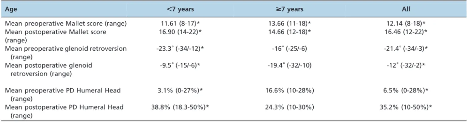
Age ,7 years $7 years All
Mean preoperative Mallet score (range) 11.61 (8-17)* 13.66 (11-18)* 12.14 (8-18)*
Mean postoperative Mallet score 16.90 (14-22)* 14.66 (12-18)* 16.46 (12-22)*
(range)
Mean preoperative glenoid retroversion (range)
-23.3˚(-34/-12)* -16˚(-25/-6) -21.4˚(-34/-3)*
Mean postoperative glenoid retroversion (range)
-9.5˚(-15/-6)* -19.4˚(-32/-10) -12˚(-32/-2)*
Mean preoperative PD Humeral Head (range)
3.1% (0-27%)* 16.6% (10-28%) 6.5% (0-28%)*
Mean postoperative PD Humeral Head (range)
38.8% (18.3-50%)* 24.3% (10-30%) 35.2% (10-50%)*
*
p,0.05.
Table 3 -Pre- and postoperative radiographic evaluations.
Radiographic evaluation
Preoperative (patients n)
Postoperative (patients n)
Reduced 0 28
Posterior Subluxation 15 7
Posterior Dislocation 20 0
Table 4 -Pre- and postoperative CT evaluations.
CT evaluation
Preoperative (patients n)
Postoperative (patients n)
Waters I 0 2
Waters II 0 10
Waters III 6 4
Waters IV 1 4
Waters V 3 0
obstetric palsy patients (19) but is frequently considered unimportant during treatment.
The humeral head internal-rotation osteotomy presented in this study, which is also known as a centering osteotomy, is analogous to the procedure that is performed in newborns with dysplastic hips. The aim is to keep the humeral head contained in the glenoid cavity, enabling reciprocal stimula-tion to form a congruent and funcstimula-tionally efficient joint (18). We obtained a mean improvement of 4.32 points in the Mallet score, similar to that reported in other studies (8,14,15). If only patients younger than 7 years of age are considered, our improvement is 5.29 points. In our group of patients, we obtained significant reductions of the glenoid retroversion, which decreased from 23.3 to 9.5 degrees, and of the humeral head subluxation, which increased from 3.13 to 38.8%. Among the patients older than 6 years of age, we did not achieve the same success; specifically, reversal of the dysplastic abnormalities of the glenoid and humeral head was not obtained, and there were only very slight gains in shoulder function. We have ceased to indicate this surgical procedure for patients aged 7 years and older.
Kambhampati et al. (15) reported worse results in older patients. They described 20 cases of persistent humeral head dislocation in older patients compared with younger patients in whom successful glenohumeral joint reductions were achieved. We agree with these authors; performing the centering osteotomy at an early stage is necessary in patients with internal rotation and adduction contracture, particularly in those with posterior subluxation or disloca-tion of the humeral head. We further recommend that this technique should be used in children up to the age of 6 years who do not present any major bone deformities of the glenohumeral joint because the potential for remodeling of the glenoid in children of this age group is greater than that in older children (18).
For patients younger than 7 years of age without signs of subluxation of the humeral head, we recommend that Sever’s procedure be used. This procedure may be performed in association with muscle transfer. We would indicate an external rotation osteotomy (18) for patients older than 6 years of age or for patients with major dysplastic abnormalities.
The main limitations of this study are its non-comparative design and the lack of a CT evaluation before and after surgery in some cases. In recent years, CT and magnetic resonance imaging have become the best modalities for conducting radiographic analysis. Our study began in 1996, when these imaging modalities were not readily available; while approximately 20 patients had pre- and postoperative CT scans, all patients were examined by conventional radiography exams. The modified Mallet scale is the score used in most clinical studies, but it has not been previously validated for the Portuguese language. A number of studies have assessed this scale and found good intra- and inter-observer reliability (20,21).
Despite significant improvements in external rotation and elevation, some patients continued to exhibit diminished strength in the contralateral shoulder. To address this problem, we are performing muscle transfers involving the teres major and latissimus dorsi in selected patients in a subsequent surgical procedure.
Humeral head internal rotation osteotomy in association with shoulder internal rotator stretching significantly improved the shoulder function of obstetric palsy sequelae patients. Glenoid retroversion was achieved in patients younger than 7 years of age, with significant improvements obtained in the relationship of the humeral head within the glenoid cavity.
& AUTHOR CONTRIBUTIONS
Assunc¸a˜o JH designed the study, wrote the text, and participated as a surgeon. Ferreira Neto AA guided the project and participated as a surgeon. Benegas E participated as a surgeon. Neto RB conducted the statistical analysis. Prada FS compiled the bibliography and participated as a surgeon. Malavolta EA served as a reviewer and participated as a surgeon. Gracitelli MEC wrote the manuscript, formatted the text and photographs, and participated as a surgeon. Camanho GL served as a reviewer.
& REFERENCES
1. Tachdjian MO. Obstetrical brachial plexus palsy: In: Tachdjian pediatric orthopedics. 2nd ed. Pilhadelphia: Saunders; 1990. p. 2009-82. 2. Pearl ML. Shoulder problems in children with brachial plexus birth
palsy: evaluation and management. J Am Acad Orthop Surg. 2009;17(4):242-54.
3. Dunkerton MC. Posterior dislocation of the shoulder associated with obstetric brachial plexus palsy. J Bone Joint Surg Br. 1989;71(5):764-6. 4. Al-Qattan MM. Rotation osteotomy of the humerus for Erb’s palsy in
children with humeral head deformity. J Hand Surg Am. 2002;27(3):479-83, http://dx.doi.org/10.1053/jhsu.2002.33198.
5. Waters PM, Smith GR, Jaramillo D. Glenohumeral deformity secondary to brachial plexus birth palsy. J Bone Joint Surg Am. 1998;80(5):668-77.
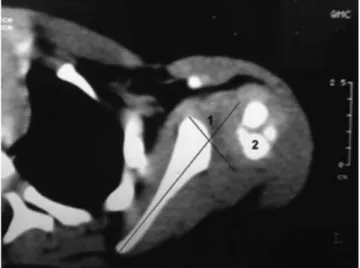
Figure 1 -Preoperative CT scan. Glenoid cavity (1) and humeral heal dislocated posteriorly (2).
6. Sever JW. The results of a new operation for obstetrical paralysis. J Bone Joint Surg Am. 1918;16(Suppl 2):248-57.
7. Kirkos JM, Kyrkos MJ, Kapetanos GA, Haritidis JH. Brachial plexus palsy secondary to birth injuries. J Bone Joint Surg Br. 2005;87(2):231-5, http://dx.doi.org/10.1302/0301-620X.87B2.14739.
8. Waters PM, Bae DS. Effect of tendon transfers and extra-articular soft-tissue balancing on glenohumeral development in brachial plexus birth palsy. J Bone Joint Surg Am. 2005;87(2):320-5, http://dx.doi.org/10. 2106/JBJS.C.01614.
9. Hale HB, Bae DS, Waters PM. Current concepts in the management of brachial plexus birth palsy. J Hand Surg Am. 2010;35(2):322-31, http:// dx.doi.org/10.1016/j.jhsa.2009.11.026.
10. Chen L, Gu YD, Wang H. Microsurgical reconstruction of obstetric brachial plexus palsy. Microsurgery. 2008;28(2):108-12, http://dx.doi. org/10.1002/micr.20459.
11. Waters PM, Bae DS. The effect of derotational humeral osteotomy on global shoulder function in brachial plexus birth palsy. J Bone Joint Surg Am. 2006;88(5):1035-1042, http://dx.doi.org/10.2106/JBJS.E.00680. 12. Hui JH, Torode IP. Changing glenoid version after open reduction of
shoulders in children with obstetric brachial plexus palsy. J Pediatr Orthop. 2003;23(1):109-13.
13. Pedowitz DI, Gibson B, Williams GR, Kozin SH. Arthroscopic treatment of posterior glenohumeral joint subluxation resulting from brachial plexus birth palsy. J Shoulder Elbow Surg. 2007;16(1):6-13, http://dx.doi. org/10.1016/j.jse.2006.04.008.
14. Sibin´ski M, Synder M. Soft tissue rebalancing procedures with and without internal rotation osteotomy for shoulder deformity in children with persistent obstetric brachial plexus palsy. Arch Orthop Trauma Surg. 2010;130(12):1499-504, http://dx.doi.org/10.1007/s00402-010-1067-6. 15. Kambhampati SB, Birch R, Cobiella C, Chen L. Posterior subluxation and
dislocation of the shoulder in obstetric brachial plexus palsy. J Bone Joint Surg Br. 2006;88(2):213-9, http://dx.doi.org/10.1302/0301-620X.88B2.17185. 16. Gilbert A, Tassin JL. Re´paration chirurgicale du plexus brachial dans la
paralysie obste´tricale. Chirurgie. 1984;110(1):70-5.
17. Friedman RJ, Hawthorne KB, Genez BM. The use of computerized tomography in the measurement of glenoid version. J Bone and Joint Surg Am. 1992;74(7):1032-7.
18. Ferreira Filho AA, Ferreira Neto AA, Benegas E, Bolliger Neto R, Malavolta EA, Prada FS, et al. Humeral head centering osteotomy for treatment of shoulder obstetric palsy sequelae: justification and descrip-tion of the technique. Tech Shoulder Elbow Surg. 2011;12(1):1-5. 19. van der Sluijs JA, van Ouwerkerk WJ, de Gast A, Wuisman P, Nollet F,
Manoliu RA. Retroversion of the humeral head in children with an obstetric brachial plexus lesion. J Bone Joint Surg Br. 2002;84(4):583-7. 20. Bae DS, Waters PM, Zurakowski D. Reliability of three classification
systems measuring active motion in brachial plexus birth palsy. J Bone Joint Surg Am. 2003;85-A(9):1733-8.