Effect of an intravitreal dexamethasone device on ocular inflammation
after phacoemulsification in dogs
Efeitos de um dispositivo intravítreo de dexametasona na inflamação ocular após facoemulsificação em cães
Tiago Barbalho LimaI Ivan Martinez PaduaI Karina Kamachi KobashigawaI Marcela AldrovaniI
Flor Diana Yocoay Claros ChacaltanaI Paloma do Espírito SantoI Lorena Carla VieiraII
Alexandre Augusto Franchi Barros SobrinhoI Armando Silva Cunha JuniorII José Luiz LausI* ISSN 1678-4596
ABSTRACT
This study examined the efficacy of an intravitreal dexamethasone-loaded device for the control of postoperative ocular inflammation in dogs following phacoemulsification. Twenty dogs with bilateral mature senile cataracts were prepared for surgery using routine protocols. A biodegradable poly (lactic-co-glycolic acid) copolymer device was inserted through the pars plana
into the vitreous chamber immediately before phacoemulsification (device group [DG], n=20). Following surgery, a conventional group (CG) received local and systemic steroids, mydriatics, and antibiotic therapy. The same treatment protocol was adopted in DG, except for steroids. All eyes were examined before surgery and at various times after phacoemulsification. Ultrasonography showed gradual device shrinkage, with only remnants remaining at postoperative day (POD) 60. Signs of uveitis were observed in 35% of the DG on POD 7, but by POD 14, 50% of eyes showed signs of uveitis and these eyes required local steroid therapy. The intraocular pressure (IOP) was higher in the DG than in the CG immediately after surgery. IOP did not differ on POD 7 and POD 14, but was lower during the late postoperative period (POD 30 to 90). Flare values were greater in the DG than in the CG immediately following surgery, but showed no subsequent differences. In summary, the intravitreal dexamethasone device did not adequately control intraocular inflammation in dogs undergoing phacoemulsification.
Key words: dog, cataract surgery, inflammation, PLGA, dexamethasone.
RESUMO
O estudo examinou a eficácia de um dispositivo intravítreo de liberação de dexametasona para o controle da inflamação ocular em cães, após facoemulsificação. Um dispositivo de copolímero poli (ácido lático-co-glicólico) foi
implantado via pars plana na câmera vítrea, imediatamente antes
da facoemulsificação (grupo dispositivo [GD], n=20). Após a cirurgia, o grupo controle (GC) recebeu terapia esteroide, midriático e antibiótico. O mesmo protocolo de tratamento foi adotado no GD, exceto pelos esteroides. Todos os olhos foram examinados antes do procedimento e em diferentes tempos após a facoemulsificação. A ultrassonografia mostrou que o dispositivo diminuiu em tamanho, sendo observado, apenas, remanescentes aos 60 dias de pós-operatório (DPO). Sinais de uveíte foram observados em 35% do GD no DPO 7, entretanto, no DPO 14, 50% dos olhos tiveram sinais de uveíte e requereram terapia esteroide local. A pressão intraocular (PIO) foi maior no GD, comparativamente ao GC, imediatamente após a cirurgia. A PIO não diferiu no DPO 7 e no DPO 14, entretanto foi menor nos momentos pós-operatórios mais tardios (DPO 30 a 90). Valores de flare foram maiores no GD que no GC, imediatamente após a cirurgia, mas não mostraram diferenças nos momentos subsequentes. Em suma, o dispositivo intravítreo de dexametasona não controlou adequadamente a inflamação intraocular em cães submetidos à facoemulsificação.
Palavras-chave: cão, cirurgia de catarata, inflamação, PLGA,
dexametasona.
INTRODUCTION
Anti-inflammatory eye drops are the most common postoperative medication used to control uveitis after phacoemulsification (WEINER & GILGER, 2010). Although such drugs are able to control the signs of the anterior uveitis induced by the surgery, one may consider that owner compliance
IDepartamento de Medicina e Cirurgia, Faculdade de Ciências Agrárias e Veterinárias, Universidade Estadual Paulista (UNESP),
14884-900, Jaboticabal, SP, Brasil. E-mail: jllaus@fcav.unesp.br. *Corresponding author.
IILaboratório de Farmácia e Tecnologia Faculdade de Farmácia, Universidade Federal de Minas Gerais (UFMG), Belo Horizonte,
MG, Brasil.
is extremely important to achieve good results, once the frequency of administration of topical medications is high in the first days following the procedure (WEINER & GILGER, 2010). To solve this problem, researchers have attempted to develop a device that gradually releases anti-inflammatory drugs at therapeutic levels. This type of treatment allows a specific tissue or cell type to be targeted while minimizing exposure to other tissues, virtually eliminating adverse systemic effects (BEHAR-COHEN, 2002).
The poly (lactic-co-glycolic acid) (PLGA) copolymer has been successfully used to fabricate devices for the sustained release of anti-inflammatory drugs, such as dexamethasone (LEE et al., 2003; CHANG-LIN et al., 2011). These devices can be placed in the anterior or posterior chamber and have been shown to be more effective than topical treatments (TAN et al., 2001; LEE et al., 2003). Previous studies have shown that sustained dexamethasone release devices are biocompatible, safe, and effective for controlling ocular inflammation after phacoemulsification in humans (VIANNA et al., 2013). In this respect, a Brazilian research group has developed a new device for the treatment of posterior segment inflammation that slowly releases anti-inflammatory drugs. This device is not toxic to human or rabbit eyes and can release dexamethasone at therapeutic concentrations for 21 days (FIALHO et al., 2007; SIQUEIRA et al., 2013).
The uveitis observed immediately after phacoemulsification is generally more severe in dogs than in humans. Therefore, we have carried out a study to evaluate the efficacy of an intravitreal dexamethasone-loaded device for the treatment of postoperative inflammation following phacoemulsification in dogs. To the best of our knowledge, this is the first study of its kind.
MATERIALS AND METHODS
The cylindrical devices measured 1.5 ± 0.2mm in length and 0.4 ± 0.03mm in diameter, were manufactured with 50:50 PLGA copolymer as a matrix to carry, and release dexamethasone at a concentration of 25% w/w (patent number WO 2008/025111 A2).
Twenty dogs of different breeds [Poodle (n=11), mixed breed (n=4), Schnauzer (n=1), Beagle (n=1), Lhasa apso (n=1), Yorkshire terrier (n=1), Cocker spaniel (n=1)], being 6 males, and 14 females, with ages ranging from 7 to 12 years, were included in the study. The dogs were referred to our veterinary
ophthalmology service and were considered to be healthy after a full clinical, hematological, and biochemical evaluation (alanine aminotransferase, creatinine, and blood glucose). On the basis of the ocular findings, only dogs with mature cataracts and with a functional retinal response (assessed by electroretinography) were included.
Preoperative treatment period began 7 days before phacoemulsification. During this period, dexamethasone/tobramycin combination eye drops (Tobradex, Alcon, São Paulo, Brazil) were instilled every 6h. Additionally, dogs were allowed to adapt to wearing an Elizabethan collar before surgery. Topical atropine sulfate (1% Atropine; Allergan, Guarulhos, Brazil) was administered 30min before the beginning of the surgical procedures. A bolus of flunixin meglumine (1mg kg-1; Banamine, Schering Plough, São Paulo, Brazil) was intramuscularly administered 30min prior to surgery.
Animals were randomly assigned to either the conventional group (CG, n = 20 eyes of 10 dogs) or to the device group (DC, n = 20 eyes of 10 dogs). Dogs in the CG were treated according to a traditional protocol (described in detail later) while those in the DG had a dexamethasone-loaded device implanted in
the vitreous.
Patients from both groups were prepared for surgery using routine anesthetic and antiseptic protocols. To avoid eventual complications related to ocular hypotony, the sustained-release dexamethasone devices were inserted before the phacoemulsification procedures. An applicator (25G 4mm trocar cannula, Alcon) was used to insert the device through the pars planna into the vitreous chamber at a position 5mm from the limbus.
Bilateral bimanual phacoemulsification (Facoemulsificador Universal II; Alcon) was performed using the “divide-and-conquer” technique. Following cataract extraction, a foldable acrylic lens was implanted (Intraocular implant; Medicontur, Geneva, Switzerland). The same surgeon performed
all the procedures.
systemic steroids therapy. However, complementary steroid eye drops were administered to patients in the DG if their eyes showed signs of persistent or worsening uveitis.
Signs of uveitis were evaluated before,
at the end of each procedure, 7, 14, 30, 60, and 90
days after surgery. Uveitis was graded assessing conjunctival hyperemia, corneal edema, and anterior chamber fibrin using the following subjective quali-quantitative criteria, as proposed by ANDRADE
et al. (2011): absent, mild, moderate, and severe.
Intraocular pressure (IOP) was measured at each time point. The amount of flare was evaluated using laser flaremetry (FM600, Kowa, Nagoya, Japan). Vitreoretinal changes were monitored using both A-scan and B-scan ultrasound biomicroscopy, and tests for assessment of visual perception and deviation of objects were considered. The same veterinarian evaluated all the animals.
Qualitative or categorical variables were analyzed using the Friedman and the Wilcoxon tests. Quantitative variables were evaluated using one-way analysis of variance for repeated measures with Tukey
or Bonferroni post-hoc testings. Data are expressed as
mean ± standard error. The probability of developing uveitis was determined using Kaplan-Meier analysis. Statistical significance was defined as p≤0.05.
RESULTS AND DISCUSSION
The research protocol used here was adopted from the methods of TAN et al. (1999), which compared device efficacy to that of traditional treatment methods without the use of a placebo implant. We believe that the use of placebo (free of topical anti-inflammatory agents to treat postoperative uveitis) does not provide a fair comparison of the device to conventional treatments.
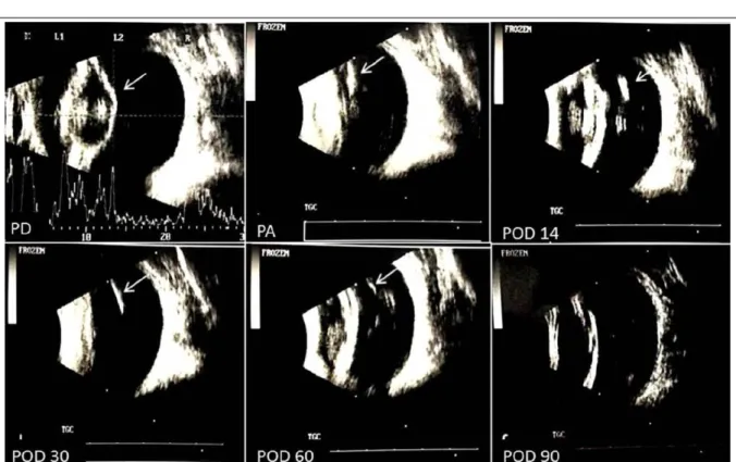
Ultrasonography performed on
postoperative day 7 (POD 7) (Figure 1) revealed a rod-shaped hyperechoic structure with no acoustic shadowing in the superior temporal quadrant in all DG eyes. This finding is consistent with the expected appearance of the device on ultrasound. On POD 30, the rod-like hyperechoic areas were thinner than observed immediately after surgery. On POD 60, only small hyperechoic points were visible, indicating that only device remnants remained. Devices were no longer visible on POD 90.
In the present study, Kaplan-Meier estimator analysis showed that 65% of eyes in the DG had no signs of uveitis on POD 7. By POD 14, this number had dropped to 50% (Figure 2).
The eyes in the DG with uveitis had more severe inflammation than eyes in the CG with uveitis and requiring complementary local steroid therapy. KLEIN et al. (2011) considered uveitis to be significant when signs persisted for at least 3 weeks. On other the hand, WILKIE & COLITZ (2013) recognized that uveitis-related complications can occur late and the early detection and treatment of ocular inflammation can have a significant effect on long-term therapeutic success.
Hyperemia was observed in the CG immediately following surgery in 40% of eyes (p =
0.21 vs. DG), but had resolved by POD 30 (p = 0.00
vs. DG). In contrast, approximately 90% of the eyes
in the DG group showed conjunctival hyperemia 14
days after surgery (p = 0.00 vs. GC). This condition
was most severe in the superior bulbar conjunctiva (the site of the device insertion), and persisted in some eyes (15%) until POD 60 (p = 0.07). We considered that device insertion was the most likely cause of this hyperemia because it has been shown to be associated with a high likelihood to cause inflammation (RADI & RENDER, 2008) and the conjunctiva was highly vascularized. However, it is important to mention that inflammation was not reported by FIALHO et al. (2006) after device insertion.
Immediately after surgery, moderate corneal edema was more common in the DG (70% of the eyes) than in the CG (15%, p<0.001); this could be related to the greater flare observed in the DG (Figure 2), and the increased possibility of endothelial decompensation. Edema (both groups) subsequently decreased in severity and no significant differences were observed between DG and GC (p = 0.73, POD 90). In general, edema can be attributed
to ultrasound, use of irrigating solutions, presence of
anterior chamber air bubbles, or surgical manipulation (HAYASHI et al., 1996). However, no previous studies have reported endothelial complications associated with the use of similar devices in the posterior segment (FIALHO et al., 2006). A case report related these complications for one human patient, where due to intravitreal device migrated on into the anterior chamber (VELA et al., 2012).
The dorsocaudal portion of the retina of a DG patient became detached near the device insertion site. In a previous rabbit study, a similar detachment was observed in 1 of 46 eyes that had received intravitreal implants (FIALHO et al., 2006). Another study (JAGER et al., 2004) found that this problem was uncommon (0.9%) following intravitreal injections and generally occurred later as a result of a
Baseline IOP was 12.2 ± 0.86mmHg in the CG and 9.4 ± 0.72mmHg in the DG (p<0.01). Immediately after surgery, the IOP was lower in the CG (6.0 ± 0.42mmHg) than in the DG (13.50 ± 2.14, p<0.001). However, IOP was not significantly different between the groups on POD 7 (CG: 11.20 ± 1.45mmHg, DG: 8.70 ± 6.63mmHg, p = 0.13) or on POD 14 (CG: 11.20 ± 1.08mmHg, DG: 8.60 ± 0.74mmHg, p = 0.06). From POD 30 until POD 90, IOP values were lower in the DG in comparison to the CG [POD 30: 12.50 ± 0.95mmHg (CG), 8.20 ± 0.93mmHg (DG), p = 0.003; POD 60: 14.20 ± 0.92mmHg (CG), 7.10 ± 0.56mmHg (DG), p<0.001). The ultrasound times to emulsify the lenses were
272.9 ± 20.9 seconds for the CG and 282.8 ± 19.8
seconds for the DG (p = 0.29).
Phacoemulsification can induce pressure peaks in the first few hours after surgery. A previous study showed that intravitreal injection increases the risk of transitory IOP elevations (HOLLANDS et al., 2007; MOLLEDA et al., 2008). However, this was not observed in the study, where the IOP remained within normal limits. The size of implant used in
this study (lower compared with other devices) may partly explain our results of IOP, because ocurrence of changes in this variable seem to depend on the dimensions and the volume of implanted components (REGNIER, 2013). Additionally, implantation of the device can cause a small loss of vitreous amount, which act as a compensatory mechanism able to maintain lower values of IOP. Other intraocular dexamethasone-releasing devices used in previous studies have not been found to cause a significant IOP changes (FIAHO et al., 2006, 2007; CHENNAMANENI et al., 2013). However, a device containing fluocinolone acetonide has been reported to induce increase in IOP (BOLLINGER et al., 2011).
The lower IOP values observed on POD 30 in the DG may be considered an adverse outcome from previous uveitis, despite this, there were no clinical consequences. However, the values remained similar to baseline prior to intervention. A survey of hypotony cases in human revealed that the condition can be associated with cataract surgery and previous uveitis (DANIEL et al., 2012). Further studies should be conducted to clarify this result findings.
Anterior chamber flare count measured immediately after surgery was higher in the DG (105.9 ± 13.9ph/ms) than in the CG (63.8 ± 22.2ph/ms; p<0.01). In the subsequent evaluations no differences in anterior chamber flare counts were observed between the groups (p>0.05). Anterior chamber fibrin was not observed in the CG at any time point. In contrast, mild-to-moderate fibrin formation was observed in the DG from POD 7 until POD 14. Such findings originated from uveitis in the early stages in the DG.
The vitreous plays an important role in ocular metabolism, and differences in its composition can affect drug delivery to the posterior segment (BALAZS & DENLINGER, 1982). Furthermore, small differences
in vitreous-device surface interactions can have an
influence on drug release and diffusion (FIALHO et al., 2006). This may explain the mixed results seen in the DG. Drug release can occur in bursts during 4 weeks
after device insertion (both in vivo and in vitro; FIALHO
et al., 2006) and it is likely that such an event may have occurred in our study. Therefore, the device-released steroid is likely to enhance the effect of topical steroids used to control uveitis during postoperative weeks
2 and 3.Following phacoemulsification, the ocular
environment appears to accelerate degradation in eyes with uveitis, leadings to faster drug release.
The inappropriate control of the intraocular inflammation in the DG may have been due to an irregular distribution of the drug within the polymer matrix, which would have resulted in inconsistent drug release. However, this possibility needs to be
tested since other researchers reported that the drug is
homogeneously distributed within the device matrix (FIALHO & SILVA-CUNHA, 2005).
Studies have shown that the device releases dexamethasone at therapeutic concentrations and that these concentrations are maintained in the vitreous (FIALHO et al., 2007). We believe that the drug diffuses from the vitreous to the anterior segment, therefore appropriate concentrations could not be maintained due to the high flow of renewal of the aqueous humor.
The need for complementary topical steroid therapy did not require systemic steroid therapy. This consideration is clinically important because systemic steroid use is associated with many adverse effects. When an intraocular device is used, the systemic exposure to dexamethasone is extremely low (CHENNAMANENI et al., 2013). In fact, it is lower than when topical or systemic steroids are used (WEIJTENS et al., 2002).
The Ozurdex is a device made from the same polymer of the device examined here; it can release dexamethasone for approximately 5 weeks. In clinical trials, the Ozurdex was shown to be effective in controlling inflammation following phacoemulsification with no apparent side effects (VIANNA et al., 2013). The ineffectiveness of our device for controlling postoperative inflammation could be attributed to the lower concentration of dexamethasone used (250µg) in comparison to the Ozudex device (700µg).
CONCLUSION
The intravitreal dexamethasone device could not adequately control intraocular inflammation in dogs following phacoemulsification.
ACKNOWLEDGEMENTS
This study was supported by the Conselho Nacional de Desenvolvimento Científico e Tecnológico (CNPq Proc. 300833/2010-5).
BIOETHICS AND BIOSSECURITY COMMITTEE APPROVAL
The study protocol was submitted to and approved by the Committee of Ethics and Animal Welfare of the College of Agricultural and Veterinarian Sciences, Jaboticabal (Protocol 11890/14).
REFERENCES
ANDRADE, A. L. et al. Microscopia especular de não contato e flarefotometria à laser no pré e pós-operatórios imediatos de cães submetidos à facoemulsificação pela técnica v-prechop. In: CONGRESSO BRASILEIRO DA ANCLIVEPA, 32., 2011,
Goiânia. Anais... Goiânia: Associação Nacional de Clínicos
Veterinários de Pequenos Animais – GO, 2011.
BEHAR-COHEN, F. Systèmes de délivrance des médicaments pour le segment antérieur: bases fondamentales et applications cliniques.
Journal Francais D’ Ophtalmologie, Issy les Moulineaux, v. 25, n. 5, p. 537-544, 2002. Available from: <http://dx.doi.org/JFO-05-2002-25-5-0181-5512-101019-ART13>. Accessed: Nov. 18, 2014. doi: JFO-05-2002-25-5-0181-5512-101019-ART13.
BALAZS, E. A.; DENLINGER, J. L. Aging changes in the vitreous. In: SEKULER, R.; DISMUKES, K.; KLINE, D. Aging
and human visual function. New York: A. R. Liss, 1982. 350 p.
BOLLINGER, K. et al. Intraocular pressure outcome of patients with fluocinolone acetonide intravitreal implant for noninfectious uveitis.
Ophthalmology, New York, v. 118, n. 10, p. 1927–1931, 2011. Available from: <http://dx.doi.org/10.1016/j.ophtha.2011.02.042>. Accessed: Feb. 15, 2015. doi: 10.1016/j.ophtha.2011.02.042.
CHANG-LIN, J. E. et al. Pharmacokinetics and pharmacodynamics of the sustained-release dexamethasone intravitreal implant.
Investigative Ophthalmology & Visual Science, Rockville, v. 52,
n. 1, p. 80-86, 2011. Available from: <http://dx.doi.org/10.1167/ iovs.10-5285>. Accessed: Jan. 9, 2015. doi: 10.1167/iovs.10-5285.
CHENNAMANENI, S. R. et al. Development of a novel bioerodible dexamethasone implant for uveitis and postoperative cataract inflammation. Journal of Controlled Release, Amsterdam, v. 167, p. 53-59, 2013. Available from: <http://dx.doi.org/10.1016/j. jconrel.2013.01.007>. Accessed: Jan. 20, 2014. doi: 10.1016/j. jconrel.2013.01.007.
DANIEL, E. et al. Risk of Hypotony in Non-infectious Uveitis.
Ophthalmology, v. 119, n. 11, p. 2377-2385, 2012. Available from: <http://www.ncbi.nlm.nih.gov/pmc/articles/PMC3475753/ pdf/nihms-380917.pdf>. Accessed: Apr. 17, 2016. doi: 10.1016/j.
ophtha.2012.05.032.
FIALHO, S. L.; SILVA-CUNHA, A. Manufacturing techniques of biodegradable implants intended for intraocular application. Drug
Delivery, New York, v. 12, n. 2, p. 109-116, 2005. Available from: <http://dx.doi.org/10.1080/10717540590921432>. Accessed: Oct.
5, 2014. doi: 10.1080/10717540590921432.
FIALHO, S. L. et al. Safety and pharmacokinetics of intravitreal biodegradable implant of dexamethasone acetate in rabbit eyes. Current Eye Research, Mortimer, v. 31, n. 6, p. 525-534, 2006. Available from: <http://www.tandfonline.com/doi/ pdf/10.1080/02713680600719036#.VeI-mPZViko>. Accessed:
Jul. 30, 2014. doi: 10.1080/02713680600719036.
FIALHO, S. L. et al. Biodegradable implants for ocular delivery of anti-inflammatory drugs. Journal of Drug Delivery Science and Technology, Paris v. 17, n. 1, p. 93-97, 2007. Available from: <http://dx.doi.org/10.1016/S1773-2247(07)50013-4>. Accessed: Sept. 5, 2014. doi: 10.1016/S1773-2247(07)50013-4.
JAGER, R. D. et al. Risks of intravitreous injection: a comprehensive review. Retina, London, v. 24, n. 5, p. 676-698,
2004. Available from: <http://ovidsp.ovid.com/ovidweb.cgi?T= JS&CSC=Y&NEWS=N&PAGE=fulltext&D=&AN=00006982-200410000-00002&PDF=y>. Accessed: Mar. 15, 2015. doi:
10.1097/00006982-200410000-00002.
KLEIN, H. E. et al. Postoperative complications and visual outcomes of phacoemulsification in 103 dogs (179 eyes): 2006–2008. Veterinary Ophthalmology, West Sussex, v. 14, n. 2, p. 114-120, 2011. Available from: <http:// onlinelibrary.wiley.com.sci-hub.org/doi/10.1111/j.1463-5224.2010.00853.x/pdf>. Accessed: Dec. 5, 2014. doi: 10.1111/j.1463-5224.2010.00853.
LEE, S. Y. et al. Surodex in a paediatric cataract surgery. British Journal of Ophthalmology, London, v. 87, n. 11, p. 1424-1426,
2003. Available from: <http://www.ncbi.nlm.nih.gov/pmc/articles/ PMC1771875/pdf/bjo08701424.pdf>. Accessed: Apr. 4, 2015. doi: 10.1136/bjo.87.11.1424.
HAYASHI, K. et al. Risk factors for corneal endothelial injury during phacoemulsification. Journal of Cataract and
Refractive Surgery, Philadelphia, v. 22, n. 8, p. 1079–1084, 1996b. Available from: <http://dx.doi.org/10.1016/S0886-3350(96)80121-0>. Accessed: Mar. 3, 2015. doi: 10.1016/ S0886-3350(96)80121-0.
HOLLANDS, H. et al. Short-term intraocular pressure changes after intravitreal injection of bevacizumab. Canadian Journal
of Ophthalmology, Montreal, v. 42, n. 6, p. 807-811, 2007.
Available from: <http://www.canadianjournalofophthalmology.ca/ article/S0008-4182(07)80042-1/pdf>. Accessed: Feb. 5, 2015. doi:
10.3129/i07-172.
MOLLEDA, J. M. et al. The ocular effects of intravitreal triamcinolone acetonide in dogs. The Veterinary Journal, v. 176,
n. 3, p. 326-332, 2008. Available from: <http://ac.els-cdn.com/ S1090023307000901/1-s2.0-S1090023307000901-main.pdf?_ tid=6cae53b8-04e7-11e6-ad27-00000aab0f26&acdnat=1460930 448_1c2cf6945006ac566fb70c771fa6e3d4>. Accessed: Apr. 17, 2016. doi: 10.1016/j.tvjl.2007.02.031.
RADI, Z. A.; RENDER, J. A. The pathophysiologic role of cyclooxygenases in the eye. Journal of Ocular Pharmacology
and Therapeutics, New Rochelle, v. 24, n. 2, p. 141-148, 2008. Available from: <http://online.liebertpub.com/doi/pdf/10.1089/ jop.2007.0078>. Accessed: Apr. 18, 2015. doi: 10.1089/ jop.2007.0078.
REGNIER, A. Drug delivery and pharmacokinetics. In: GELATT, K. N.; GILGER, B. C.; KERN, T. J. Veterinary Ophthalmology.
SIQUEIRA, R. C. et al. Retinal safety and efficacy of a dexamethasone biodegradable implant to treat macular edema
associated to retinal vein occlusion: a phase I/II clinical trial. Investigative Ophthalmology Visual Science, Rockville, v. 54, n.
15, 2013. E-Abstract 5066. Available from: <http://abstracts.iovs. org/cgi/content/short/54/6/5066 >. Accessed: Apr. 15, 2015.
TAN, D. T. et al. Randomized clinical trial of a new dexamethasone delivery system (Surodex) for treatment of post-cataract surgery inflammation. Ophthalmology,
Philadelphia, v. 106, n. 2, p. 223-231, 1999. Available from: <http://ac.els-cdn.com/S016164209990060X/1- s2.0-S016164209990060X-main.pdf?_tid=9bb79926-c766-11e5-9241-00000aab0f02&acdnat=1454168101_ e11c74ac34543c5a7960927c15389870>. Accessed: Jan. 30, 2016. doi: 10.1016/S0161-6420(99)90060-X.
TAN, D. T. et al. Randomized clinical trial of Surodex steroid drug delivery system for cataract surgery: anterior versus
posterior placement of two Surodex in the eye. Ophthalmology,
Philadelphia, v. 108, n. 12, p. 2172-2181, 2001. Available from: <http://www.aaojournal.org/article/S0161-6420(01)00839-9/pdf>. Accessed: Sept. 14, 2014. doi: S0161-6420(01)00839-9.
VELA, J. I. et al. Repositioning of dexamethasone intravitreal implant (Ozurdex) migrated into the anterior chamber.
International Ophthalmology, v. 32, p. 583-584, 2012. Available
from: <http://link.springer.com/article/012-9604-7/fulltext.html>. Accessed: Jan. 30, 2016. doi:
10.1007/s10792-012-9604-7.
VIANNA, L. M. M. et al. Intracapsular dexamethasone implant in patients undergoing phacoemulsification and intraocular lens implantation. Arquivos Brasileiro de Oftalmologia, São Paulo, v. 76, n. 4, p. 226-228, 2013. Available from: <http://dx.doi. org/10.1590/S0004-27492013000400007>. Accessed: Apr. 25,
2015. doi: 10.1590/S0004-27492013000400007.
WEIJTENS, O. et al. Intraocular penetration and systemic absorption after topical application of dexamethasone disodium
phosphate. Ophthalmology, New York, v. 109, p. 1887–1891,
2002. Available from: <http://www.aaojournal.org/article/6420(02)01176-4/pdf>. Accessed: Oct. 20, 2014. doi: S0161-6420(02)01176-4.
WEINER, A. L.; GILGER, B. C. Advancements in ocular drug delivery. Veterinary Ophthalmology, West Sussex, v. 13, n. 3, p. 395-406, 2010. Available from: <http://onlinelibrary.wiley.com/ doi/10.1111/j.1463-5224.2010.00835.x/pdf>. Accessed: Feb. 2, 2015. doi: 10.1111/j.1463-5224.2010.00835.x.