Myo cardial antio xidant and o xidative
stre ss change s due to se x ho rm o ne s
1Laboratório de Fisiologia Cardiovascular, Departamento de Fisiologia,
Instituto de Ciências Básicas da Saúde, Universidade Federal do Rio Grande do Sul, Porto Alegre, RS, Brasil
2Catedra de Q uímica General e Inorgánica, Facultad de Farmacia y Bioquimica,
Universidad de Buenos Aires, Buenos Aires, Argentina
3St. Boniface Hospital Research Centre, University of Manitoba,
Winnipeg, MB, Canada J. Barp1,
A.S.R. Araújo1,
T.R.G. Fernandes1,
K.V. Rigatto1,
S. Llesuy2,
A. Belló-Klein1
and P. Singal3
Abstract
The purpose of the present study was to examine myocardial antioxi-dant and oxidative stress changes in male and female rats in the presence of physiological sex hormone concentrations and after cas-tration. Twenty-four 9-week-old Wistar rats were divided into four groups of 6 animals each: 1) sham-operated females, 2) castrated females, 3) sham-operated males, and 4) castrated males. When testosterone and estrogen levels were measured by radioimmunoas-say, significant differences were observed between the castrated and control groups (both males and females), demonstrating the success of castration. Progesterone and catalase levels did not change in any group. Control male rats had higher levels of glutathione peroxidase (50%) and lower levels of superoxide dismutase (SOD, 14%) than females. Control females presented increased levels of SOD as com-pared to the other groups. After castration, SOD activity decreased by 29% in the female group and by 14% in the male group as compared to their respective controls. Lipid peroxidation (LPO) was assessed to evaluate oxidative damage to cardiac membranes by two different methods, i.e., TBARS and chemiluminescence. LPO was higher in male controls compared to female controls when evaluated by both methods, TBARS (360%) and chemiluminescence (46%). Castration induced a 200% increase in myocardial damage in females as deter-mined by TBARS and a 20% increase as deterdeter-mined by chemilumines-cence. In males, castration did not change LPO levels. These data suggest that estrogen may have an antioxidant role in heart muscle, while testosterone does not.
Co rre spo nde nce
A. Belló-Klein Laboratório de Fisiologia Cardiovascular
Departamento de Fisiologia ICBS, UFRGS
Rua Sarmento Leite, 500 90050-170 Porto Alegre, RS Brasil
Fax: + 55-51-3316-3166 E-mail: belklein@ vortex.ufrgs.br
Presented at the IV International
Symposium on Vasoactive Peptides, Belo Horizonte, MG, Brazil, O ctober 19-21, 2001.
Research supported by CNPq, CAPES and FAPERGS.
Received January 3, 2002 Accepted August 6, 2002
Ke y wo rds
·Catalase
·Glutathione peroxidase
·Lipid peroxidation
·Superoxide dismutase
Intro ductio n
Reactive oxygen species are formed un-der both physiological and pathological con-ditions in mammalian tissues. Because of their high reactivity, they may interact with biomolecules, inducing oxidative injury. Re-active oxygen species have been implicated
its cardioprotection have received consider-able interest.
Epidemiological studies have demon-strated that premenopausal women appear to be protected from coronary artery disease as compared to men of the same age (2-4). It is also known that in premenopausal women, the incidence of myocardial infarction and other complications related to atheroscle-rotic disease is lower than in men (5). The incidence of cardiovascular diseases after menopause is similar to that observed in males. These findings suggest a protective role for endogenous estrogen.
After menopause, the coronary artery dis-ease risk is incrdis-eased by several metabolic and vascular changes, which may be related in part to estrogen deficiency. Estrogen has favorable effects on lipid profiles, endotlial cell function, vascular reactivity and he-mostatic factors. Exogenous estrogen pro-tects against atherosclerosis by modulating low-density lipoprotein oxidation, binding free radicals and lowering plasma choles-terol (6,7). Enhanced expression of endothe-lial nitric oxide synthase and its inducible isoform in the myocardium have also been observed with the administration of exog-enous estrogen (8,9).
The benefits of estrogen replacement therapy in reducing risk factors for cardio-vascular disease account for only about 50%. This implies that additional mechanisms must exist whereby estrogen exerts a cardiopro-tective action (10). Previous studies have concentrated mostly on the vascular effects of estrogen, but cardio-protection by estro-gen is not necessarily restricted to the vascu-lature. In a recent review, Babiker and col-laborators (11) stated that the benefits of estrogen gradually shift from the vascular system to the myocardium. This view is sup-ported by the fact that functional estrogen receptors have been detected in the myocar-dium. These receptors regulate the expres-sion of many genes including connexin 43 and heavy chain a-myosin (major
contrac-tile proteins in the heart) (12). Estrogen has also been shown to be a calcium channel blocker, possibly providing additional car-diovascular protection (13).
In addition to vascular and myocardial effects, estrogen may exert its protective effect via a third property, namely its ability to act as an antioxidant. All estrogens have a phenolic hydroxyl group at position 3 and a methyl group at position 13. The presence of this phenol group gives estrogen its antioxi-dant property by scavenging oxygen free radicals. Furthermore, estrogens can induce antioxidant enzyme expression by stimulat-ing the antioxidant defense system (14).
A condition of low iron, such as that caused by menstruation, is another mechan-ism proposed for cardioprotection in pre-menopausal women. Reduced levels of iron may result in decreased formation of hy-droxyl radicals via the Fenton reaction, and a consequent reduced potential for oxidative damage (15).
The purpose of the present study was to characterize myocardial pro-oxidant and an-tioxidant profiles in male and female rats to determine whether physiological differences in sex hormone production may cause changes in these parameters. We also deter-mined if the elimination of sex hormones in male and female rats could alter oxidative stress patterns in the myocardium.
Mate rial and Me tho ds
Expe rime ntal pro to co l
Baker et al. (16). All females were in the estrous phase at the time of surgery (16).
The stage of the estrous cycle was deter-mined by vaginal swab. The phases observed were: diestrus, when mucus, leukocytes and some nucleated cells were present (2 to 3 days on average); proestrus, when only nucle-ated cells were present (12 h); estrus, when only cornified cells were observed (24 h - rut phase), and metaestrus, when leukocytes, cornified cells and some nucleated cells were present (16).
Ho rm o nal m e asure m e nts
Sex hormone levels (estradiol, progeste-rone and testosteprogeste-rone) were evaluated in plasma 7 days after surgery by radioimmu-noassay using a Biomedicals kit (Biomedi-cal Technologies, Inc., Stoughton, MA, USA).
Pre paratio n o f tissue ho m o ge nate s
Immediately following blood collection, rats were killed by cervical dislocation and their hearts were removed. The hearts were cooled in ice and homogenized in 1.15% (w/v) KCl containing 1 mM PMSF. The homogenates were centrifuged at 700 g for 10 min to discard nuclei and cell debris and the supernatant fraction obtained was frozen at -70ºC for further measurements (18).
Antio xidant e nzyme activitie s
Superoxide dismutase (SOD) activity was determined in heart homogenates by rate inhibition of pyrogallol auto-oxidation at 420 nm. This activity was determined from a standard curve of commercially available SOD (percentage inhibition of pyrogallol auto-oxidation). The enzyme activity was reported as U SOD/mg protein (19).
Glutathione peroxidase activity was meas-ured in heart homogenates by monitoring NADPH oxidation at 340 nm. The reaction medium consisted of 30 nM sodium
phos-phate buffer, pH 7.0, 0.17 mM reduced glu-tathione, 0.2 U/ml glutathione reductase, and 0.5 mM tert-butyl hydroperoxide. Glutathi-one peroxidase activity is reported as nmol min-1 mg protein-1 (20).
Catalase activity was measured in the homogenates after treatment with Triton X-100 by monitoring the decrease in absorp-tion at 240 nm. The reacabsorp-tion medium con-sisted of 50 mM sodium phosphate buffer, pH 7.2, and 10 mM H2O2 (21). The results are reported as nmol/mg protein (22).
Lipid pe ro xidatio n m e asure m e nts
Lipid peroxidation (LPO) was measured by two methods: tert-butyl hydroperoxide-initiated chemiluminescence and thiobarbi-turic acid reactive substances (TBARS).
In the chemiluminescence method, light emitted from the reaction between tert-butyl hydroperoxide and lipids is measured with a liquid scintillation counter. This counter was adapted to count light emission using a tri-tium channel (LKB Rack Beta Liquid Scin-tillation Spectrometer, model 1215, LKB-Produkter AB, Bromma, Sweden). The pro-tein content was adjusted to 1 mg/ml of reaction medium (120 mM KCl, 30 mM sodium phosphate buffer, pH 7.4) and added to 3 mM tert-butyl hydroperoxide in a final volume of 4 ml. The results are reported as counts per minute (cpm)/mg protein (23).
malon-aldehyde was used as a standard (24).
Pro te in m e asure m e nt
The protein content of the homogenate was measured by the method of Lowry et al. (25) using bovine serum albumin as a stan-dard.
Statistical analysis
The data were compared by one-way ANOVA followed by the Student-Newman-Keuls multiple comparison test. Results are reported as means ± SEM and differences were considered to be significant when P<0.05.
Re sults
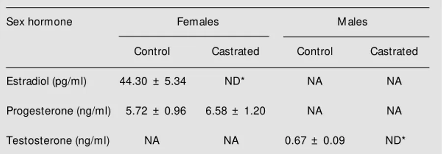
Sex hormone data are shown in Table 1. As expected, testosterone levels in the cas-trated male group and estrogen levels in the castrated female group were undetectable. No significant differences in progesterone levels were detected between control and castrated female groups (Table 1). These data confirm the efficacy of surgical castration.
Enzymatic antioxidant defense mechan-isms were evaluated in the myocardium of male and female rats. The activity of SOD, which detoxifies the superoxide anion, was lower (14%) in the male control group than in the female control group (P<0.05). After castration, SOD activity was decreased by 29% (P<0.001) in the female group and by 14% (P<0.05) in the male group when com-pared to their respective controls (Table 2). Glutathione peroxidase, an enzyme that me-tabolizes H2O2 and organic peroxides, was 50% higher in the male control group than in the female control group (P<0.05). Castra-tion induced a small nonsignificant increase in glutathione peroxidase activity in females and a similar decrease in males (Table 2). The male control group also showed a 20% nonsignificant reduction in the activity of catalase, an enzyme responsible for metabo-lizing hydrogen peroxide. No significant dif-ferences were found in catalase activity when comparing the male and female castrated groups (Table 2).
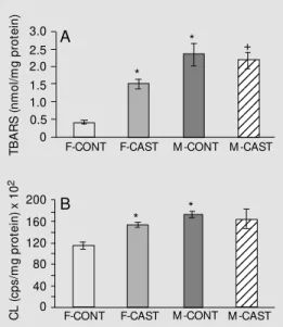
In order to detect increased oxidative damage to cardiac tissue, LPO was assessed by two methods, i.e., TBARS and chemilu-minescence. LPO was significantly higher in male controls than in female controls both by the TBARS (360%) and chemilumines-cence (46%) methods (Figure 1A,B; P<0.001 and P<0.01, respectively).
In females, castration caused a 200% increase in LPO (P<0.01) as determined by TBARS (Figure 1A) and a 20% increase (P<0.05) as determined by chemilumines-cence (Figure 1B). Male LPO levels did not
Table 2. Antioxidant activity in cardiac muscle homogenates from different groups of rats.
Antioxidant Females M ales
enzymes
Control Castrated Control Castrated
SOD (U/mg protein) 38.82 ± 0.83 27.60 ± 1.34* 33.38 ± 0.87* 28.51 ± 1.33* (F = 26.20)
GPx (nmol min-1 mg 67.10 ± 5.64 78.28 ± 5.71 100.59 ± 6.31* 94.09 ± 11.06
protein-1) (F = 4.07)
CAT (pmol/mg 31.60 ± 1.44 25.50 ± 1.76 25.40 ± 1.39 28.10 ± 2.76 protein) (F = 2.30)
Data are reported as means ± SEM for six animals in each group. SOD, superoxide dismutase; GPx, glutathione peroxidase; CAT, catalase.
* P<0.05 compared to the control group of the same sex (ANOVA). Table 1. Plasma sex hormone levels in different groups of rats.
Sex hormone Females M ales
Control Castrated Control Castrated
Estradiol (pg/ml) 44.30 ± 5.34 ND* NA NA
Progesterone (ng/ml) 5.72 ± 0.96 6.58 ± 1.20 NA NA
Testosterone (ng/ml) NA NA 0.67 ± 0.09 ND*
Data are reported as means ± SEM for six animals in each group. NA, not analyzed; ND, not detected.
change after castration (Figure 1A,B).
D iscussio n
While there have been many studies on the relationship between sex hormones, LPO and antioxidants, the present study is the first to examine these parameters in the pres-ence of physiological hormone concentra-tions in myocardial tissue. Most of the previ-ous experiments were conducted on post-menopausal women or castrated female ani-mals on estrogen replacement therapy (26). One of the aims of the present study was to evaluate the oxidative stress and antioxi-dant defenses in the myocardium of male and female control rats. Oxidative stress was assessed by LPO. A wide variety of proce-dures have been used to measure LPO, but they are indirect and lack sensitivity, selec-tivity as well as practicability. Furthermore, there is no ideal method to measure LPO, and the literature suggests using more than one to improve accuracy. Therefore, if the results obtained with the use of different techniques are concordant, then the results are acceptable. In the present study, the re-sults obtained by TBARS and chemilumi-nescence were in agreement. Lower levels of TBARS and chemiluminescence were ob-served in the female control group (Figure 1A,B), which presented higher estrogen lev-els, as compared to the male control group. The reduction in LPO in the presence of estrogen confirms the antioxidant action of this hormone and its scavenger properties, which minimize free radical damage to mem-brane lipids (27).
Myocardial catalase was not different between male and female control groups in terms of antioxidant activity (Table 2). This finding may mean that hydrogen peroxide production in the heart is not influenced by sex hormones, since this enzyme is specific for hydrogen peroxide. However, with re-spect to superoxide dismutase, female rat hearts have a higher activity than male
con-trol hearts (Table 2). Since females have enhanced SOD activity (which is an impor-tant antioxidant enzymatic defense) and in view of the non-enzymatic defense offered by estrogen, oxidative stress was kept under control, as shown by the LPO data. Although glutathione peroxidase activity was decreased in females as compared to male controls, the pro-oxidant/antioxidant balance was in the favor of the antioxidants and the oxidative damage was reduced in females.
Glutathione peroxidase enhancement in male hearts, that do not have estrogen pro-tection, is not enough to balance free radical production. This appears to be an example of oxidative stress-induced up-regulation of an antioxidant enzyme (28).
The antioxidant potential of various ste-roid hormones (estriol, estradiol, estrone, progesterone, testosterone, androstenedione, cortisol and others) have been evaluated and it was shown that estrogens, especially es-triol and estradiol, are natural antioxidants, while the other steroids do not present sig-nificant antioxidant activity (29).
The other aim of the present study was to determine whether the removal of sex hor-mones could affect the oxidative stress pat-tern observed under physiological conditions. Bilateral castration was performed in male
Figure 1. Lipid peroxidation.
A, Thiobarbituric acid reactive substances (TBARS) in heart muscle homogenates from different groups (F = 18.53).
B, Chemiluminescence (CL) applied to the heart muscle homogenates of different ex-perimental groups (F = 6.46). * P<0.05 compared to the fe-male control group. +P<0.05
compared to the castrated fe-male group (ANOVA). M = male, F = female, CONT = control, CAST = castrated. Data are reported as means ± SEM for six animals in each group. 1234 1234 1234 1234 1234 1234 1234 1234 1234 1234 1234 1234 1234 1234 1234 1234 1234 1234 1234 1234 1234 TB A R S (n m ol /m g pr ot ei n) 3.0 C L (c ps /m g pr ot ei n) x 1 0 2 2.5 2.0 1.5 1.0 0.5 0 F-CONT
F-CONT F-CAST M -CONT M -CAST 160 120 80 40 0 200 A B * *
F-CAST M -CONT M -CAST *
and female rats, and 7 days after surgery, hormonal levels were quantified. A marked increase in LPO was observed in females after castration, thus approaching male val-ues (Figure 1A,B). This finding suggests that the antioxidant potential of estrogen may be an important contributor to the cardiovascu-lar protection of the hormone, in addition to its regulatory effects on LDL oxidation, en-dothelium-derived relaxing factors and anti-oxidant enzymes (30). Seven days post-castration were enough to reduce sex hor-mones to undetectable levels; however, this period may not be long enough to change enzyme activity. This may be the case for catalase, which is not affected by castration. Another possibility is that catalase activity in the heart is so low that small changes were not detected (28).
SOD seems to be more sensitive to hor-monal changes. In the present study, we found a decrease in SOD activity when both estrogen and testosterone were removed. Others have reported changes in SOD activ-ity in the presence of variations in estrogen (26,30,31). Most of these studies demon-strated a decrease in SOD activity when exogenous estradiol was administered (32, 33). This apparent discrepancy may be due to the fact that some of those experiments were performed on cell cultures or on previ-ously castrated animals on hormone replace-ment therapy with estrogen concentrations
much higher (micromolar) than physiologi-cal levels (nanomolar). However, there was also an increase in SOD activity when exog-enous 17ß-estradiol was administered (34). We have demonstrated that long-term deple-tion of ovarian hormones (21 and 28 days) induces an increase in myocardial SOD ac-tivity (Barp J, Morgan-Martins MI, Marroni C, Vercelino R, Fernandes TRG and Belló-Klein A, unpublished data). Decreased su-peroxide anion (O2
·
-) production by theen-dothelium in response to estrogen is consid-ered to contribute to the vascular protective properties of estrogen (31). This is because O2
·
- can inactivate nitric oxide and thus leadto endothelial dysfunction (35). In this study, SOD activity was also found to be decreased after short-term depletion of the male sex hormone, suggesting that testosterone could also influence the enzymatic defense sys-tem. There is some information on oxidative stress in men showing a pro-oxidant effect of testosterone (36).
Previous studies from our laboratory have indicated the need to consider seasonal var-iations in antioxidant enzyme activities evalu-ated by LPO (37). The present data empha-size the fact that gender differences should be considered when studying oxidative stress. Our results showed apparent changes in myocardial oxidative stress and in antioxi-dants between male and female controls as well as after castration.
Re fe re nce s
1. Singal PK, Khaper N, Palace V & Kumar D (1998). The role of oxidative stress in the genesis of heart disease. Cardiovascular Research, 40: 426-432.
2. Wenger NK, Speroff L & Packard B (1993). Cardiovascular health and disease in w omen. New England Journal of M edi-cine, 329: 247-256.
3. M atthew s KA, M eilahn E, Kuller LH, Kelsey SF, Caggiula AW & Wing RR (1989). M enopause and risk factors for coronary heart disease. New England Journal of M edicine, 321: 641-646. 4. Nabulsi AA, Folsom AR & White A (1993).
Association of hormone replacement therapy w ith various cardiovascular risk factors in postmenopausal w omen. New England Journal of M edicine, 328: 1069-1075.
5. Ganong WF (1999). Review of M edical Physiology. 19th edn. Appleton & Lange, East Norw alk, CT, USA.
6. Kuhl H (1994). Cardiovascular effects and estrogen/gestagen substitution therapy.
Therapeutische Umschau, 51: 748-754. 7. M affei S & De Caterina R (1996).
Hor-mone replacement therapy and cardiovas-cular risk. Giornale Italiano di Cardiologia,
26: 899-940.
8. Farhat M Y, Lavigne M C & Ramw ell PW (1996). The vascular protective effect of estrogen. FASEB Journal,10: 615-624. 9. Nuedling S, Kahlert S, Loebbert K,
Doevendans PA, M eyer R, Vetter H & Grohé C (1999). 17ß-Estradiol stimulates expression of endothelial and inducible NO synthase in rat myocardium in vitro
and in vivo. Cardiovascular Research, 43: 666-674.
41: 524-531.
11. Babiker FA, De Windt LJ, van Eickels M , Grohé C, M eyer R & Doevendans PA (2002). Estrogenic hormone action in the heart: regulatory netw ork and function.
Cardiovascular Research, 53: 709-719. 12. Grohé C, Kahlert S, Loebbert K, Stimpel
M , Karas RH, Vetter H & Neyses L (1997). Cardiac myocytes and fibroblasts contain functional estrogen receptors. FEBS Let-ters, 416: 107-112.
13. Collins P, Beale CM & Rosano GM C (1996). Oestrogen as a calcium channel blocker. European Heart Journal, 17 (Suppl D): 27-31.
14. M assafra C, De Felice C, Gioia D & Buonocore G (1998). Variations in erythro-cyte antioxidant glutathione peroxidase activity during the menstrual cycle. Clini-cal Endocrinology, 49: 63-67.
15. Sullivan M J, Green HJ & Cobb FR (1991). Altered skeletal muscle metabolic re-sponse to exercise in chronic heart fail-ure: relation to skeletal muscle aerobic enzyme activity. Circulation, 84: 1597-1607.
16. Baker HJ, Lindsey JR & Weisbroth SH (1979). The Laboratory Rat. Vol. I, Biology and Diseases. Academic Press, New York, NY, USA.
17. Genuth SM (1993). The endocrine sys-tem. In: Berne RM & Levy M N (Editors),
Physiology. 3rd edn. M osby-Year Book, Inc., St. Louis, M O, USA.
18. Llesuy SF, M ilei J, M olina H, Boveris A & M ilei S (1985). Comparison of lipid peroxi-dation and myocardial damage induced by adriamycin and 4'-epiadriamycin in mice.
Tumori, 71: 241-249.
19. M arklund S (1985). Pyrogallol autoxida-tion. In: Greenw ald RA (Editor), Handbook of M ethods for Oxygen Radical Research.
CRC Press, Boca Raton, FL, USA, 243-247.
20. Flohé L & Gunzler WA (1984). Assay of glutathione peroxidase. M ethods in
Enzy-mology, 105: 114-121.
21. Chance B, Sies H & Boveris A (1979). Hydroperoxide metabolism in mammalian organs. Physiological Review s, 59: 527-625.
22. Boveris A & Chance B (1973). The mito-chondrial generation of hydrogen perox-ide. Biochemical Journal, 134: 707-716.
23. Gonzalez Flecha B, Llesuy S & Boveris A (1991). Hydroperoxide-initiated chemilu-minescence: an assay for oxidative stress in biopsies of liver, heart and muscle. Free Radical Biology and M edicine, 10: 41-47.
24. Buege JÁ & Aust SD (1978). M icrosomal lipid peroxidation. M ethods in Enzymol-ogy, 52: 302-309.
25. Low ry OH, Rosebrough AL, Farr AL & Randall R (1951). Protein measurement w ith the Folin phenol reagent. Journal of Biological Chemistry, 193: 265-275. 26. Akcay T, Dincer Y, Kayali R, Colgar U, Oral
E & Cakatay U (2000). Effects of post-menopausal w omen. Journal of Toxicol-ogy and Environmental Health, Part A, 59: 1-5.
27. Niki E & Nakano M (1990). Estrogens as antioxidants. M ethods in Enzymology, 186: 330-333.
28. Halliw ell B & Gutteridge JCM (1999). Free Radicals in Biology and M edicine. 3rd edn. Oxford University Press, New York, NY, USA.
29. M ooradian AD (1993). Antioxidant proper-ties of steroides. Journal of Steroid Bio-chemistry and M olecular Biology, 45:
509-511.
30. Dantas APV, Scivoletto R, Fortes ZB, Nigro D & Carvalho M HC (1999). Influ-ence of female sex hormones on endo-thelium-derived vasoconstrictor prosta-noid generation in microvessels of spon-taneously hypertensive rats. Hyperten-sion,34: 914-919.
31. Barbacanne M A, Rami J, M ichel JB, Souchard JP, Philippe M , Besombes JP, Bayard F & Arnal JF (1999). Estradiol
in-creases rat aorta endothelium-derived re-laxing factor (EDRF) activity w ithout changes in endothelial NO synthase gene expression: possible role of decreased endothelium-derived superoxide anion production. Cardiovascular Research, 41: 672-681.
32. Kenaley JA & Ji LL (1991). Antioxidant enzyme activity during prolonged exer-cise in amenorrheic and eumenorrheic athletes. M etabolism: Clinical and Experi-mental, 40: 88-92.
33. Arnal JF, Clamens S, Pechet C, Negre SA, Allera C, Girolami JP, Salvayre R & Bayard F (1996). Ethinylestradiol does not en-hance the expression of nitric oxide syn-thase in bovine endothelial cells but in-creases the release of bioactive nitric ox-ide by inhibiting superoxox-ide anion produc-tion. Proceedings of the National Acade-my of Sciences, USA, 93: 4108-4113.
34. Oberley TD, Shultz JL & Oberley LW (1994). In vivo modulation of antioxidant enzyme levels in normal hamster kidney and estrogen-induced kidney tumor. Free Radical Biology and M edicine, 16: 741-751.
35. Nakazono K, Watanabe N, M atsuno K, Sasaki J, Sato T & Inoue M (1991). Does superoxide underlie the pathogenesis of hypertension? Proceedings of the Na-tional Academy of Sciences, USA, 88: 10045-10048.
36. Dincer Y, Ozen E, Kadioglu P, Hatemi H & Akcay T (2001). Effect of sex hormones on lipid peroxidation in w omen w ith poly-cystic ovary syndrome, healthy w omen, and men. Endocrine Research, 27: 309-316.