University of São Paulo Medical School and Institute of Mathematics and Statistics
– University of São Paulo - São Paulo - Brazil.
Mailing address: Clovis de Carvalho Frimm - Faculdade de Medicina da USP, LIM
51 - Av. Dr. Arnaldo, 455 - 01246-903 - São Paulo, SP, Brasil - E-mail:
[email protected].
Research supported by Fundação E.J. Zerbini and LIM-51-HC/FMUSP.
Arq Bras Cardiol, volume 80 (nº 5), 515-20, 2003
Clovis de Carvalho Frimm, Marcia Kiyomi Koike, Mariana Cúri
São Paulo, SP - Brazil
Subendocardial Fibrosis in Remote Myocardium Results from
Reduction of Coronary Driving Pressure During Acute
Infarction in Rats
Cardiac remodeling following myocardial infarction
(MI) is characterized by scar formation with wall thinning at
the site of myocyte fiber loss and progressive left
ventricu-lar (LV) dilatation with collagen deposition within
non-infarcted myocardium (non- MI)
1.
Ventricular dilatation is a primary consequence of MI
lo-cation and size
2, but other factors influence fibrosis
accumu-lation, such as the cardiac renin-angiotensin system,
endo-thelins, catecholamines, and inflammatory mediators
3,4.
However, the role of hemodynamic changes occurring during
ongoing infarction have not been investigated
5. It is still also
unclear where fibrosis deposition actually takes place,
because subendocardial (SE) regions of non-MI have rarely
been contemplated in post-MI morphometric studies
6,7.
The aim of this study was to investigate, using the
expe-rimental MI model in the rat, the role of hemodynamic
changes taking place acutely after left coronary artery
ligature in subsequent fibrosis accumulation within non-MI.
Methods
Twenty-one male Wistar rats weighing 275±5g were
used for the experiments. All procedures were carried out in
accordance with the norms of the Brazilian College of
Ani-mal Experiments and conformed to the “Guide for the Care
and Use of Laboratory Animals.” Our Institutional Ethical
Committee approved the protocol.
Animals were anesthetized with Ketamine chloride,
50 mg.kg
-1i.p. and Pentobarbital sodium, 25 mg.kg
-1i.p. and
put under mechanical ventilation with a rodent ventilator
(Model 683, Harvard Apparatus Inc., MA USA). Systemic
and LV blood pressures were obtained from femoral and
carotid arteries, respectively. The catheters were connected
to pressure transducers and coupled to a calibrated
pre-amplifier (General Purpose Amplifier 4 - model 2, Stemtech
Inc. WI, USA). The pressure tracings were recorded by
using a computerized system processor (AT/Codas, Dataq
Instruments Inc., OH, USA).
Objective -
To investigate the role of hemodynamic
changes occurring during acute MI in subsequent fibrosis
deposition within non-MI.
Methods -
By using the rat model of MI, 3 groups of 7
rats each [sham, SMI (MI <30%), and LMI (MI >30%)] were
compared. Systemic and left ventricular (LV) hemodynamics
were recorded 10 minutes before and after coronary artery
ligature. Collagen volume fraction (CVF) was calculated in
picrosirius red-stained heart tissue sections 4 weeks later.
Results -
Before surgery, all hemodynamic variables
were comparable among groups. After surgery, LV
end-dias-tolic pressure increased and coronary driving pressure
decreased significantly in the LMI compared with the sham
group. LV dP/dt
maxand dP/dt
minof both the SMI and LMI
groups were statistically different from those of the sham
group. CVF within non-MI interventricular septum and right
ventricle did not differ between each MI group and the sham
group. Otherwise, subendocardial (SE) CVF was statistically
greater in the LMI group. SE CVF correlated negatively with
post-MI systemic blood pressure and coronary driving
pressure, and positively with post-MI LV dP/dt
min. Stepwise
regression analysis identified post-MI coronary driving
pressure as an independent predictor of SE CVF.
Conclusion -
LV remodeling in rats with MI is
charac-terized by predominant SE collagen deposition in non-MI
and results from a reduction in myocardial perfusion
pres-sure occurring early on in the setting of MI.
non-MI interventricular septal wall (IVS), and the right
ven-tricular myocardium (RV).
MI rats were classified into groups SMI and LMI,
according to the presence of small (
≤
30%) or large MI
(>30%), respectively.
ANOVA, complemented by the Bonferroni
t
test, was
used for comparing quantitative structural variables
bet-ween MI groups and the sham group. Repeated-measures
analysis of variance, complemented by the Wald test, was
used to evaluate the effects of MI on hemodynamic
varia-bles. Normality and equal variance were verified in all
analy-ses. Data were expressed as mean ± S.E.M. Statistical
signi-ficance was established at a p <0.05.
The potential relationships between postexperiment
hemodynamic variables and subsequent fibrosis
deposi-tion within non-MI was assessed by using Pearson’s
corre-lation coefficient. Multiple linear regression analysis was
performed for detecting among the hemodynamic variables
which were the best predictors for subsequent fibrosis
deposition. The stepwise selection method was used, with
p-values of 0.10 and 0.05 considered significant for entering
a variable into or removing it from the model, respectively.
The statistical analysis was performed with SAS (
Statisti-cal Analysis System
) software
11.
Results
The 3 study groups comprised 7 rats each. Infarct size
was 20.4±1.5% (range: 15.1 to 25.3%) in the SMI group and
50.5 ± 2.4% (range: 40.8 to 58%) in the LMI group.
Heart, lungs, and liver to body weight ratios were
sig-nificantly greater in the LMI than in the sham group (table I).
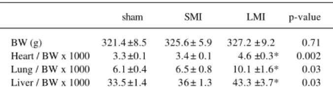
Figure 1 illustrates the hemodynamic variables
compu-ted before and after the experiment. Before surgery, all 3
groups were statistically comparable. Surgery did not affect
any of the hemodynamic variables in the sham group.
LVEDP increased fivefold in the LMI group, resulting
in significantly higher values than in the sham group. The
twofold increase in LVEDP observed in the SMI group was
not statistically different from that in the sham group.
CDP significantly decreased only in the LMI group
(-30%). Postexperiment CDP was comparable among all 3
groups.
A statistically significant decrease in LV dP/dt
max(-36%) was demonstrated for the LMI group alone, and LV
After hemodynamic stabilization, pressure tracings
were recorded for 10 minutes before opening the chest wall
and continued throughout the entire surgical procedure.
The thoracic cage was closed and residual air drained from
the pleural space. When new steady hemodynamic signals
were observed, an additional 10-minute period of
hemody-namic recording was performed. The average of beat to beat
hemodynamic measures recorded during the 2
aforementio-ned 10-minute periods was analyzed.
Heart rate (beats.min
-1), LV systolic pressure (LVSP,
mm Hg), LV end-diastolic pressure (LVEDP, mm Hg), LV dP/
dt
max, LV dP/dt
min(mm Hg.s
-1) and systemic systolic blood
pressure (SBP, mmHg), and diastolic blood pressure (DBP,
mmHg) were recorded. To obtain an estimate of coronary
blood flow, coronary-driving pressure (CDP) was calculated
as the difference between DBP and LVEDP
8.
MI was produced by ligature of the left coronary
arte-ry, by using a modification of a previously described
techni-que
9. Sham-operated rats were operated on similarly except
for not tying the ligature around the coronary artery.
During recovery, after observation of spontaneous
respiration, mechanical ventilation was concluded and
arte-rial catheters were withdrawn. The animals were returned to
their individual cages.
Four weeks after the experiment, animals were
anesthe-tized with Pentobarbital sodium, 30 mg.kg
-1i.p. The heart,
lungs, and liver were removed, cleaned, and weighed. Atria
and large vessels were removed before weighing the heart.
MI was demonstrated by grossly visible scarring of the LV
free wall.
A coronal slice of the heart including both ventricles
was obtained at the equatorial plane where the largest
sur-face of infarction was detected. Tissue fixation was
perfor-med in 10 percent buffered formalin.
Six-micron paraffin embedded sections were cut and
stai-ned with Sirius Red 3BA in saturated picric acid solution
10.
By using an image analysis system (Leica Q500 iW, Leica
Imaging Systems ltd., Cambridge, UK), these sections were
analyzed morphometrically. Fibrillar collagen was identified
in the picrosirius-stained sections by its red colored
appearance.
A videocamera equipped with a macro lens permitting
the visualization of the entire coronal section of each heart
was used to identify MI and non-MI regions and to obtain
infarct size. The ratios between endocardial infarct surface
length and endocardial total LV circumference and the ratios
between epicardial infarct surface length and epicardial
total LV circumference were calculated and averaged to
obtain infarct size
2.
Using a microscopic x10 objective, fibrillar collagen
within MI and non-MI was estimated as a collagen volume
fraction (CVF, %). CVF was determined as the percentage of
red-stained connective tissue areas per total myocardial
area, excluding perivascular collagen. Non-MI CVF was
addressed separately in 3 distinct regions of each tissue
section examined: the inner third of the non-MI
correspon-ding to the LV subendocardium (SE), the medium third of the
Table I - Body weight and organs to body weight ratios at 4 weeks of
follow-up in sham, SMI, and LMI groups.
sham
SMI
LMI
p-value
BW (g)
321.4 ±8.5
325.6 ± 5.9
327.2 ± 9.2
0.71
Fig. 1 - Systemic and left ventricular (LV) hemodynamic measurements of the sham, SMI, and LMI groups computed before (1) and following (2) the experiment. * = p<0.05 vs. sham;
† = p<0.05, 2 to 1difference in comparison with 2 to 1 difference found in sham.
LV dP/dt
maxLV dP/dt
minsham
SMI
LMI
sham
SMI
LMI
Systemic Systolic Blood Pressure
sham
SMI
LMI
Systemic Diastolic Blood Pressure
sham
SMI
LMI
LV end-diastolic pressure
sham
SMI
LMI
Coronary Driving Pressure
sham
SMI
LMI
dP/dt
mindid not change significantly. Despite these
nonuni-form and not always significant changes, after surgery LV
dP/dt
maxand LV dP/dt
minturned out to be statistically
diffe-rent between each the MI groups and the sham group.
SBP and DBP had a tendency to decrease, particularly
in the LMI group (5% and 7%, 9% and 7%, 23% and
-19% for sham, SMI, and LMI groups, respectively) but
remained comparable among groups after surgery.
Surgery did not change heart rate in any group (sham,
345±17 to 330±18; SMI 323±16 to 317±14; LMI 353±14 to
328±16 beats.min
-1).
Table II depicts CVF determined within MI and
non-MI regions. non-MI CVF was comparable between the Snon-MI and
LMI groups. Non-MI CVF in IVS and in RV was comparable
among the 3 groups. Otherwise, LMI showed a greater
non-MI SE CVF than that in the sham group.
Table II - Collagen volume fraction of sham, SMI, and LMI groups in
myocardial infarction and in 3 different noninfarcted myocardial
regions 4 weeks after surgical experiment
CVF (%)
sham
SMI
LMI
p-value
Infarcted region
58.4 ± 3.3
52.8 ±2.5
0.210
LV subendocardium
1.7 ± 0.4
2.2 ± 0.7
5.6 ±1.2*
0.008
Interventricular septum
0.7 ± 0.1
0.6 ± 0.1
0.5 ±0.1
0.240
Right ventricle
1 ± 0.2
1.3 ± 0.2
0.7 ±0.2
0.120
SMI indicates small myocardial infarction; LMI- large myocardial infarction;
CVF- collagen volume fraction; LV- left ventricle; *p<0.05 vs. sham.
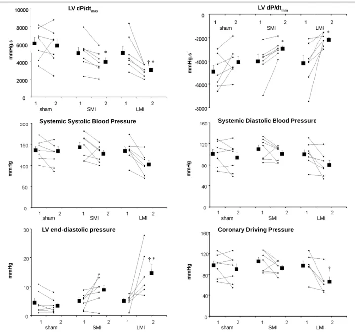
Fig. 2 - Picrosirius red-stained sections of a coronal heart tissue section from rat with a large myocardial infarction. The (0) represents a picture obtained under a macro lens and
shows the entire tissue section encompassing both the left and the right ventricles. The subsequent panels represent endomyocardial layers obtained under progressive
microscopic magnification: 1) corresponds to x2.5, 2) to x10 and 3) x20 magnification objectives.
0
1
2
3
3
3
Fig. 3 - Relationship between subendocardial (SE) collagen volume fraction (CVF)
in noninfarcted myocardium at 4 weeks and coronary driving pressure (CDP)
measured immediately after the surgical experiment.
SE CVF (%)
CDP (mmHG)
LMI SMI sham
r = -0.52, p = 0.02
observed in collagen fibers revealed as a thick layer of
fibro-sis scattered within the non-MI SE LV.
As CVF was predominantly found within the SE layer of
non-MI LV, the amount of collagen fibers found at this
loca-tion was used for further statistical analyses. Of the
hemody-namic variables investigated, non-MI SE CVF correlated
inversely with SBP (r = -0.51, p = 0.02), DBP (r = -0.44, p =
0.04), and CDP (r = -0.52, p = 0.02), and directly with LV dP/
dt
min(r = 0.44, p =0.04).
CDP was the only hemodynamic variable identified as
independently related to non-MI SE CVF (parameter
esti-mate = -0.061, standard error = 0.02, Rsqr = 0.27, p = 0.02).
Fi-gure 3 graphically represents the linear correlation found
between non-MI SE, CVF, and CDP.
Discussion
The present study confirmed previous findings
sho-wing that experimental left coronary artery ligature in rats
produces immediate systemic and LV hemodynamic
chan-ges
12. These changes occurred predominantly in animals
with the largest MI and corresponded to decreases in both
LV dP/dt
maxand CDP and to increases in both LVEDP and LV
dP/dt
min.
content was found rather typically at the SE region. Recently,
experiments using the isolated rat heart preparation have
shown that baseline flow may be reduced by as much as 28%
immediately after coronary artery ligature
13.
The in vivo nature of the present study permitted us
to establish a potential connection between acute
hemody-namic changes jeopardizing blood flow to non-MI and late
development of SE fibrosis.
As hemodynamics was more severely impaired in the
LMI group, we speculate that the acute impairment of LV
systolic performance
2chiefly contributed to the reduction
in coronary perfusion pressure and blood flow
8. Ventricular
diastolic dysfunction would further limit coronary
perfu-sion and aggravate ischemia.
We recognize that although CDP has been
demonstra-ted to be an independent predictor of subsequent SE
fibro-sis, the associated r-square value was of not so great a
mag-nitude. In part, this may be due to the relatively early
hemo-dynamic measurements undertaken in the present study.
Although studies performed in humans
14and in dogs
15suggest that an initial transitory improvement may occur in
post-MI hemodynamics, in rats these parameters usually
deteriorated during the first 24 hours
12and beyond
16-19.
Further ischemia at the SE region may occur during the
pro-gression of remodeling as a consequence of reduced
coro-nary blood flow reserve
20-22.
Controversy regarding the pathogenesis of collagen
accumulation within non-MI, involving different local and
systemic mediators
3,4,23-26, still remains. The present
fin-dings indicate that underperfusion to non-MI should also
be taken into account as a potential mechanism, particularly
to explain SE collagen fiber accumulation.
The predominance of SE over interstitial fibrosis has
been reported before
27,28. This pattern of collagen
deposi-tion is rather in support of being representative of a
repara-tive scarring process in response to impeding myocardial
perfusion. It may also explain reports of remote wall motion
abnormalities occurring early on and persisting for as long
as 2 months after MI
29,30.
Ventricular remodeling has been characterized by
thin-ning and expansion of the infarcted wall, on the one hand,
and by enlargement of the ventricular cavity, on the other,
occurring as early as 2 days
31after MI, or even earlier
29. It
has been attributed to the occurrence of side to side
slippa-ge of remnant myocyte fibers
32. In disagreement with this
assumption is the fact that LV epicardial circumference
length remains unchanged up to 21 days after MI
31.
Alterna-tively, it is quite conceivable to suppose that early
enlarge-ment of the ventricular cavity may be, in part, the result of
loss of myocyte cells jeopardized by poor coronary
perfu-sion taking place under unfavorable acute hemodynamic
conditions, such as those reported in the present study.
Based on the present findings, we speculate that
impeding ischemia of non-MI occurs acutely after MI,
par-ticularly affecting the SE region, and may result from
hemodynamic changes interfering with CDP. This appears
to play a chief role in subsequent fibrosis found in this
region. To be confirmed, this hypothesis deserves further
investigation.
Acknowledgments
We are grateful to Dr Maria de Lourdes Higuchi for her
technical assistance and for providing the facilities for
processing and analyzing the tissue sections. We also thank
Dr Irineu Tadeu Velasco for providing the facilities of the
experimental laboratory of the University of São Paulo
Medical School (LIM-51) where this study was performed.
References
1.
Anversa P, Li P, Zhang X, Olivetti G, Capasso JM. Ischaemic myocardial injury
and ventricular remodelling. Cardiovasc Res 1993; 27: 145-57.
2.
Pfeffer MA, Pfeffer JM, Fishbein MC, et al. Myocardial infarct size and ventricular
function in rats. Circ Res 1979; 44: 503-12.
3.
Frimm CC, Sun Y, Weber KT. Wound healing following myocardial infarction in
the rat: role for bradykinin and prostaglandins. J Mol Cell Cardiol 1996; 28:
1279-85.
4.
Nicoletti A, Michel JB. Cardiac fibrosis and inflammation: interaction with
hemodynamic and hormonal factors. Cardiovasc Res 1999; 41: 532-43.
5.
Walsh JT, Batin PD, Hawkins M, McEntegart D, Cowley AJ. Ventricular
dilata-tion in the absence of ACE inhibitors: influence of haemodynamic and
neurohor-monal variables following myocardial infarction. Heart 1999; 81: 33-9.
6.
Van Kerckhoven R, Kalkman EA, Saxena PR, Schoemaker RG. Altered cardiac
collagen and associated changes in diastolic function of infarcted rat hearts.
Cardiovasc Res 2000; 46: 316-23.
7.
Marijianowski MM, Teeling P, Becker AE. Remodeling after myocardial
infarction in humans is not associated with interstitial fibrosis of noninfarcted
myocardium. J Am Coll Cardiol 1997; 30: 76-82.
8.
Cross C, Riechen P, Salisbury P. Coronary driving pressure and vasomotor tonus
as determinants of coronary blood flow. Circulation Research 1961; 9: 589-600.
9.
Fishbein MC, Maclean D, Maroko PR. Experimental myocardial infarction in the
rat: qualitative and quantitative changes during pathologic evolution. Am J
Pathol 1978; 90: 57-70.
10. Junqueira LC, Bignolas G, Brentani RR. Picrosirius staining plus polarization
microscopy, a specific method for collagen detection in tissue sections.
Histo-chem J 1979; 11: 447-55.
11. SAS Institute Inc. SAS. SAS/STAT User’s Guide. 6 ed. Cary, NC: SAS Institute,
1989.
12. Schoemaker RG, Urquhart J, Debets JJ, Struyker Boudier HA, Smits JF. Acute
hemodynamic effects of coronary artery ligation in conscious rats. Basic Res
Cardiol 1990; 85: 9-20.
13. Nelissen-Vrancken HJ, Debets JJ, Snoeckx LH, Daemen MJ, Smits JF. Time-related
normalization of maximal coronary flow in isolated perfused hearts of rats with
myocardial infarction. Circulation 1996; 93: 349-55.
14. Ginzton LE, Conant R, Rodrigues DM, Laks MM. Functional significance of
hypertrophy of the noninfarcted myocardium after myocardial infarction in
humans. Circulation 1989; 80: 816-22.
15. Gibbons EF, Hogan RD, Franklin TD, Nolting M, Weyman AE. The natural
his-tory of regional dysfunction in a canine preparation of chronic infarction.
Circu-lation 1985; 71: 394-402.
angiotensin-conver-ting enzyme inhibition in experimental chronic heart failure: effects on survival,
hemodynamics, and cardiovascular remodeling. Circulation 1997; 95: 1314-9.
19. Mulder P, Richard V, Derumeaux G, et al. Role of endogenous endothelin in
chronic heart failure: effect of long-term treatment with an endothelin antagonist
on survival, hemodynamics, and cardiac remodeling. Circulation 1997; 96:
1976-82.
20. Schoemaker RG, Saxena PR, Kalkman EA. Low-dose aspirin improves in vivo
hemodynamics in conscious, chronically infarcted rats. Cardiovasc Res 1998;
37: 108-14.
21. Uren NG, Crake T, Lefroy DC, de Silva R, Davies GJ, Maseri A. Reduced
coro-nary vasodilator function in infarcted and normal myocardium after myocardial
infarction. N Engl J Med 1994; 331: 222-7.
22. Kalkman EA, Bilgin YM, van Haren P, van Suylen RJ, Saxena PR, Schoemaker
RG. Determinants of coronary reserve in rats subjected to coronary artery
liga-tion or aortic banding. Cardiovasc Res 1996; 32: 1088-95.
23. Frimm CC, Sun Y, Weber KT. Angiotensin II receptor blockade and myocardial
fibrosis of the infarcted rat heart. J Lab Clin Med 1997; 129: 439-46.
24. Sun Y, Cleutjens JP, Diaz-Arias AA, Weber KT. Cardiac angiotensin converting
enzyme and myocardial fibrosis in the rat. Cardiovasc Res 1994; 28: 1423-32.
25. Sun Y, Weber KT. Angiotensin II receptor binding following myocardial
infarction in the rat. Cardiovasc Res 1994; 28: 1623-8.
26. Weber KT. Targeting pathological remodeling: concepts of cardioprotection and
reparation. Circulation 2000; 102: 1342-5.
27. Beltrami CA, Finato N, Rocco M, et al. Structural basis of end-stage failure in
ischemic cardiomyopathy in humans. Circulation 1994; 89: 151-63.
28. Michel JB, Lattion AL, Salzmann JL, et al. Hormonal and cardiac effects of
conver-ting enzyme inhibition in rat myocardial infarction. Circ Res 1988; 62: 641-50.
29. Solomon SD, Greaves SC, Rayan M, Finn P, Pfeffer MA, Pfeffer JM. Temporal
dissociation of left ventricular function and remodeling following experimental
myocardial infarction in rats. J Card Fail 1999; 5: 213-23.
30. Kramer CM, Lima JA, Reichek N, et al. Regional differences in function within
noninfarcted myocardium during left ventricular remodeling. Circulation 1993;
88: 1279-88.
31. Roberts CS, Maclean D, Maroko P, Kloner RA. Early and late remodeling of the
left ventricle after acute myocardial infarction. Am J Cardiol 1984; 54: 407-10.
32. Olivetti G, Capasso JM, Sonnenblick EH, Anversa P. Side-to-side slippage of
myocytes participates in ventricular wall remodeling acutely after myocardial
infarction in rats. Circ Res 1990; 67: 23-34.
Bula resumida – MICARDIS® Telmisartam - Uso adulto - Forma farmacêutica e apresentações: Comprimidos de 40 mg: embalagens com 14 e 28 comprimidos. Comprimidos de 80 mg: embalagens com 14 e 28 comprimidos. Composição: Cada comprimido contém 40 mg ou 80 mg de telmisartam. Excipientes q.s.p. 1 comprimido. Indicações: Tratamento da hipertensão arterial, como monoterapia ou em associação com outros agentes anti-hipertensivos. Contra-indicações: Hipersensibilidade ao ingrediente ativo ou aos excipientes. Gravidez e lactação. Obstrução biliar. Disfunção hepática ou renal grave. Intolerância hereditária à frutose. Precauções: Hipertensão renovascular: pacientes com estenose arterial renal bilateral ou estenose da artéria com um único rim funcionando: risco aumentado de hipotensão grave e insuficiência renal.
Disfunção renal ou transplante hepático: monitoração periódica dos níveis séricos de potássio e creatinina. Não há experiência em pacientes com transplante renal recente. Desidratação: hipotensão
sintomática, especialmente após a primeira dose, pode ocorrer em pacientes que têm volemia e/ou sódio depletado, o que deve ser corrigido antes do início da terapêutica com MICARDIS. Outras condições de
estimulação do SRAA e condições dependentes da atividade SRAA (insuficiência cardíaca congestiva grave): hipotensão aguda, hiperazotemia, oligúria ou, raramente, insuficiência renal aguda.
Hiperaldosteronismo primário: não se recomenda o uso de MICARDIS. Estenose valvar aórtica e mitral e cardiomiopatia hipertrófica obstrutiva: Recomenda-se precaução especial. Hipercalemia:
recomenda-se monitoração adequada dos níveis séricos de potássio em pacientes de risco. Diuréticos poupadores de potássio, suplementos de potássio, sais de potássio ou outros medicamentos que podem aumentar
os níveis de potássio, como a heparina: podem levar a um aumento da potassemia. Portanto, nestas situações MICARDIS deve ser administrado com cautela. Distúrbios hepatobiliares: pode-se esperar redução
da depuração em pacientes com disfunções obstrutivas do sistema biliar ou insuficiência hepática, pois a eliminação da droga é principalmente biliar. Intolerância à frutose: os comprimidos de MICARDIS contém sorbitol; portanto, é inadequado para pacientes com intolerância hereditária à frutose. Outros: menor eficácia na redução da pressão arterial na população negra do que na população não-negra. Cardiopatia isquêmica ou doença cardiovascular isquêmica pode resultar em infarto do miocárdio. Interações medicamentosas: MICARDIS pode aumentar o efeito hipotensor de outros agentes anti-hipertensivos. Observou-se um aumento de 20% da concentração plasmática média de digoxina. Relataram-Observou-se aumentos reversíveis das concentrações séricas de lítio e de toxicidade; portanto, recomenda-Observou-se cuidadosa monitoração do uso concomitante com lítio. Gravidez e lactação: Contra-indicado. Reações adversas: As reações adversas à droga obtidas a partir de todos os estudos clínicos com telmisartam foram: Infecções do trato urinário, infecções do trato respiratório superior, ansiedade, visão anormal, vertigem, dor abdominal, diarréia, boca seca, dispepsia, flatulência, dor de estômago, eczema, aumento de suor, artralgia, dor nas costas, cãibras nas pernas ou dores nas pernas, mialgia, sintomas de tendinite, dor no peito, sintomas de gripe. Além disso, desde a introdução de telmisartam no mercado, relataram-se casos raros de eritema, prurido, desmaio, insônia, depressão, vômito, hipotensão, bradicardia, taquicardia, dispnéia, eosinofilia, trombocitopenia, fraqueza e perda de eficácia. Relataram-se casos isolados de angioedema, urticária e outros eventos relacionados. Investigações: Raramente, observaram-se diminuição na hemoglobina ou aumento no ácido úrico. Observaram-se aumentos na creatinina ou nas enzimas hepáticas. Efeitos
na habilidade de dirigir e utilizar máquinas: Ainda não se realizaram estudos específicos. Contudo, ao dirigir ou operar máquinas, pode ocasionalmente ocorrer tontura ou sonolência. Posologia: A dose
recomendada é de 40 mg uma vez ao dia. Alguns pacientes podem apresentar benefício com dose diária de 20 mg. Em casos em que a pressão arterial pretendida não seja atingida, a dose de MICARDIS pode ser aumentada para no máximo 80 mg uma vez ao dia. Alternativamente, MICARDIS pode ser usado em combinação com diuréticos tiazídicos, como a hidroclorotiazida, para se obter uma redução maior da pressão arterial. Quando se considerar um aumento da dose, deve-se levar em conta que o máximo efeito anti-hipertensivo é geralmente atingido quatro a oito semanas após o início do tratamento. MICARDIS pode ser administrado com ou sem alimento. Insuficiência renal: Não há necessidade de ajustes de dose em pacientes com insuficiência renal leve a moderada. Telmisartam não é removido do sangue por hemofiltração. Insuficiência hepática: Nos pacientes portadores de insuficiência hepática leve a moderada, não se deve exceder a dose diária de 40 mg. Pacientes idosos: Não são necessários ajustes de doses.
Crianças e adolescentes: Não há dados de segurança e eficácia de MICARDIS em crianças e adolescentes. VENDA SOB PRESCRIÇÃO MÉDICA - MS - 1.0367.0110 - Boehringer Ingelheim do Brasil Química
e Farmacêutica Ltda.
Bula resumida – MICARDIS® HCT – Telmisartam/Hidroclorotiazida - Uso adulto - Composição: Cada comprimido de MICARDIS HCT contém 40 mg/12,5 mg ou 80 mg/12,5 mg de telmisartam/hidroclorotiazida.