Instituto Nacional de Cardiologia Laranjeiras – MS – Universidade Gama Filho Mailing address: Paulo Roberto Benchimol Barbosa - Rua Pompeu Loureiro, 36/ 702 – 22061-000 - Rio de Janeiro, RJ, Brazil - e-mail:[email protected] English version by Stela Maris C. e Gandour
Arq Bras Cardiol, volume 80 (nº 5), 551-7, 2003
Paulo Roberto Benchimol Barbosa, José Barbosa Filho, Ivan Cordovil
Rio de Janeiro, RJ - Brazil
Effect of Oscillatory Breathing on the Variability of the RR
Intervals and its Prognostic Importance in Individuals with
Left Ventricular Global Systolic Dysfunction
In healthy individuals, the duration of RR intervals in tha surface electrocardiogram varies cyclically from beat to beat. This variation is more significant in youngsters, is modulated by breathing movements, and is called respira-tory sinus arrhythmia.
The characteristics of the breathing movements can alter the cyclic variations observed between successive electrocardiographic R waves (RR intervals). These altera-tions result both from direct influences on the modulation of the parasympathetic nervous system and the indirect influences of chemoreceptor mechanisms 1.
In approximately 50% of the individuals with left ventricular global systolic dysfunction, the presence of abnormal patterns of breathing can be identified. In these individuals, oscillatory patterns of breathing (oscillatory breathing) can be observed both during sleep and during wakefulness, and are characterized by successive periods of tachy/hyperpnea followed by brady/hypopnea, and, eventually, apnea 2-10.
Of the types of oscillatory breathing of clinical signi-ficance, Cheyne-Stokes respiration and periodic breathing stand out. Cheyne-Stokes respiration is the breathing pat-tern in which periods of tachy/hyperpnea are followed by regular periods of apnea. In periodic breathing, however, the periods of tachy/hyperpnea are cyclically followed by periods of brady/hypopnea with no apnea.
Although some authors have reported that oscillatory breathing, especially when observed during wakefulness, may be associated with more severe clinical findings and a more reserved prognosis 2,6-8, the subject remains contro-versial.
This study aimed at analyzing the effects of oscillatory breathing on the behavior of the cyclic variations of the normal electrocardiographic RR intervals (V-RR) during wakefulness in individuals with left ventricular global systo-lic dysfunction and in sinus rhythm, and at assessing the in-fluence of oscillatory breathing on clinical and laboratory findings, and on the mortality rate of the individuals studied.
Objective – To assess the effect of the oscillatory breathing on the variability of RR intervals (V-RR) and on prognostic significance after one year follow-up in sub-jects with left ventricular global systolic dysfunction.
Methods – We studied 76 subjects, whose age ranged from 40 to 80 years, paired for age and gender, divided into two groups: group I - 34 healthy subjects; group II - 42 sub-jects with left ventricular global systolic dysfunction (ejec-tion frac(ejec-tion ≤ 0.40). The ECG signals were acquired du-ring 600s in supine position, and analyzed the variation of the thoracic amplitude and the V-RR. Clinical and V-RR variables were applied into a logistic multivariate model to foretell survival after one year follow-up.
Results – Oscillatory breathing was detected in 35.7% of subjects in vigil state of group II, with a concen-tration of the spectral power in the very low frequency band, and was independent of the presence of diabetes, functional class, ejection fraction, cause of ventricular dys-function and survival after one year follow-up. In the logis-tic regression model, ejection fraction was the only inde-pendent variable to predict survival.
Conclusion - 1) Oscillatory breathing pattern is fre-quent during wakefulness in the left ventricular global systolic dysfunction and concentrates spectral power in the very low band of V-RR; 2) it does not relate to severity and cause of left ventricular dysfunction; 3) ejection frac-tion is the only independent predictive variable for survi-val in this group of subjects.
Methods
The study protocol was designed according to the principles of the Declaration of Helsinki and pertinent Brazilian legislation and was submitted to and approved by the Committee on Medical Ethics of the Instituto Nacional de Cardiologia Laranjeiras.
In the present study, 76 individuals of a consecutive cohort were included. They were referred for assessment in the sector of noninvasive electrocardiology of the Division of Arterial Hypertension of the Instituto Nacional de Cardio-logia Laranjeiras from January 1998 to January 2001. These individuals belonged to the database of signal-averaged electrocardiographic signals, which was compiled in the same sector of the Instituto Nacional de Cardiologia Laran-jeiras from September 1997 onwards. They were divided into two groups, adjusted by age, sex, and body mass index. Thirty-four individuals were healthy (group I) and 42 had left ventricular global systolic dysfunction (group II). All group II individuals were regularly taking diuretics, angio-tensin-converting enzyme inhibitors, and digitalis, prescri-bed based on criteria of primary clinical evaluation. They were in sinus rhythm and their NYHA functional classes ranged from II to IV.
The individuals in each group were divided according to two age brackets as follows: subgroup a - from 40 to 59 years; subgroup b - from 60 to 80 years. Group I comprised 21 individuals between 40 and 59 years (group Ia – mean ± standard deviation (SD) [median] – 48.7±5.2 [48.0] years, 10 males), and 13 individuals between 60 and 80 years (group Ib – 71.3±5.9 [71.0] years, 8 males). The clinical history, physical examination, laboratory tests, radiological exami-nation, and electrocardiography at rest were normal in all group I individuals.
Group II comprised 18 individuals between 40 and 59 years (group IIa – 48.7±6.2 [48.0] years, 14 males) and 24 individuals between 60 and 80 years (group IIb – 69.3±5.7 [69.0] years, 8 males).
All group II individuals had left ventricular ejection fraction ≤ 40% assessed on one-dimensional echocardio-graphy. Twenty-two individuals had dilated heart disease of undetermined origin (cine coronary angiography with nor-mal coronary arteries), 12 individuals had ischemic heart disease (previous history of myocardial infarction; presen-ce of segmentary contractility changes on two-dimensional echocardiography; electrocardiographic changes compati-ble with necrosis in at least two leads exploring the same region and in accordance with the echocardiographic chan-ges), and 8 individuals had hypertensive heart disease [myocardial mass index (MMI) > 130 g⋅m-2, and MMI > 150g⋅m-2 for females assessed by the Devereux formula divided by the body surface area]. Six individuals had non-insulin-dependent diabetes mellitus (DM2) and kept glyce-mia under control with diet or oral antidiabetic drugs, or both. Smoking, alcoholic ingestion, and the degree of sedentary lifestyle were evaluated in the groups and sho-wed no differences in their distribution.
All individuals underwent directed anamnesis, physi-cal examination, and complementary tests (glycemia, cho-lesterolemia, renal and hepatic functions, electrocardiogra-phy at rest, chest radiograelectrocardiogra-phy, and echocardiograelectrocardiogra-phy). The following parameters were assessed: heart rate, respiratory rate and amplitude, body weight, and height. Blood pressure was measured in both arms with a mercury-column sphyg-momanometer. The echocardiographic measurements were performed according to the protocols of the echocardiogra-phy department of the Instituto Nacional de Cardiologia Laranjeiras. The electrocardiographic signals were acquired for analyzing V-RR as described below. All consultations and examinations were performed from 10 AM to 6 PM. In group II, 20 individuals were in functional class III-IV.
The electrocardiographic signals were taken during 600 s, at a sampling rate of 1 kHz, with the A/D CAD-1232 converter of 14 bits coupled to the AECG03 amplifier (both Lynx Tecnologia Ltda., São Paulo, Brazil) and analyzed using the ECGAR® system 11. The signals were acquired with the individuals lying in the dorsal decubitus position and breathing at their natural rate in an isolated environment at a constant temperature of 25oC, during the period from 10 AM to 6 PM. The examinations were performed at intervals of at least 4 hours after the last meal, and in the smokers, at least 30 minutes after the last tobacco load.
The analysis of the breathing movements was perfor-med with the individuals lying in the dorsal decubitus posi-tion on a tilt table and breathing spontaneously, and, after a 5-minute rest, capture of the signals was initiated. For the group II individuals who could not tolerate the dorsal decu-bitus position at zero degree, the table was tilted between 30 and 45 degrees, for the examination to be comfortably per-formed.
The respiratory rate and amplitude of the breathing movements were analyzed by using the protocol reported by Moody et al 12, and the behavior of oxygen (O
2) satura-tion was analyzed by using external oximetry. Breathing was considered normal when the respiratory rate was between 12 and 24 breaths per minute, and the amplitude of breathing movements and O2 saturation varied little. The presence of oscillatory breathing was identified when the amplitude of breathing, the respiratory rate, and the values of O2 satura-tion varied cyclically, characterizing Cheyne-Stokes respira-tion and periodic breathing. The oscillatory pattern was confirmed with the graphic recording of the breathing movements.
All group I and II individuals had good quality electro-cardiographic signals, which allowed an adequate evalua-tion of the breathing movements.
cy regions, between 0.05 Hz and 0.15 Hz, and high frequen-cy regions, between 0.15 Hz and 0.40 Hz.
The variation in thoracic amplitude was analyzed through synchronous comparison with the cardiac beats, applying the function of quadratic spectral coherence (r2) between both variables, and analysis of the values in the very low, low, and high frequency bands. The confidence intervals of r2 were calculated by using a modified method reported by Miranda de Sá et al 14. In practice, r2 values > 0.6 were considered significant.
The variables of the frequency domain were presented in absolute values (very low frequency, low frequency, and high frequency) and percentages relative to the total power of modulation (pVLF, pLF, and pHF, respectively), and expressed as means±SD. The absolute values were trans-formed into their natural logarithms (LnT) for normalization before the analysis, because of the strong asymmetry in its function of density of probability 15,16.
The comparisons of the frequency domain variables, both in percentage and absolute values between groups I and II were performed by using the 2-tailed Student t test for the same age brackets in each group. The study of the V-RR variables in different age brackets was carried out by Barbo-sa et al 11,15 and Barbosa-Filho et al 13, but was not performed in the present study. Analysis of the relative distribution of the power spectrum in the frequency bands was performed by using the χ2 adherence test against the hypothesis of uniform distribution of power. The variables expressing counting values were analyzed through the χ2 test with the Yates correction or the Fisher comparison for small samples. For analyzing diagnostic significance in group II, the variables pVLF, pLF, and pHF were dichotomized between individuals with normal and oscillatory breathing accor-ding to their age bracket. For this, the method that analyzes the function defined by the product of the functions of probability distribution in each group was used. The values
of specificity and sensitivity were identified at the points that maximize this function 13. Odds ratio and the respective 95% confidence intervals (95% CI) were calculated for each variable alone, and the statistical significances were evaluated through the χ2 test applied to the comparison of proportions.
The results were used for the construction of a multi-variate logistic regression model aiming at analyzing the presence of variables with independent predictive values for survival after 1 year of evolution. The exploratory varia-bles pHF, pLF, pVLF, DM2, left ventricular ejection fraction, functional class, and Cheyne-Stokes respiration were adjus-ted to the model to foretell survival and were selecadjus-ted by the stepwise process. Adjustment to the regression model was evaluated through the χ2 adherence test with 3 degrees of freedom, and the adjusted predictive variables were analy-zed by using the χ2 test of maximal likelihood ratio. The odds ratio values for each variable were calculated after the best adjustment to the model with the respective 95% confi-dence intervals.
Data were analyzed with the statistical module Stat-graphics 5.1 Plus (Statistical Graphics Corporation, USA) and MS Excel 2000 (Microsoft, USA). The alpha significan-ce level was fixed in 0.05 for all tests.
Results
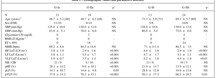
The values and distribution of the several frequency bands of the power spectrum in the groups studied are shown in table I. The absolute values of HF LnT, LF LnT, and VLF LnT in group II were significantly lower (p<0.001) than those observed in group I.
In regard to intragroup analysis, the values of HF LnT, LF LnT, and VLF LnT were significantly different between subgroups Ia and Ib (p<0.001), but not between subgroups IIa and IIb.
Table I – Demographic values and parameters assessed
G-Ia G-IIa p* G-Ib G-IIb p*
N 21 18 13 24
Age (years)** 48.7 ± 5.2 [48] 48.7 ± 6.2 [48] NS 71.3 ± 5.9 [71] 69.3 ± 5.7 [69] NS
Sex (F/M) 11/10 4/14 NS 5/8 16/8 NS
SBP (mm Hg) 125.0 ± 10.0 128.0 ± 12.0 NS 138.0 ± 10.6 130.0 ± 13.0 NS
DBP (mm Hg) 83.0 ± 5.1 78.0 ± 6.0 NS 80.0 ± 5.4 75.0 ± 8.0 NS
Glycemia>120 mg/dL 0 2 - 0 4
-BMI>25 Kg×m-2 0 0 - 0 0
-EF < 40% 0 18 - 0 24
-MHR (bpm) 69.2 ± 8.6 84.5 ± 14.0 NS 71 ± 111.4 86.5 ± 15 NS
HF LnT (LN ms2) 5.8 ± 1.0 2.9 ± 1.6 <0.001 4.4 ± 1.0 2.9 ± 1.0 <0.001
LF LnT (Ln ms2) 5.8 ± 1.1 3.3 ± 1.7 <0.001 4.7 ± 1.7 3.1 ± 1.1 <0.001
VLF LnT (Ln ms2) 5.9 ± 0.7 3.5 ± 1.4 <0.001 5.2 ± 1.0 4.4 ± 1.8 <0.01
NB / OB 21 / 0 8 / 10 <0.001 13 / 0 19 / 5 NS
pHF (%) 28.1 ± 14.2 9.0 ± 10.1 <0.001 21.0 ± 13.7 13.9 ± 12.1 NS
pLF (%) 34.1 ± 18.7 14.6 ± 9.8 0.05 28.8 ± 12.8 19.6 ± 13.6 NS
pVLF (%) 37.8 ± 14.2 76.3 ± 15.1 <0.001 50.3 ± 17.3 66.3 ± 18.7 0.01
*- level of statistical significance; **- values of age expressed as mean±SD [median], the remaining values are expressed as mean±SD; HF - high frequency; LF - low
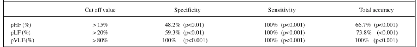
tory types analyzed had no statistically significant associa-tion with the funcassocia-tional class or with the cause of the left ventricular global systolic dysfunction. In group II, a left ventricular ejection fraction ≤ 25% was associated with nor-mal breathing with an odds ratio equal to 0.17 (95% CI [0.03 to 0.88]; p=0.03). The values for sensitivity and specificity are shown in table II. The variable pVLF in the cut off > 80% had a predictive accuracy of 100% and identified all indivi-duals with the oscillatory breathing pattern. The compa-risons of the clinical variables and V-RR between indivi-duals with normal and oscillatory breathing are shown in table III.
Twelve months after the initial assessment, 42.2% of the group II individuals had died. Of these patients, in the initial assessment, 21.4% had oscillatory breathing and 78.6% had normal breathing (odds ratio = 0.34, 95% CI [0.06 to 1.56]; NS), 78.6% were in functional class III-IV (odds ratio = 4.07; 95% CI [0.71 to 28.91]; NS), and 71.4% had left ventricular ejection fraction ≤ 25% (odds ratio = 7.00, 95% CI [1.21 to 43.91]; p=0.03). The age and sex of these individuals who died after 12 months of evolution (60.6±15.5 years, 71.4% of males) did not significantly differ from those of the survivors in the same period (59.8±12 years, NS, 68.4% of males, NS). In the frequency domain, using the cut off values of pLF in 20% and of pHF in 15% to dichotomize the individuals who died and who survived, an odds ratio of 0.06 (95% CI [0.01 to 0.48]; p = 0.004) was observed for pLF and an odds ratio of 0.38 (95% CI [0.07 to 2.09]; NS) was ob-served for pHF.
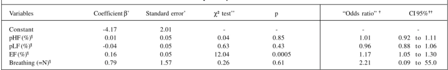
The logistic model to foretell survival after 1 year of evo-lution was adequately adjusted to the variables selected (χ2
3 = 1.09; NS). The variables DM2, functional class, and pVLF had no statistical significance in the model and were withdrawn during the process of stepwise selection, all with
χ² values < 0.01 in the test of maximal likelihood ratio (p ≈ 0.99). The results of the adjusted model are shown in table IV.
Discussion
Respiratory disorders of the oscillatory breathing type are frequent in individuals with left ventricular global systo-lic dysfunction, especially those recorded with the indivi-dual awake. Some studies have shown that these disorders may act unfavorably on the prognosis of individuals with left ventricular global systolic dysfunction 2,8,9,17.
When the electrocardiographic RR intervals are analyzed by using the power spectrum and recorded in The behavior of the variables pVLF, pLF, and pHF in
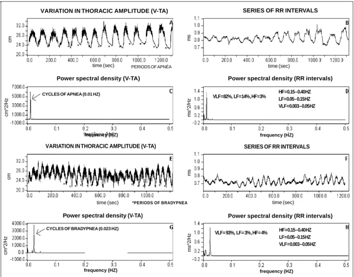
group II depended on the breathing pattern. Therefore, in the 27 individuals with normal breathing, the distribution of power in the different bands was similar to that found in control individuals. For the 15 individuals with oscillatory breathing, Cheyne-Stokes respiration or periodic breathing, the power was concentrated in the very low frequency band (fig. 1).
In group II, considering individuals with and without DM2, the means of the following variables showed no sta-tistically significant differences: V-RR (respectively, for pHF 10.4%±10.2% x 16.4%±16.3%; for pLF 18.3%± 11.9% x 9%±8%; for pVLF 71.1%±16.6 x 74.5%±23.4% ), left ventricular ejection fraction (respectively, 28.8%±10.5% x 36.8%±9.4%), distribution of the individuals in functional class III-IV (respectively, 66% x 50%), individuals with oscillatory breathing (respectively, 41% x 50%), and survival after 1 year of evolution (respectively, 55% x 76%). The analysis of r2 between V-RR and the variation in thoracic amplitude showed that, both in group I and group II with normal breathing, a significant coherence was obser-ved in all bands [very low frequency (p<0.05) and high fre-quency (p<0.05)]. In group II, the value of r2 was significant only in the individuals with oscillatory breathing, in whom the V-RR power was concentrated in the very low frequen-cy (p<0.05) (fig. 2).
The analysis of the odds ratio showed that the
respira-BREATHING PATTERNS LEFT VENTRICULAR GLOBAL
HEALTHY NB
(N = 27 [ 63,4%] )
OB (N = 15 [ 35,7%] )
pVLF 54.0%
pVLF 58.3%
pVLF
89.0 pHF
3.4% pLF 7.6% pHF
17.6% pLF
24.1% pHF
23.7% pLF
22.3%
HEALTHY: N=34, HEART FAILURE: N=42
Fig. 1 – Percentage distribution of the frequency bands in the series of RR intervals in healthy individuals, in individuals with left ventricular global systolic dysfunction and normal breathing, and individuals with left ventricular global systolic dysfunction and oscillatory breathing. Note that in individuals with left ventricular global systolic dysfunction – oscillatory breathing (OB) – the modulating energy concentrates in the very-low-frequency band (pVLF). LVGSD = left ventricular glo-bal systolic dysfunction, pVLF = percentage of energy concentrated in the very-low-frequency region, NB= normal breathing.
SYSTOLIC DYSFUNCTION
Table II – Analysis of the diagnostic value of the variables analyzed in group II in regard to the presence of oscillatory breathing
Cut off value Specificity Sensitivity Total accuracy
pHF (%) > 15% 48.2% (p<0.01) 100% (p<0.001) 66.7% (p<0.001)
pLF (%) > 20% 59.3% (p<0.01) 100% (p<0.001) 73.8% (<0.001)
pVLF (%) > 80% 100% (p<0.001) 100% (p<0.001) 100% (p<0.001)
different frequency bands, these intervals are observed to be modulated by respiration in a frequency band ranging from 0.01 Hz to 0.40 Hz (unpublished data). In that band, the effect of the breathing movements on the power spectrum of RR intervals can be predominantly felt by the cyclic variations in the tonus of the autonomic nervous system on the sinus node, mainly mediated by baroreflexes 18.
Analyzing the behavior of the power spectrum of the RR intervals and comparing it with that observed in healthy individuals, two distinct patterns are observed in indivi-duals with left ventricular global systolic dysfunction. In 15 (35.7%) individuals with oscillatory breathing, a mean of 89.2% of the total spectral power was concentrated in the very low frequency band, despite having Cheyne-Stokes respiration or periodic breathing, showing that the RR inter-vals are mainly modulated by low frequency breathing movements (figs. 1 and 2). For the other 27 (64.3%) indivi-duals with no great cyclic variations in the amplitude of brea-thing movements, the behavior of the power spectrum had a normal distribution (fig. 1).
In the forms of oscillatory breathing described in the
Table III – Analysis of the breathing pattern in group II in regard to functional parameters *
NB OB p
N 27 15
-Age (years) 63.9 ± 13.0 55.2 ± 12.1 NS
Sex (F/M) 11 / 16 2 / 13 NS
FC III-IV (%) 63.2% 53.3% NS
DM2 (%) 10.5% 20% NS
EF (%) 25.1 ± 9.5 36.2 ± 8.6 <0.01 HF LnT (Ln ms) 2.9 ± 1.4 2.7 ± 1.6 NS LF LnT (Ln ms2) 3.3 ± 1.4 3.2 ± 1.6 NS
VLF LnT (Ln ms2) 3.7 ± 1.5 5.9 ± 1.8 <0.001
pHF (%) 17.6 ± 8.8 3.4 ± 8.8 <0.001 pLF (%) 24.1 ± 8.4 7.6 ± 8.4 <0.001 pVLF (%) 58.3 ± 8.1 89 ± 8.1 <0.001 % of death after 12 months 57.9% 21.4% 0.01
Cause 52.6% 40%
DHD –ISC –HYP** 36.8% - 10.5% 33.3% - 26.7% NS
* NB- normal breathing; OB- oscillatory breathing; **DHD- dilated heart disease; ISC- ischemic heart disease; HYP- hypertensive heart disease; FC-NYHA functional class; HF- high frequency; LF- low frequency; VLF- very low frequency; LnT- logarithmic transformation (refer to text for details); pHF- percentage of high frequency; pLF- percentage of low frequency; pVLF-percentage of very low frequency; EF- left ventricular ejection fraction.
Fig. 2 – Simultaneous recording of the variation in thoracic amplitude and the RR intervals in individuals with Cheyne-Stokes respiration (A-B-C-D tracings) and periodic breathing (E-F-G-H tracings). Note, in both types of breathing, the spectral coherence in the VLF band (C-D tracings → r2=0.72; G-H tracings → r2=0.89), between V-RR and V-TA.
V-RR = variability in the RR intervals. V-TA = variability in thoracic amplitude. VARIATION IN THORACIC AMPLITUDE (V-TA)
time (sec) PERIODS OF APNEA
SERIES OF RR INTERVALS
time (sec)
Power spectral density (V-TA)
CYCLES OF APNEA (0.01 HZ)
frequency (HZ)
Power spectral density (RR intervals)
HF = 0.15 – 0.40 HZ LF = 0.05 – 0.15 HZ VLF = 0.003 – 0.05 HZ VLF = 82%, LF = 14% , HF = 3%
frequency (HZ)
VARIATION IN THORACIC AMPLITUDE (V-TA)
time (sec) *PERIODS OF BRADYPNEA
SERIES OF RR INTERVALS
time (sec)
Power spectral density (V-TA)
CYCLES OF BRADYPNEA (0.023 HZ)
frequency (HZ) frequency (HZ)
HF = 0.15 – 0.40 HZ LF = 0.05 – 0.15 HZ VLF = 0.003 – 0.05 HZ VLF = 93%, LF = 3% , HF = 4%
Power spectral density (RR intervals)
cm
cm
A2/Hz
cm
A2/Hz
cm
ms
ms
A2/Hz
ms
ms
literature, the energy reduction in the low and high frequen-cy bands of the power spectrum, which represent the modulation of the efferent controls of the sympathetic-vagal system, has been attributed to the reduction in the regulato-ry capacity of the baroreflexes. This regulatoregulato-ry capacity may be due to the sensitivity loss in the peripheral baroreceptors or to the reduction in the rhythmicity of the efferent autono-mic impulses originating in the cerebral nuclei 19,20. This fact was very evident in the present study considering that the values of the energy in the high and low frequency bands were significantly lower in group II (p<0.001).
On the other hand, in left ventricular global systolic dysfunction, an increase in the sensitivity of the peripheral chemoreceptors and in the time of circulation between the lung and the regulatory cerebral breathing centers has been observed. Therefore, while the hypersensitivity of the chemoreceptors intensifies the response of the systems, which control gain (increased sensitivity to the alterations in O2 and CO2), and reduces the response of the systems that reduce it (a reduction in the O2 and CO2 reserves), the increase in the time of circulation delays the process of information transference. These alterations, on the other hand, cause instability of the feedback systems that control breathing, therefore determining the two forms of oscilla-tory breathing 21-23. Considering that V-RR is predominantly modulated by breathing movements, which, because of the already cited facts, are constituted by oscillations of very low frequency, the characteristics of the spectral power modulating V-RR are consistently modified. A deviation to the left in the frequency bands of the power spectrum is observed with a significant power concentration in the very low frequency band.
The importance of the recognition of these breathing types and of the V-RR behavior in left ventricular global systolic dysfunction during sleep and wakefulness, lies in the fact that they can change the quality of life and impair cardio-pulmonary performance, making the prognosis of left
ventri-cular global systolic dysfunction more reserved, regardless of the degree of impairment of clinical and laboratory para-meters. These facts, especially those related to the develop-ment of arrhythmias and the deterioration of ventricular func-tion could have as pathophysiological bases the autonomic imbalance, hypoxia, hypercapnia, and the increase in the sympathetic amines, therefore aggravating neurohormonal disorders, especially in regard to the elevated levels of cate-cholamines 10,24. In regard to the clinical, laboratory, and evo-lutional findings of the individuals studied, the breathing patterns and the characteristics of V-RR were independent of functional class, the cause of left ventricular global systolic dysfunction, and the magnitude of the changes in the echo-cardiographic parameters. However, its arrhythmogenic capacity could not be assessed. Although other researchers reported that oscillatory breathing during wakefulness increases mortality in individuals with left ventricular global systolic dysfunction 8, this fact was not observed in our study. Using a logistic regression model with the stepwise type of selection of the variables adjusted, the multivariate analysis showed that only left ventricular ejection fraction has an independent predictive value to foretell survival after a 1-year evolution (p=0.0005).
In conclusion: 1) oscillatory breathing is a common breathing type in individuals with left ventricular global systolic dysfunction observed in 35.7% of individuals with that disorder during wakefulness and characterized by a typical V-RR pattern; 2) oscillatory breathing has no corre-lation with the severity and cause of left ventricular global systolic dysfunction, the presence of DM2, and is not an independent predictive marker of mortality after 12 months of evolution; 3) the multivariate analysis reveals that the ejection fraction is the only variable with an independent predictive value for survival after a 1-year evolution in individuals with left ventricular global systolic dys-function.
Table IV – Results of the multivariate logistic regression model for predicting survival of group II individuals after 1 year of evolution with 4 exploratory variables
Variables Coefficient β* Standard error* χ² test** p “Odds ratio”g CI 95%gg
Constant -4.17 2.01 - - -
-pHF (%)¶ 0.01 0.05 0.04 0.85 1.01 0.92 to 1.11
pLF (%)¶ -0.04 0.05 0.63 0.43 0.96 0.88 to 1.06
EF (%)¶ 0.16 0.05 12.04 0.0005 1.17 1.05 to 1.30
Breathing (=N)¶ 0.79 1.57 0.26 0.61 2.21 0.09 to 55.0
*- represents the weight of each variable in the model and the respective standard error of the estimate; **- χ² test for the maximal similarity ratio (1 degree of freedom);
g- controlling other variables in the model for each 1-unit increase in a certain variable, the probability of an event occurring increases proportionally to the respective
odds ratio value; gg- 95% confidence interval; ¶- pHF- percentage of total power corresponding to the high-frequency region of the power spectrum; pLF– percentage
Referências
1. Henry RA, Beightol LA, Eckber DL. Interaction between CO2 chemoreflexes and
arterial baroreflexes. Am J Physiol Heart Circ Physiol 1998; 43: H2177-H87. 2. Ponikowski P, Anker SD, Chua TP, et al. Oscillatory breathing patterns
du-ring wakefulness in patients with chronic heart failure. Clinical implication and role of augmented peripheral chemosensitivity. Circulation 1999; 100: 2418-24.
3. Feld H, Priest S. A cyclic breathing pattern in patients with poor left ventricular function and compensated heart failure: a mild form of Cheyne-Stokes respira-tion? J Am Coll Cardiol 1993; 21: 971-4.
4. Mortara A, Sleight P, Pinna GD, et al. Abnormal awake respiratory patterns are common in chronic heart failure may prevent evaluation of autonomic tone by measures of heart rate variability. Circulation 1997; 96: 246-52.
5. Pinna GD, Maestri R, Montara A, La Rovere MT. Cardiorespiratory interactions during periodic breathing in awake chronic heart failure patients. Am J Physiol Heart Circ Physiol 2000; 278: H932-H41.
6. Tkacova R, Niroumand M, Lorenzi-Filho G, BradleyTD. Overnight shift from obstructive to central apneas in patients with heart failure. Role of PCO2 and
circulatory delay. Circulation 2001; 103: 238-43.
7. Lorenzi-Filho G, Rankin F, Bies I, et al. Effects of inhaled carbon dioxide and oxygen on Cheyne-Stokes respiration in patients with heart failure. Am J Respir Crit Care Med 1999; 159: 1490-8.
8. Andreas S, Hagenah G, Möller C, et al. Cheyne-Stokes respiration and progno-sis in congestive heart failure. Am J Cardiol 1996; 78: 1260-4.
9. Montara A, Bernardi L, Pinna GD, et al. Alteration of breathing in chronic heart failure: clinical relevance of arterial oxygen saturation instability. Clin Sci (Colch) 1996; 91: 72-4.
10. Davies SW, John LM, Wedzicha JA, Lipkin DP. Overnight studies in severe chro-nic heart failure: arrhythmias and oxygen desaturation. Br Heart J 1991; 65: 77-83.
11. Barbosa PR, Barbosa Filho J, Morais de Sá CA. Influência da idade, sexo e doença coronária sobre a modulação autonômica do coração. Arq Bras Cardiol 1996; 67: 325-9.
12. Moody GB, Mark RG, Zoccola A, Mantero S. Derivation of respiratory signals from multi-lead ECGs. Computers in Cardiology 1985; 12: 113-6. 13. Barbosa-Filho J, Barbosa PR, Cordovil I. Modulação Autonômica do Coração
na Hipertensão Arterial Sistêmica. Arq Bras Cardiol 2002; 78: 181-8. 14. Miranda de Sá AMFL, Infantosi AFC, Simpson DM. A statistical technique for
measuring synchronism between cortical regions in the EEG during rhythmic stimulation. IEEE Trans Biomed Eng 2001; 48: 1211-5.
15. Barbosa PR, Barbosa Filho J, Morais de Sá CA. Efeito da idade sobre a modulação autonômica do coração. Revista da SOCERJ 1995; 4: 79-84.
16. Bigger JT Jr, Fleiss JL, Steiman RC, et al. Frequency domain measures of heart rate va-riability and mortality after myocardial infarction. Circulation 1992; 85: 164-71. 17. Hanly PJ, Zuberi-Khokhar NS. Increased mortality associated with Cheyne-Stokes respiration in patients with congestive heart failure. Am J Resp Crit Care Med 1996; 153: 272-6.
18. Piepoli M, Sleight P, Leuzzi S, et al. Origin of respiratory sinus arrhythmias in conscious humans. An important role of arterial carotid baroreceptor. Circulation 1997; 95: 1813-21.
19. Saul JP, Arai Y, Berger RD, et al. Assessment of autonomic regulation in chronic congestive heart failure by heart rate spectral analysis. Am J Cardiol 1988; 61: 1292-9.
20. Van de Borne P, Montano N, Pagani M, et al. Absence of low-frequency variabili-ty of sympathetic nerve activivariabili-ty in severe heart failure. Circulation 1997; 95: 1449-54.
21. Lahiri S, Hsiao C, Zhang R, et al. Peripheral chemoreceptors in respiratory oscillations. J Appl Physiol 1985; 58: 1901-8.
22. Khoo MC, Kronauer RE, Strohl KP, Slutsky AS. Factors inducing periodic breathing in humans: a general model. J Appl Physiol 1982; 53: 644-59. 23. Naughton M, Benard D, Tam A, et al. Role of hyperventilation in the pathogenesis
of central sleep apneas in patients with congestive heart failure. Am Rev Respir Dis 1993; 148: 330-8.