Arq Neuropsiquiatr 2010;68(3):410-413
410
Article
Valsalva maneuver procedures in
the diagnosis of right-to-left shunt
by contrast-enhanced transcranial
doppler using agitated saline solution
with blood as a contrast agent
Marcos Christiano Lange, Viviane Flumignan Zétola, Elcio Juliato Piovesan, Lineu César Werneck
ABSTRACT
Objective: To compare two different timings for the performance of the Valsalva maneuver (VM) using an infusion of agitated saline solution with blood as contrast agent (CA) to right-to-left shunt (RLS) screening. Method: 42 patients were submitted to a standardized contrast-enhanced transcranial doppler (cTCD) to screen for right-to-left shunt (RLS). cTCD technique was done with two different moments of the VM: [1] the CA injection during the VM (CAduringVM test); [2] the CA injection before the VM (CApreVM test). Results:
Positive MCA tests were observed in 47 (56%) CAduringVM tests and in 50 (59.5%) CApreVM tests, p=0.64. There was an almost perfect agreement for the positive tests between the CAduringVM and CApreVM test, rs=0.829 (95% CI 0.61-1.00, p<0.001). Conclusion: The
present study demonstrates that there is no significant difference in the results of RLS screening by cTCD when two different moments of VM were done.
Key words: contrast media, paradoxical embolism, microbubbles, transcranial doppler, ultrasonography, Valsalva maneuver.
Manobra de Valsalva no diagnóstico de embolia paradoxal pelo doppler transcraniano contrastado com o uso de solução salina agitada associada a sangue como meio de contraste
RESUMO
Objetivo: Comparar dois momentos diferentes da manobra de Valsalva (MV) com o uso de solução salina com sangue como meio de contraste (MC) para investigação de embolia paradoxal (EP). Método: 42 pacientes foram submetidos a protocolo padronizado de DTCc com a MV em dois momentos diferentes: [1] injeção do MC durante a MV (teste ACduranteMV); [2] injeção de MC antes da MV (teste ACpreMV). Resultados: Exames positivos foram observados em 47 (56%) ACMs testes ACduranteMV e 50 (59.5%) testes ACpreMV, p=0.64. Houve uma correlação quase perfeita entre ambos os testes, rs=0.829
(95% CI 0.61-1.00, p<0.001). Conclusão: O presente estudo demonstra que não existe diferença significativa na positividade de EP pelo DTCc quando são comparados dois momentos diferentes da MV.
Palavras-chave: meios de contraste, embolia paradoxical, microbolhas, doppler transcraniano, ultra-som, manobra de Valsalva.
Correspondence
Marcos Christiano Lange Hospital de Clínicas Serviço de Neurologia
Rua General Carneiro 181 / 4º andar 80060-900 Curitiba PR - Brasil E-mail: [email protected]
Received 20 May 2009
Receveid in final form 2 September 2009 Accepted 8 September 2009
Cerebrovascular Diseases Unit, Neurology Division, Internal Medicine Department, Hospital de Clínicas, Universidade Federal do Paraná, Curitiba PR, Brazil.
Contrast-enhanced transcranial dop-pler (cTCD) is a reliable and reproducible screening method for right-to-left shunt
(RLS) diagnosis1,2. he Valsalva maneuver
increas-Arq Neuropsiquiatr 2010;68(3)
411
Valsalva maneuver in shunt diagnosis by TCD Lange et al.
ing the right atrial pressure to facilitate or reveal an inter-mittent RLS via a patent foramen ovale (PFO)2,3.
Previous studies have demonstrated that the ideal time to perform the VM is five seconds after contrast agent (CA) infusion, but these studies were done pref-erentially with agitated saline solution (AS) or a galac-tose-based CA1,3,4. Studies used a small sample of a
pa-tient’s own blood to obtain an agitated saline solution with blood (ASb) as a means of increasing the number of
microbubbles (MBs) generated compared to AS5.
he aim of this study was to compare two diferent timings of the VM with the infusion of ASb as a CA.
Method
We evaluated 42 patients in the Laboratory of Neu-rosonology for RLS diagnosis. All of the subjects gave their written informed consent. Clinical and neurologi-cal evaluations were done in all of the cases and none pa-tient had an extracranial or an intracranial stenosis eval-uated by ultrasonography. here were 32 patients with ischemic stroke, 5 with transitory ischemic attacks, and 5 with migraine with aura. he local ethics committee ap-proved this study.
The cTCD (doppler-Box DWL, Singen, Germany) procedures were performed with the patients in a supine position. Two 2-MHz pulsed doppler transducers were ixed with a head frame (DiaMon DWL, Singen, Germa-ny) and insonated both middle cerebral artery (MCA) main stems through the temporal window at a depth of 50 to 65 mm to capture a small sample volume of 8 mm in length with two spectral gates 8 mm apart and the M-mode (32 sample gates in each channel). A 256-point fast Fourier transform analysis was used.
he CA was composed of a mixture of 8 mL saline, 1 mL air, and 1 mL of the patient’s blood. Before the infu-sion, the solution was prepared by agitating the mixture between two 10 ml syringes 10 times through a three-way tap connected to an 18 gauge intravenous catheter in-serted in a right antecubital supericial vein. he distance from the catheter to the syringes was less than 10 cm. he CA was injected in 5 seconds into the antecubital vein.
All of the patients were submitted to two different methods, and each was repeated twice with the ASb: [1] the VM was maintained for at least 10 seconds, with the CA infusion ive seconds after the beginning of the VM (CAduringVM test); [2] the VM was maintained for at least ive seconds, with the CA infusion ive seconds be-fore the beginning of the VM (CApreVM test).
he VM efectiveness was monitored by a 25% de-crease in the MCA low velocity and a mouthpiece con-nected to a manometer. he subjects were trained before the tests, and they were instructed to maintain an expi-ratory pressure of 40 mmHg. A small leak in the tubing
prevented the subjects from maintaining the pressure by closing the glottis, and care was taken to prevent deep breathing before and after the release of the strain6.
All of the tests were 60 seconds in duration, and they were recorded for later interpretation and quantiication oline. here was an interval of three minutes between each of the tests.
A test was considered positive when at least one em-bolus track (ET) with previously deined criteria was de-tected on both of the spectral gates displays and the
M-mode of at least one of the monitored MCAs1,7. he ET
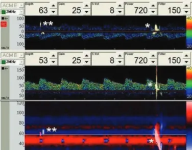
criteria were a unidirectional, typically visible and audi-ble, short duration, high-intensity signal within the dop-pler low spectrum with a movement toward the MCA as time progresses and a positively-sloped track in the M-mode image (Fig 1)1,7-9.
he ET counts were obtained individually for each MCA during an oline analysis. he highest count ob-tained with ASb in each VM procedure was considered for analysis. he studies were classiied based on previous criteria and deined as: negative=no ET observed; grade I=1-10 ETs, and grade II>10 ETs2.
he latency time, in seconds, between the CA injec-tion and the detecinjec-tion of the irst ET were analyzed in each MCA. he duration, in seconds, of the ET passage after the CA injection was evaluated in all of the tests by measuring the time between the irst and the last ET in each of the MCAs tested.
The statistical analyses were performed with SPSS 12.0 software (SPSS Inc.). Statistical signiicance was as-sessed by a Student’s t-test for the parametric variables, and the Chi-square and Mann-Whitney tests were used for the non-parametric variables. he degree of
Arq Neuropsiquiatr 2010;68(3)
412
Valsalva maneuver in shunt diagnosis by TCD Lange et al.
ment for a positive test (≥1 MB) was tested with the kap-pa test for agreement. Statistical signiicance was deter-mined at p<0.05.
Results
For the 42 patients evaluated, the mean age was 41.6±11.9 years old, 52% were female, and a total of 84 MCA tests with the CAduringVM test and 84 MCA tests with the CApreVM test, none of the patients had any ad-verse events during or after the tests. From all patients evaluated, 28 presented at least one positive CAduring VM test and 27 presented at least one CApreVM test positive (p=0.893)
Positive tests were observed in 47 (56%) of the CA-duringVM tests and in 50 (59.5%) of the CApreVM test (p=0.64). Only two tests were positive with the CAdur-ingVM test and negative with the CApreVM test, while ive tests were positive with the CApreVM test and nega-tive with the CAduringVM test. here was an almost per-fect agreement for positivity between the CAduringVM
test and the CApreVM test, with a correlation rs=0.829
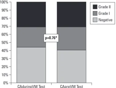
(95% CI 0.61-1.00, p<0.001). he ET grades were simi-lar in both groups: the CAduringVM test demonstrated 37 (44%) negative, 21 (25%) grade I, and 26 (31%) grade II tests, and the CApreVM test demonstrated 34 (40.5%) negative, 24 (28.6%) grade I, and 26 (31%) grade II tests (p=0.76) (Fig 2).
he mean number of ET (97.7±125.6 in the CAdur-ingVM test versus 77.3±111.2 in the CApreVM test, p=0.40), the latency time in seconds (10.6±9.0 in the CAduringVM test versus 13.5±5.3 in the CApreVM test, p=0.56), and the duration time of the ET passage in sec-onds (18.0±16.8 in the CAduringVM test versus 17.9±16.5 in the CApreVM test, p=0.98) were similar in both groups.
discussion
he present study demonstrated that the timing of the VM made no diference when ASb was used as the CA for RLS screening by cTCD. he mean number of ET, the latency time, and the duration did not difer based on the timing of the VM.
Previous studies with diferent CAs demonstrated that CA infusion during the VM and before the VM showed good sensitivity values compared to contrast-enhanced transesophageal echocardiography (cTEE) for RLS di-agnosis, preferentially for PFO diagnosis1-4,10. An
inter-national consensus suggested that the VM should start ive seconds after the beginning of the CA injection and should be maintained for at least ive seconds1. his
pro-cedure was done in the current study (CApreVM test), and the results demonstrated an almost perfect agree-ment compared to the CA injection during the VM (CA-duringVM test) for positivity of RLS with ASb.
Droste et al. demonstrated that at least two diferent moments of VM must be done if the irst procedure was
negative4, these results were conirmed by other studies
during the PFO diagnosis11. In the current study, it was
observed that a diferent procedure changed seven MCA results: two that were negative with the CApreVM test and became positive with the CAduringVM test; ive that were negative with the CAduringVM test changed to pos-itive with the CApreVM test.
his study has some limitations. he patients were not submitted to PFO diagnosis by cTEE to identify the sen-sitivity and speciicity for cardiac RLS by this technique. In a previous study, we demonstrated that standardized
cTCD is a good method for RLS screening2. Another
lim-itation was the use of only ASb; the results could be dif-ferent with other CA, and until now, no previous com-parative analysis has been done. We recently demonstrat-ed that ASb is as good as AS for RLS screening by cTCD (unpublished data).
In conclusion, we agree that the VM procedure used in the current consensus for the diagnosis of RLS by cTCD can be accepted when ASb is used as the CA, but if a negative result is observed, the patient should be sub-mitted to a diferent protocol with a diferent timing of the VM, which could involve CA injection during VM (CA duringVM test).
RefeRences
1. Jauss M, Zanette E. Detection of right-to-left shunt with ultrasound contrast agent and transcranial doppler sonography. Cerebrovasc Dis 2000;10:490-496. 2. Lange MC, Zetola VF, Souza AM, et al. Transcranial doppler for patent foramen
ovale screening: is there a good correlation with transesophageal echocar-diography? Arq Neuropsiquiatr 2008;66:785-789.
3. Schwarze JJ, Sander D, Kukla C, Wittich I, Babikian V, Klingelhöfer J. Method-ological parameters inluence the detection of right-to-left shunts by con-trast transcranial doppler. Stroke 1999;30:1234-1239.
4. Droste DW, Siling K, Stypmann J, et al. Contrast transcranial doppler ultra-0% 10% 20% 30% 40% 50% 60% 70% 80% 90% 100% Grade II Grade I Negative CApreVM Test CAduringVM Test p=0.76*
Arq Neuropsiquiatr 2010;68(3)
413
Valsalva maneuver in shunt diagnosis by TCD Lange et al.
sound in the detection of right-to-left shunts: time window and threshold in microbubble numbers. Stroke 2000;31:1640-1645.
5. Sastry S, Daly K, Chengodu T, McCollum C. Is transcranial doppler for the de-tection of venous-to-arterial circulation shunts reproducible? Cerebrovasc Dis 2007;23:424-429.
6. Pott F, van Lieshout JJ, Ide K, Madsen P, Secher NH. Middle cerebral artery blood velocity during a Valsalva maneuver in the standing position. J Appl Physiol 2000;88:1545-1550.
7. Moehring MA, Spencer MP. Power m-mode doppler (PMD) for observing ce-rebral blood low and tracking emboli. Ultrasound Med Biol 2002;28:49-57.
8. Ringelstein EB, Droste DW, Babikian VL, et al. Consensus on microembolus detection by transcranial doppler ultrasound. Stroke 1998;29:725-729. 9. Saqqur M, Dean N, Schebel M, et al. Improved detection of microbubble
sig-nals using power m-mode doppler. Stroke 2004;35:14-17.
10. Zanette EM, Mancini G, De Castro S, Solaro M, Cartoni D, Chiarotti F. Patent foramen ovale and transcranial doppler: comparison of diferent procedures. Stroke 1996;27:2251-2255.