Amaral RH et al. / Aortic lesion simulating pulmonary disease
Radiol Bras. 2014 Set/Out;47(5):320–322 320
0100-3984 © Colégio Brasileiro de Radiologia e Diagnóstico por Imagem
Case Report
Aortic lesion simulating pulmonary disease: a case report
*
Lesão de aorta simulando doença pulmonar: relato de caso
Amaral RH, Souza VVS, Nin CS, Pedraza CAA, Biegelmeyer J, Maciel AC. Aortic lesion simulating pulmonary disease: a case report. Radiol Bras. 2014 Set/Out;47(5):320–322.
Abstract
R e s u m o
The authors report the case of an elderly woman assessed for dyspnea and right costal margin pain, whose chest radiography demonstrated opacity simulating pulmonary lesion, and computed tomography revealed the vascular origin of the condition. Acute aortic syndrome due to ruptured atheromatous plaque penetrating through the elastic lamina in association with aortic hematoma and aneurysm is a relevant differential diagnosis to be considered in these cases.
Keywords: Aorta; Aortic arch; Computed tomography.
Relatamos o caso de uma paciente idosa avaliada por apresentar dispneia e dor na borda costal direita, em que a radiografia de tórax demonstrou opacidade simulando lesão pulmonar e a tomografia computadorizada revelou ser de origem vascular. A síndrome aórtica aguda por ulceração de placa ateromatosa, penetrando através da lâmina elástica, associada a hematoma aórtico e aneurisma constitui importante diagnóstico diferencial nesses casos.
Unitermos: Aorta; Arco aórtico; Tomografia computadorizada.
* Study developed at the Department of Radiology – Santa Casa de Misericórdia de Porto Alegre, Porto Alegre, RS, Brazil.
1. MDs, Residents, Department of Radiology – Santa Casa de Misericórdia de Porto Alegre, Porto Alegre, RS, Brazil.
2. MD, Resident, Department of Internal Medicine – Santa Casa de Misericórdia de Porto Alegre, Porto Alegre, RS, Brazil.
3. PhD, Head of the Department of Radiology – Santa Casa de Misericórdia de Porto Alegre, MD, Radiologist, Unit of Radiology – Hospital de Clínicas de Porto Alegre (HCPA), Porto Alegre, RS, Brazil.
Mailing Address: Dr. Ricardo Holderbaum do Amaral. Santa Casa de Misericórdia de Porto Alegre – Serviço de Radiologia. Rua Professor Annes Dias, 295, Centro Histórico. Porto Alegre, RS, Brazil, 90020-090. E-mail: [email protected].
Received June 14, 2013. Accepted after revision October 25, 2013.
chronic obstructive pulmonary disease (COPD), and heavy smoking. At physical examination, the patient presented subtle perioral cyanosis, diffuse expiratory wheezes and crack-les in the lower third of the chest, bilaterally, with no other significant alteration. The cardiac enzymes curve was nor-mal and electrocardiography indicated left ventricle hyper-trophy, with no other abnormality.
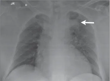
Frontal chest radiography performed with the patient in dorsal decubitus demonstrated the presence of an opacity on the projection of the left upper lobe, adjacent to the aor-tic arch that was found elongated and sinuous, with enlarge-ment of the cardiac area, diffuse and bilateral interstitial infiltrates, and presence of residual calcified micronodules (Figure 1).
Ricardo Holderbaum do Amaral1, Vinícius Valério Silveira de Souza1, Carlos Schuler Nin1, Cesar Adrian
Alvarez Pedraza1, Júlia Biegelmeyer2, Antonio Carlos Maciel3
http://dx.doi.org/10.1590/0100-3984.2013.1827
INTRODUCTION
Penetrating aortic ulcer (PAU) was first described in 1934(1), and only in 1986 was characterized as a clinical and
pathological entity(2), which, in association with its relative
rarity, explains the wide unawareness of this condition. It is caused by ulceration of an atheromatous plaque, penetrat-ing through the elastic lamina and progresspenetrat-ing to the me-dia, in association with aortic wall hematoma. It is part of acute aortic syndrome (AAS), together with aortic dissection, intramural hematoma and traumatic aortic rupture, which may present similar clinical manifestations, hence the high relevance of a rapid and accurate radiological diagnosis(2,3).
CASE REPORT
A 76-year-old woman attended the emergency unit of a university hospital complaining of sudden onset of dyspnea in association with right costal margin pain for one week. The patient presented a significant clinical history with car-diac failure secondary to hypertension, emphysematous
Amaral RH et al. / Aortic lesion simulating pulmonary disease
Radiol Bras. 2014 Set/Out;47(5):320–322 321
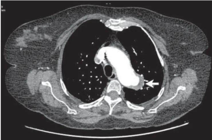
Non-contrast-enhanced and contrast-enhanced computed tomography revealed the presence of calcified atheromatous plaques and focal dilatation with 2.6 cm in diameter on the posterolateral aspect of the aortic arch, filled by contrast agent, in communication with the aortic lumen, associated with subintimal hematoma, compatible with PAU (Figures 2, 3 and 4).
Because of the clinical condition and of the prohibitive surgical risk, option was made for clinical management in agreement with the patient and her family. A maximum sys-tolic pressure of 120 mmHg was established as a goal, with management of comorbidities with emphasis on COPD sta-bilization. Until the present moment, 70 days after the first consultation, the patient is asymptomatic from the cardio-vascular point of view, with no sign of lesion progression.
DISCUSSION
As a considerably less frequent condition than dissec-tion (70%) and intramural hematoma (20%), PAU
corre-Figure 2. Contrast-enhanced computed tomography, pre-contrast phase show-ing saccular dilatation with 2.6 cm in diameter on the posterolateral aspect of the aortic arch, in association with subintimal hematoma (arrow). Axial section.
Figure 3. Contrast-enhanced computed tomography, arterial phase showing saccular dilatation with 2.6 cm in diameter on the posterolateral aspect of the aortic arch (arrow) filled by contrast agent and in communication with the aortic lumen, in association with subintimal hematoma. Axial section.
sponds to 5% of AAS. Usually, this disease involves the de-scending aorta and rarely the aortic arch (0.1% of AAS)(3–6).
PAU affects patients with advanced atherosclerosis, par-ticularly the elderly and hypertensive individuals. Initially, the lesion is asymptomatic – an atheromatous ulceration restricted to the intimal layer. Later, the ulcer deepens, pen-etrating through the elastic lamina and progressing to the media with different degrees of hematoma. The degree of distension and weakening of the aortic wall caused by the hematoma may lead to the development of a saccular aneu-rysm and rupture(3,4).
Contrast-enhanced computed tomography is the diag-nostic method of choice in most cases. Findings include focal lesion with subintimal hematoma, located under an intimal layer that is often calcified and internally displaced. Fre-quently there is thickening or contrast uptake by the aortic wall, giving the appearance of an aggressive lesion(3). De-spite the lower sensitivity for detecting small lesions, this method is satisfactory, considering that it can diagnose even-tual extraluminal diseases, and computed tomography an-giography acts as a complementary method in the evalua-tion of mural abnormalities(7). Transesophageal
echocardio-graphy has been successfully used and is highly sensitive and specific in the differential diagnosis of aortic disease(7). Rarely,
multiple ulcers may be found(3).
Scarce data are available about the natural history of this disease. However, some authors suggest a poorer prognosis than that for aortic dissection(2,4), with series reporting
rup-ture in up to 40% of the cases(3). Its management still re-mains controversial. Surgical intervention is recommended in the presence of intramural hematoma expansion, signs of imminent rupture and hemodynamic instability(3). The
Amaral RH et al. / Aortic lesion simulating pulmonary disease
Radiol Bras. 2014 Set/Out;47(5):320–322 322
ence of pain seems to be the most relevant clinical variable in such an evaluation(2).
Currently, the management by means of endovascular prosthesis is preferred considering its less invasiveness as compared with the open repair(8). Endovascular prosthesis
can be used even in cases of aortic rupture, with lower mor-bidity and mortality(4). However, in many cases the
endovas-cular treatment of the ascending aorta and aortic arch is not feasible because of technical difficulties. In such cases, sur-gical procedure is the method of choice(2). Immediate
inter-vention is not always required considering that, many times, the course of the disease is benign(9).
The authors describe the case of a lesion that simulated a pulmonary disease at conventional radiography. Further investigation with computed tomography was fundamental for the diagnosis and for guiding the appropriate clinical approach.
REFERENCES
1. Shennan T. Dissecting aneurysms. Medical Research Council Spe-cial Report Series No. 193. London: His Majesty’s Stationary Of-fice; 1934.
2. Stanson AW, Kazmier FJ, Hollier LH, et al. Penetrating atheroscle-rotic ulcers of the thoracic aorta: natural history and clinicopatho-logic correlations. Ann Vasc Surg. 1986;1:15–23.
3. Hayashi H, Matsuoka Y, Sakamoto I, et al. Penetrating atheroscle-rotic ulcer of the aorta: imaging features and disease concept. Radiographics. 2000;20:995–1005.
4. Piffaretti G, Tozzi M, Lomazzi C, et al. Endovascular repair of ab-dominal infrarenal penetrating aortic ulcers: a prospective observa-tional study. Int J Surg. 2007;5:172–5.
5. Girela A, Barbosa F, Quiroga J. Treatment of penetrating aortic ul-cer involving the aortic arch associated with lesion of the left main coronary artery. Rev Arg Cardiol. 2012;80:405–13.
6. Wells CM, Subramaniam K. Acute aortic syndrome. In: Subramaniam K, Park KW, Subramaniam B, editors. Anesthesia and perioperative care for aortic surgery. New York: Springer; 2011. p. 17–36. 7. Sommer T, Fehske W, Holzknecht N, et al. Aortic dissection: a
com-parative study of diagnosis with spiral CT, multiplanar transeso-phageal echocardiography, and MR imaging. Radiology. 1996;199: 347–52.
8. Novero ER, Metzger PB, Obregon J, et al. Tratamento endovascular das doenças da aorta torácica: análise dos resultados de um centro. Radiol Bras. 2012;45:251–8.