RESUMO
JUSTIFICATIVA E OBJETIVOS:Fibrose pleural idiopá-tica é uma doença rara e pode afetar ambos pulmões já desde uma idade precoce. O achado mais comum na ibrose pleural idiopática é uma restrição pulmonar grave que pode levar a um quadro de falência respira-tória e hipoxemia.
RELATO DO CASO: Paciente do sexo masculino, 26 anos, internado com reagudização de insuiciência respiratória crônica e submetido à ventilação mecâni-ca prolongada. Após intensa investigação e uma apre-sentação clínica atípica, foi estabelecido o diagnóstico de ibrose pleural idiopática associado à ibrose pul-monar.
CONCLUSÕES: O prognóstico de pacientes com i-brose pleural idiopática é extremamente ruim, particu-larmente em fase avançada da doença. Recomenda-se o tratamento precoce com corticosteróides ou decorti-cação pleural cirúrgica.
Failure to Wean Caused by Cryptogenic Fibrosing
Pleuritis and Bilateral Lung Trapping. Case Report*
Falência do Desmame em Paciente com Fibrose Pleural
Idiopática e Trapping Pulmonar Bilateral. Relato de Caso
Elsemiek Verweel1, Jos le Noble, PhD1, Christine Groeninx-van Zoelen1,
Alex Maat2, Willy Thijsse1, Patricia Gerritsen1, Jan Bakker, PhD3
1. Médica Assistente do Departamento de Terapia Intensiva 2. Médico Assistente do Departamento de Cirurgia Torácica 3. Médico Chefe do Departamento de Terapia Intensiva
*Received from Department of Intensive Care, Department of Pulmo-nary Medicine and Cardiovascular Centre Rotterdam Erasmus Medi-cal Centre, Location Dijkzigt
Presented in August, 23, 2007 Accepted in October, 08, 2007 Author for correspondence: Jos le Noble, M.D. PhD
Department of Intensive Care, H-620 Erasmus Medical Centre, Location Dijkzigt Dr. Molewaterplein 50
3015 GD Rotterdam, the Netherlands Phone: +31 630097795
E-mail: [email protected]
©Associação de Medicina Intensiva Brasileira, 2007
Unitermos: decorticação pleural, ibrose pleural, ibro-se pulmonar, Fibrotórax Idiopático.
SUMMARY
BACKGROUND AND OBJECTIVES: Cryptogenic i - i-brosing pleuritis is an extremely rare disease, which can affect both lungs from a very young age. The most common inding is severe lung restriction resulting in both hypoxemic and ventilatory failure.
CASE REPORT:Male patient, 26 year old with
acu-te deacu-terioration of chronic respiratory failure. Follo-wing admission prolonged mechanical ventilation was necessary. An atypical clinical presentation made the diagnosis difficult, but eventually cryp-togenic fibrosing pleuritis and lung fibrosis were established.
CONCLUSIONS: The prognostic outcome of patients
with the inal diagnosis of cryptogenic i brosing pleuri- ibrosing pleuri-tis is extremely poor, especially in an advanced phase of this disease. We recommend early treatment with corticosteroids or surgical pleural decortication.
Key Words: cryptogenic, Fibrothorax, lungibrosis, pleural decortication, pleural ibrosis.
INTRODUCTION
Fibrothorax is usually associated with bacterial, fungal or tuberculosis infection, asbestos exposure, vasculi-tis or exposure to speciic drugs. However, a prima-ry cause is not always obvious. This has led to a re-latively new entity, cryptogenic ibrosing pleuritis. This syndrome was irst described in 1988 by Buchanan et al.1. Cryptogenic ibrosing pleuritis is characterized by
We report the clinical course and outcome of a patient admitted to our ICu with respiratory failure in whom the diagnosis was cryptogenic ibrosing pleuritis and lung ibrosis. We discuss the importance of recognizing this rare syndrome at an early stage as treatment with corticosteroids or surgical decortication could prevent development of respiratory failure and may increase life-expectancy.
CASE REPORT
A 26-year old male was admitted to the ICu because of type 2 respiratory failure requiring mechanical ventila-tion.
The medical history revealed a gradual psychomotor retardation. The family history was non-contributory and the patient had no siblings. There was no con-sanguinity. As a child a discrepancy between length and skull circumference has been noted. A hydro-cephalus was absent. Our patient did not smoke and there was no history of chronic obstructive pulmonary disease. His irst symptoms were revealed in 2002 when he complained of fatigue and dyspnoea fol-lowing mild exercise. A pleural effusion was present. Laboratory indings were not helpful in establishing the diagnosis and analysis of the pleural luid did not reveal tumor cells. Blood cultures, anti-DNA an-tibody, anti-cardiolipine antibody and tests for tu-berculosis were all negative. At that time, a biopsy of the pleura parietalis did reveal inlammation with ibrosis without signs of asbestosis. Despite symp-tomatic treatment with diuretics and bronchodilators thedyspnoea gradually worsened. No systemic ster-oids were prescribed. In 2004 he was referred to our hospital for re-evaluation of the disease and further treatment. Because of macrocephaly, severe kypho-scoliosis and enlargement of the ascending aorta (Ø 50 mm) genetic screening was performed, but no known genetic syndromes were found. However, during the work-up the patient’s condition deterio-rated. In January 2005 blood gas analysis showed: pH 7.42, pCO2 7.0 kPa, pO2 8.3 kPa, BE + 8 mmol/L, satO2 91%. Lung function: VC 0.58 l (12%), FEV1 0.49 l (13%) and FEV1/ VC 84% (102%), compatible with severe restriction. A computer tomography (CT) scan showed severe entrapment of the right lung and to a lesser extent of the left lung without evidence of pulmonary ibrosis. The outpatient clinic physicians prescribed steroids in a low dose (10 mg/day). The patient continued his work as lift truck operator.
Admission
In June 2005 the patient was admitted to the ICu because of progressive respiratory failure. Physi-cal examination revealed a frail young man with respiratory distress with a respiratory rate of 30-40 breaths per minute. Body weight was 53 kg and length 175 cm. Vital signs revealed a blood pressure of 110/60 mmHg, a pulse of 66 beats/min. His neck veins were engorged. On inspection macrocephaly, high palate, and an abnormal thorax with evidence of a kyphoscoliosis and dystrophic appearance were seen. Auscultation of the lungs revealed bi-lateral coarse crackles and rhonchi, cardiovascular examination revealed a right-sided cardiac impulse, and no abnormal murmurs were heard. Abdominal examination was unremarkable except a slightly enlarged liver. The lower extremities showed no (lymph) oedema. On neurological examination there was no muscle weakness, focal deficits or other ab-normalities. Blood gas analysis showed: pH 7.46, pCO2 7.0 kPa, pO2 9.5 kPa. High dose steroids were started (1 mg/kg/day) because of clinical suspicion of pulmonary fibrosis.
On day 2 mechanical ventilation was required be-cause of carbon dioxide retention due to progressive exhaustion of the patient (pH 7.16, pCO2 16.2 kPa, pO2 9.5 kPa). The patient received pressure control-led mechanical ventilation after intubation with an expiratory tidal volume (VT) of 300 ml (5.7 mL/kg), a respiration rate of 20 breaths/min and with an initial positive end-expiratory pressure (PEEP) setting of 5 cmH2O. Peak inspiratory pressure and plateau pres-sure were 30 and 18 mmHg respectively. Fraction of inspired oxygen (FiO2) was weaned down to 0.6, and inspiration-expiration (I:E) ratio adjusted to 1:1. Lungs were poorly compliant, (calculated) dynamic and static compliance were 12 and 23 ml/kPa re-spectively.
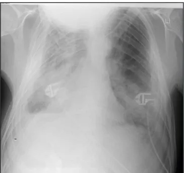
Figure 1 - X-Thorax on Day of Admittance to the ICu
Chest radiograph showing increased markings in all lung ields, a density on the right side without signs of cardiomegaly and in ad-dition bilateral pleural thickening with effusions most prominent on the right side.
Figure 2 - CT scan of the Thoracic Cavity
Computed tomography of the thorax demonstrating bilateral pleural thickening and effusions (arrow). Lung parenchyma showed areas of patchy iniltrates and progression of sub pleural atelectasis, due to pulmonary ibrosis.
Echocardiography of the heart revealed dilatation of the aorta ascendens (50 mm), tricuspid incompetence without signs of restrictive cardiomyopathy or valvu-lar vegetations. Laboratory examination: sodium 140 mmol/L, potassium 5.2 mmol/L, calcium 2.37 mmol/L, bilirubin 8 umol/L, LD 222 u/L, ureum 4.9 mmol/L, cre-atinine 54 umol/L, glucose 5.6 mmol/L, hemoglobin 9.1
mmol/L,hematocrit 0.48 l/L, platelets 400 10 E9/l, WBC 9.3 10E9/l, CRP 69 mg/L. Insertion of a pulmonary ar-tery catheter revealed: Pulmonary arar-tery pressures of 47/33 (23) mmHg, CVP 15 mmHg, cardiac index 4.6 L/min/m2 and pulmonary artery occlusion pressure
(PAOP) 19 mmHg.
Pleural decortication was planned. After induction of anesthesia and positioning of the patient in left lateral position it was extremely dificult to ventilate the pa-tient adequately. A mini right-sided thoracotomy was made. Due to the rooing-tile position of the ribs it was not possible to reach the pleura through an intercostal space. Two ribs were partially resected. A full thickness quadrangular specimen of pleura was resected and we noticed that adhesions between pleura and lung were so extensive that a serious attempt to decorticate the whole lung would result in massive blood loss. Given these problems pleural decortication was considered impossible. The full thickness biopsy of the pleura showed extended ibrotic tissue with focal reactive me-sothelium. There were no signs of mesothelioma. Our patient died despite full supportive treatment on day 14 due to ventilator associated pneumonia with sepsis. Permission for autopsy was refused by the parents.
Final Diagnosis
Respiratory failure due to cryptogenic ibrosing pleuri-tis (ibrothorax), lungibrosis and bilateral lung trapping complicated by a ventilator associated pneumonia with sepsis.
DISCUSSION
Respiratory failure in patients with ibrothorax is clini-cally characterized by ibrosing pleuritis, bilateral lung trapping and parenchymal disease. It encompasses a broad spectrum of diagnostic possibilities, which in-clude empyema (bacterial, tubercular or fungal infec-tions) and non-infectious causes such as hemothorax, autoimmune disease, medication or asbestos expo-sure. In most patients a primary cause can be establi-In most patients a primary cause can be establi-shed1,2. There are few cases of ibrothorax with
persis-tent pleural effusions such as a yellow nail syndrome or lymphangiectasis3.
However, in a very small subset of patients no primary cause can be established despite thorough clinical and laboratory investigations, as was the case in our pa-tient. In a landmark article published by Buchanan et al. in 19881 he described 4 patients with similar
intro-duced the term cryptogenic ibrosing pleuritis. Our pa-tient showed typical roentgenographic indings without history of trauma or infection. Radiologically, a peel of uniform thickness surrounded both lungs as well as ex-tensive parenchymal involvement. Routine pulmonary functions test revealed a severe restrictive ventilatory dysfunction. Cryptogenic ibrosing pleuritis is classiied as a form of idiopathic lung disease based on histo-logical appearance, which may exist as an idiopathic non-malignant entity. Cryptogenic ibrosing pleuritis may typically present at an early age, however it can present at any age.In our patient histological biopsy of the pleura showed inlammatory disease through dep-osition of ibrous tissue on the visceral pleural surface without signs of asbestosis or granulomas resulting in the diagnosis of cryptogenic ibrosing pleuritis. The vol-ume of the involved right sided hemithorax was dimin-ished and in our patient respiratory failure with carbon dioxide retention developed for which mechanical ven-tilation was necessary. since 1988 less than 10 articles have been published dealing with patients with similar clinical presentations of cryptogenic ibrosing pleuritis. There has been a renewed interest in this disease due to its recognized association with several metabolic di-seases4.
Our patient did not have obvious metabolic abnorma-lities as has been described by Hayes et al.4, but also
died at young age. In addition patients with cryptoge-nic ibrosing pleuritis may have an increased incidence of histocompatibility antigen HLA B441,4 which
predis-poses for congenital abnormalities. Although there was clinical suspicion in our patient for congenital abnor-mality this could not be conirmed by genetic consul-tation.
The pathogenesis of pleural ibrosis and abnormal pleu-ral space remodeling is incompletely understood. Data suggest a primary role for the visceral pleura during the development of pleural ibrosis5. Changes in the
for-mation of ibrinous intrapleural matrix may be the key feature in the development of respiratory impairment. Cytokines (TGF-beta and TNF-alpha) facilitate ibrin matrix formation and could be therapeutically inluen-ced by corticosteroids5.
Although the number of patients with cryptogenic i-brosing pleuritis treated with corticosteroids described in the literature is small, the disease may undergo re-mission following immunosuppressive therapy1,6. Our
patient with an advanced phase of the disease did not show any remission despite treatment with high dose corticosteroids. It cannot be excluded that treatment
in a very early phase with high dose corticosteroids or the addition of azathioprine would have any beneicial effect on prognosis. Impairment in the manifestations of cryptogenic ibrosing pleuritis can furthermore be achieved by surgical treatment, but because decortica- decortica-tion is a major surgical procedure it should not be per- is a major surgical procedure it should not be per-formed in patients debilitated by other disease1,7,8. One
major condition to perform this surgery is that there is a plane separating the visceral pleura from the adjacent lung8. Our patient showed extensive pleural thickening,
parenchymal disease in an advanced stage and severe deformation of the thorax making a surgical decortica-tion technically not feasible.
successful lung allograft transplantation in a patient with bilateral pleural ibrosis has only been reported by Azoulay et al.9. Due to progressive clinical deterioration
of our patient lung transplantation was not a serious therapeutic option.
Once all clinical hallmarks of cryptogenic ibrosing pleu-ritis are present and a critically ill patient is admitted to the ICu for mechanical ventilation the prognosis dete-riorates. If in doubt about the exact diagnosis a pleural biopsy is warranted. Treatment with high dose corti-costeroids or surgical exploration proved to be ineffec-tive and not feasible in our patient. several attempts to wean the patient from the ventilator were unsuccessful. spontaneous ventilation aggravated respiratory failure due to chest wall and parenchymal disease, indicating increased work of breathing.
Based on our observations, patients with cryptogenic ibrosing pleuritis should be aggressively treated with corticosteroids in an early phase of the disease on an outpatient basis. surgical decortication of pleural i -surgical decortication of pleural i - decortication of pleural i - of pleural i-brous tissue should be considered once ventilatory function decreases in combination with clinical symp-tomatology and exertional dyspnea. The prognostic outcome of patients with the inal diagnosis of crypto-genic ibrosing pleuritis is extremely poor, especially in an advanced phase of this disease.
Abbreviation list
BE Base excess
CBF Cryptogenic ibrosing pleuritis
Cm Centimeter
CT-scan Computer tomography scan CVP Central venous pressure
FEV1 Forced expiratory volume (in 1 second) FiO2 Fractional concentration of inspired oxygen ICu Intensive Care unit
Mg Milligram
Min Minute
Ml Milliliter
Mm Millimeter
Mmol Millimol
PEEP Positive end-expiratory pressure PAOP Pulmonary artery occlusion pressure sat. saturation
TGF Tumor growth factor TNF Tumor necrosis factor
umol Micromol
VC Vital capacity
VT Tidal volume
REFERENCES
01. Buchanan DR, Johnston ID, Kerr IH et al - Cryptogenic bilateral ibrosing pleuritis. Br J Dis Chest, 1988;82:186-193.
02. Ghoshal AG, saha AK, Roy DJ et al – Fibrothorax--problem, proile and prevention. J Indian Med Assoc, 1997;95:610-612.
03. Cohen M, sahn sA - Resolution of pleural effusions. Chest, 2001;119:1547-1562.
04. Hayes JP, Wiggins J, Ward K et al - Familial cryptogenic ibrosing pleu-ritis with Fanconi’s syndrome (renal tubular acidosis). A new syndrome. Chest, 1995;107:576-578.
05. Huggins JT, sahn sA - Causes and management of pleural ibrosis. Res-pirology, 2004;9:441-447.
06. O’Connor TM, Haider W, Crotty T et al – Immunosuppressant-responsive idiopathic lymphocytic pleuritis. Respiration, 2005;72:202-204. 07. sharma s, smith R, Al-Hameed F - Fibrothorax and severe lung
restric-tion secondary to lupus pleuritis and its successful treatment by pleurec-tomy. Can Respir J, 2002;9:335-337.
08. Baram D, Degene A, Amin M et al - A case of hypercapnic respiratory failure. Chest, 2004;126: 1994-1999.