338
Radiol Bras. 2017 Set/Out;50(5):338–348 Letters to the Editor0100-3984 © Colégio Brasileiro de Radiologia e Diagnóstico por Imagem
Letters to the Editor
Nonketotic hyperglycemia with involuntary movements
Dear Editor,
A 61-year-old woman who had been using insulin irregularly for the treatment of type II diabetes presented with hemichorea-hemiballism that had appeared suddenly in the left arm and left leg two weeks prior. Blood tests showed a blood glucose level of 450 mg/dL, a creatinine level of 0.9 mg/dL, and a urea level of 38 mg/dL. The complete blood count showed no abnormali-ties. The cerebrospinal luid glucose concentration was 350 mg/ dL. Magnetic resonance imaging (MRI) revealed a right-sided lesion, showing a hyperintense signal on T1-weighted images and a slightly hyperintense signal on T2-weighted images, lo-cated in the region of the caudate nuclei and putamen, with no enhancement, no evidence of bleeding in the magnetic sus-ceptibility-weighted sequences, and no restricted diffusion on diffusion-weighted imaging (Figure 1). These imaging indings, together with the clinical and biochemical history, conirmed the diagnosis of hemichorea–hemiballism due to nonketotic hy-perglycemia.
Nonketotic hyperglycemia, also known as diabetic striatopa-thy, is a rare cause of involuntary movements as a primary mani-festation of diabetes mellitus; it mainly affects elderly
individu-als, presenting as the triad of hemichorea-hemiballism, hypergly-cemia, and a lesion in the basal nuclei showing a hyperintense signal on T1-weighted images(1). Clinical and imaging indings
are typically unilateral, although they can be bilateral in up to 11.4% of cases(2), being potentially reversible and usually
resolv-ing within 2–12 months after the treatment of hyperglycemia(3,4).
Although the pathophysiology of nonketotic hyperglycemia is unknown, potential mechanisms include metabolic changes such as the deposition of proteins and of degradation prod-ucts of myelin, blood, calcium, or other minerals, which tend to decrease as serum glucose is controlled(5). Another accepted
theory is that a hyperglycemia-induced change in perfusion re-sults in reduced Krebs cycle activity, inducing anaerobic me-tabolism, causing the brain to use alternative sources of energy, and metabolizing the gamma-aminobutyric acid (GABA) inhibi-tory neurotransmitter. In nonketotic hyperglycemia, GABA and acetate levels drop rapidly, leading to a decrease in acetylcho-line synthesis. It has therefore been speculated that the reduced levels of acetylcholine and GABA in the basal nuclei leads to dysfunction of those nuclei, thus producing involuntary move-ments such as those seen in chorea-hemiballism(6,7).
For the evaluation of central nervous system diseases, the imaging method of choice is MRI(8–14). In
hemichorea-hemi-ballism due to nonketotic hyperglycemia, MRI indings are characterized by lesions in the region of the caudate nucleus or lenticular nucleus, showing hyperintense signals in T1-weighted sequences and discretely hyperintense signals in T2-weighted sequences, without enhancement or diffusion restriction, such lesions typically being unilateral(1,3,4), as in the case presented
here. The diagnosis of lesions with high signal intensity in T1-weighted sequences of the region of the basal nuclei is broad; the following can be cited as some of the main causes(1,4):
he-patic encephalopathy; prolonged exposure to manganese; pro-longed parenteral nutrition; Wilson’s disease; subacute intra-cerebral hemorrhage; exogenous carbon monoxide toxicity; and exogenous methanol toxicity. Correlation with the clinical and biochemical data is fundamental to making the deinitive diag -nosis(1,4).
In conclusion, although the occurrence of hemichorea-hemiballism as a complication of uncontrolled diabetes is un-common, the diagnosis should be considered when the clinical and MRI indings are characteristic of the disease. Thus, delays in the initiation of appropriate treatment can be avoided.
REFERENCES
1. Bekiesinska-Figatowska M, Romaniuk-Doroszewska A, Banaszek M, et al. Lesions in basal ganglia in a patient with involuntary movements as
a irst sign of diabetes – case report and review of the literature. Pol J
Radiol. 2010;75:61–4.
2. Krishna S, Sodhi KS, Saxena AK, et al. Hyperdense basal ganglia in
non-ketotic hyperglycemia. J Emerg Med. 2015;49:e57–8.
3. Bekiesinska-Figatowska M, Mierzewska H, Jurkiewicz E. Basal ganglia lesions in children and adults. Eur J Radiol. 2013;82:837–49.
4. Chokshi FH, Aygun N, Mullins ME. Imaging of acquired metabolic and
toxic disorders of the basal ganglia. Semin Ultrasound CT MR. 2014; 35:75–84.
5. Hegde AN, Mohan S, Lath N, et al. Differential diagnosis for bilateral abnormalities of the basal ganglia and thalamus. Radiographics. 2011; 31:5–30.
6. Aggarwal A, Bansal N, Aggarwal R. Nonketotic hyperglycemia presenting
as monoballism. J Emerg Med. 2016;50:e133–4.
7. Hansford BG, Albert D, Yang E. Classic neuroimaging indings of nonke -totic hyperglycemia on computed tomography and magnetic resonance
Figure 1. A,B: T1-weighted MRI showing a right-sided lesion with a hyperin-tense signal in the caudate nuclei and putamen (arrows). C: T2-weighted MRI showing a slightly hyperintense signal in the same regions. D: T2*-weighted MRI showing that there was no blood deposition in those regions.
A
B
339
Radiol Bras. 2017 Set/Out;50(5):338–348Letters to the Editor
http://dx.doi.org/10.1590/0100-3984.2015.0253
Tiago Medina Salata1, Lívia de Oliveira Antunes1, Bruno Niemeyer
de Freitas Ribeiro2, Rafael Silveira Borges1, Diogo Goulart Corrêa1
1. Hospital Casa de Portugal, Rio de Janeiro, RJ, Brazil. 2. Instituto Estadual do Cérebro Paulo Niemeyer, Rio de Janeiro, RJ, Brazil. Mailing address: Dr. Tiago Medina Salata. Rua do Bispo, 72, Rio Comprido. Rio de Janeiro. RJ, Brazil, 20261-064. E-mail: [email protected].
imaging with absence of typical movement disorder symptoms
(hemi-chorea-hemiballism). J Radiol Case Rep. 2013;7:1–9.
8. Machado VS, Silva Junior NA, Queiroz LS, et al. Central nervous system
involvement in sarcoidosis. Radiol Bras. 2015;48:334–5.
9. Dultra AHA, Noro F, Melo ASA, et al. Primary intercavernous lymphoma
of the central nervous system. Radiol Bras. 2015;48:337–8.
10. Ribeiro BNF, Lima GA, Ventura N, et al. Chronic kernicterus: magnetic
resonance imaging indings. Radiol Bras. 2016;49:407–8.
11. Langer FW, Suertegaray G, Santos D, et al. Hemichorea-hemiballism: the role of imaging in diagnosing an unusual disorder in patients with nonketotic hyperglycemia. Radiol Bras. 2016;49:267–8.
12. Ribeiro BNF, Salata TM, Borges RS, et al. Posterior reversible encepha -lopathy syndrome following immunoglobulin therapy in a patient with Miller-Fisher syndrome. Radiol Bras. 2016;49:58–9.
13. Campos LG, Trindade RAR, Faistauer A, et al. Rhombencephalitis: pic-torial essay. Radiol Bras. 2016;49:329–36.
14. Georgeto SM, Zicarelli CAM, Gariba MA, et al. T1-weighted
gradient-echo imaging, with and without inversion recovery, in the identiication
of anatomical structures on the lateral surface of the brain. Radiol Bras. 2016;49:382–8.
Intestinal perforation: an unusual complication of barium enema
Dear Editor,
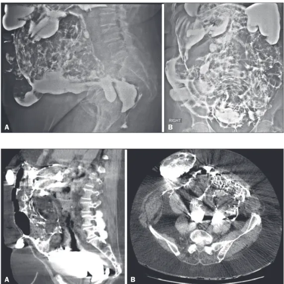
An 83-year-old female patient complaining of constipation was referred to our institution for elective enema with barium contrast, which showed diffuse irregularity in the mucosal folds of the colonic loops and signs of extravasation of the contrast medium into the abdomen and pelvic cavity (Figure 1). After the examination, the patient remained stable, without additional complaints. However, she did not agree to being hospitalized, signing a waiver. Despite being informed of the risks, she re-mained resolute, promising to return if there were any
symp-toms. She subsequently returned to the hospital with an acute abdomen, at which time she underwent computed tomography of the abdomen for preoperative evaluation, which demonstrated abdominal wall hernias, diverticulosis of the sigmoid colon, and a large amount of contrast material distributed diffusely throughout the peritoneal cavity and the hernias (Figure 2). The main hypothesis was perforation of the wall of the gastroin-testinal tract by the enema. The patient underwent exploratory laparotomy, with an inventory of the abdominal cavity, which conirmed the tomography indings and identiied a laceration at the rectosigmoid junction. After 14 days in the intensive care unit, the patient died.
Figure 2. Computed tomography scans of the abdomen, in the axial (A)
and sagittal (B) planes.
A
B
Figure 1. Images acquired during bar-ium enema examination, in lateral (A)
and anteroposterior (B) views.