Expression of the Immunohistochemical
Markers p16 and Ki-67 and Their Usefulness in
the Diagnosis of Cervical Intraepithelial
Neoplasms
Reprodutibilidade do diagnóstico das neoplasias
intraepiteliais cervicais e a in
fl
uência dos marcadores
imuno-histoquímicos p16 e Ki-67 como ferramentas
auxiliares
Fernanda Lopes Pontes de Melo
1Carmen Lúcia Penteado Lancellotti
2Maria Antonieta Longo Galvão da Silva
31Master’s Program in Health Sciences, School of Medical Sciences,
Santa Casa de São Paulo, São Paulo, SP, Brazil
2Pathological Anatomy Course, School of Medical Sciences, Santa
Casa de São Paulo, São Paulo, SP, Brazil
3Pathological Anatomy Course, School of Medical Sciences, Santa
Casa de São Paulo, São Paulo, SP, Brazil
Rev Bras Ginec Obst 2016;38:82–87.
Address for correspondence Fernanda Lopes Pontes de Melo, MD, Pathological Anatomy Service, Irmandade da Santa Casa de Misericórdia de São Paulo Central Hospital, Rua Doutor Cesário Mota Júnior, 112 - Vila Buarque, São Paulo, SP 01221-020, Brazil
(e-mail: [email protected]).
Keywords
►
cervical
intraepithelial
neoplasia
►
HPV
►
p16
►
K
i-67
►
kappa
Abstract
Objective
The aim of this study was to determine the expression of the
immunohis-tochemical markers p16 and K
i-67 in cervical intraepithelial neoplasms and their
in
fl
uence on the level of agreement among different observers and for the same
observer.
Methods
The study included 184 patients with cervical intraepithelial neoplasms
previously con
fi
rmed through biopsies performed between 2005 and 2006. Three
pathologists reviewed the biopsies by using hematoxylin-eosin staining to reach a
consensus on the diagnosis. Subsequently, an immunohistochemical study analyzed
the expression of p16 and K
i-67 in such cases.
Results
The comparison among the reviewing pathologists revealed only moderate
agreement (kappa
¼
0.44). The agreement improved when the differentiation of
high-grade lesions (cervical intraepithelial neoplasm
–
CIN
–
3) was analyzed (kappa
¼
0.59). p16 staining exhibited a high negative predictive value and sensitivity;
however, the speci
fi
city was low. Overall, both qualitative and quantitative analyses
of p16 and a quantitative analysis K
i-67 exhibited low accuracy. The agreement among
diagnoses before immunohistochemistry was 0.47. The use of immunohistochemistry
increased the agreement to 0.68.
received July 10, 2015 accepted
November 19, 2015 published online February 3, 2016
DOI http://dx.doi.org/ 10.1055/s-0036-1571470. ISSN 0100-7203.
Introduction
Every year, there are500,000 new cases of uterine cervical cancers in women worldwide. In 2012, 265,000 deaths were reported. In Brazil, uterine cervical cancer is the third most frequent cancer among women (not considering non-mela-noma skin cancer).1Approximately 80% of deaths could be prevented by screening for precursor lesions in women 25 to 65 years of age.2
Histopathological examination is the gold standard for a proper intraepithelial neoplasm diagnosis, and this tech-nique is used to determine the best treatment for uterine cervical cancer patients. The reproducibility of the diagnosis is crucial. However, clinical studies have shown that the reproducibility of cervical biopsy interpretations is, at most, moderate.3,4 Multiple factors not related to the human papilloma virus (HPV), such as atrophy, immature metapla-sia, and reactive/inflammatory atypia can change cervical mucus. Indeed, these conditions can simulate cervical squa-mous intraepithelial neoplasms and cause discrepancies even among experienced pathologists.5–7
The literature suggests that the regular use of immuno-histochemical markers, such as p16 and Ki-7, can improve
diagnostic reproducibility.7–12
Thus, the present study investigated the relationship between the expression of the immunohistochemical markers p16 and Ki-67 and the grade of cervical
intraepi-thelial neoplasms. In addition, we determined the usefulness of these markers as auxiliary pathologist tools to detect high-risk cases with an improved degree of agreement.
Methods
We retrospectively analyzed surgical uterine cervical sam-ples obtained at the Pathological Anatomy Service of Santa Casa de Misericórdia de São Paulo, São Paulo, Brazil, between 2005 and 2006. These samples were previously diagnosed as positive for cervical intraepithelial neoplasia. The material used was derived from tissue removed by incisional and excisional biopsies (loop electrosurgical excision procedure (LEEP) and hysterectomies),fixed in 10% formalin and em-bedded in paraffin. Three pathologists reviewed the cases first independently and then jointly by using only hematox-ylin-eosin staining. We used the independent diagnoses from each pathologist to analyze inter-observer agreement. The result of the joint analysis by the three pathologists was defined as the consensus diagnosis and considered to be the gold standard. Subsequently, samples stained with
Conclusion
Our study showed that the agreement among observers using traditional
diagnostic criteria of cervical intraepithelial lesions can improve with the use of
immunohistochemistry.
Resumo
Objetivo
Observar a expressão dos marcadores imuno-histoquímicos p16 e K
i-67 em
neoplasias intraepiteliais cervicais e sua in
fl
uência na concordância entre observadores
diferentes e, entre o mesmo observador.
Métodos
Foram incluídas no estudo 184 pacientes com neoplasias cervicais
intrae-piteliais con
fi
rmadas por biópsia realizadas durante os anos de 2005 e 2006. As biópsias
foram revistas, primeiramente, por três patologistas utilizando-se apenas a coloração
de Hematoxilina-Eosina. Foi realizado um consenso acerca do diagnóstico.
Posterior-mente, foi realizado o estudo imuno-histoquímico e analisada a expressão de p16 e K
i-67 nesses casos.
Resultados
A comparação entre os patologistas revisores mostrou uma concordância
apenas regular (k
¼
0,44. A concordância foi melhor quando analisada apenas a
capacidade de diferenciar lesões de alto grau (NIC 3) (k
¼
0,59). A marcação de p16
mostrou alto valor preditivo negativo e sensibilidade, porém baixa especi
fi
cidade. Em
geral, tanto p16 qualitativo, quanto p16 quantitativo e K
i-67 quantitativo mostraram
baixa acurácia geral. A concordância entre os diagnósticos antes da
imuno-histoquí-mica obteve k
¼
0,47, e após o auxílio da imuno-histoquímica houve um aumento do
Kappa para 0,68. A marcação de p16 mostrou alto valor preditivo negativo e
sensibilidade, porém baixa especi
fi
cidade. Em geral, tanto p16 qualitativo, quanto
p16 quantitativo e K
i-67 quantitativo mostraram baixa acurácia geral.
Conclusão
Nosso estudo mostrou que a concordância no diagnóstico tradicional de
lesões intraepiteliais cervicais é regular e que pode ser melhorada como o auxílio da
imuno-histoquímica.
Palavras -chave
►
neoplasia
intraepitelial
cervical
►
HPV
►
p16
►
K
i-67
hematoxylin-eosin and samples that underwent immuno-histochemistry were analyzed together.
The paraffin blocks were manually cut using a microtome, and sections were stained with hematoxylin-eosin. Histo-logical sections were placed on silanized slides for immuno-histochemistry with the markers p16 (p16 INK4a, clone G175–405, Zeta) and Ki-67 (clone MIB-1, Dako).
Immunohis-tochemistry was performed using the streptavidin-biotin-peroxidase technique following the manufacturer’s instruc-tions and routine immunohistochemistry protocols of the Pathological Anatomy Service of Santa Casa de São Paulo.
We assessed p16 staining using two methods: qualitative and quantitative. The qualitative method was based on Lesni-kova et al.8The sample was considered positive if at least 10% of the epithelial cells surrounding the lesion showed nuclear and/ or cytoplasmic expression (starting from the basal/parabasal layer and variably extending to the intermediate and superfi -cial layers). The sample was defined as negative if the p16 expression was less than 10%. The quantitative method was based on Nam et al.13Samples were defined as Grade 0 when less than 1% of the epithelial cells surrounding the lesion were positive for p16, Grade 1 when 1 to 5% of the epithelial cells were positive for p16, Grade 2 when 5 to 25% of the cells were positive for p16, and Grade 3 when over 25% of the cells surrounding the lesion were positive.
Ki-67 staining was evaluated quantitatively according to
Nam et al.13The samples were defined as Grade 1 when less than 5% of the epithelial cell nuclei stained positive for Ki-67,
Grade 2 when 5 to 30% of the epithelial cell nuclei stained positive for Ki-67, and Grade 3 when the nuclear positivity
was greater than 30%.
Statistical Analysis
To determine the rates of agreement (original diagnosis, reviewing pathologists, and consensus), we used the Kappa test to examine the results of the same diagnostic test across different individuals and diagnoses provided by the same individual at different times. The data are described as a simple agreement rate (the percentage of diagnoses that were similar), which we evaluated using the kappa statistic and its confidence interval and p-value. The confidence interval was 95%, and p-values<0.05 were considered
statistically significant.
The consensus diagnosis was considered the gold standard for all analyses. The Kappa test requires symmetric contingen-cy tables; thus, the cases diagnosed as metaplasia were excluded from the analysis between the original diagnosis and that of each reviewing pathologist or the consensus.
To evaluate the agreement among observers, comparisons between cervical intraepithelial neoplasia (CIN) groups in-cluded metaplasia versus CIN 1, CIN 2, or CIN 3 and CIN 3 versus the other diagnoses. There were no asymmetry prob-lems with the contingency tables after grouping.
We assessed the value of immunohistochemical markers as independent diagnostic criteria by calculating the sensi-tivity, specificity, positive predictive value, negative predic-tive value, and accuracy of each marker to properly diagnose CIN 3 when compared with the consensus diagnosis.
The influence of the immunohistochemical markers on the rate of agreement between each pathologist and the consensus was calculated by directly comparing the kappa agreement rates. We then compared the diagnoses reported by the pathologist with and without the use of immunohis-tochemistry. The cases without immunohistochemical data or previous and consensus diagnoses were not included in thefinal analysis. The calculations were performed using SPSS 15.0 for Windows.
The Ethics Committee of the Hospital Santa Casa de Misericórdia de São Paulo approved this study.
Results
A total of 184 cases of cervical intraepithelial neoplasms were included in the study. According to the original diagnosis, 41 cases were CIN 1, 59 cases were CIN 2, and 84 cases were CIN 3. The age of patients ranged from 16 to 81 years, and the mean and median ages were 36 and 38 years, respectively. The age distribution of the group consisted of 17% of patients between 16 and 25 years of age, 47% of patients between 26 to 40 years of age, 28% of patients between 41 to 60 years of age, and 8% of patients over 60 years of age. The analyzed material was a biopsy in 61% of cases, an electrosurgical excision in 37% of cases, and a surgical specimen (cervix and uterus) in 2% of cases. Immunohistochemical reactions were performed in 153 cases. Total 31 cases were excluded from the analysis because the material was insufficient to complete the experiment. Because of these limitations, we only calcu-lated the agreement rate before and after access to the immunohistochemical markers for Pathologist 1.
For Pathologist 1, 18% of the cases were CIN 1, 17% of the cases were CIN 2, 38% of the cases were CIN 3, and 27% of the cases were metaplasia. Pathologist 2 defined 11% of the cases as CIN 1, 28% of the cases as CIN 2, and 21% of the cases as CIN 3. Pathologist 2 classified 37% of the cases as metaplasia. Pathologist 3 characterized 15% of the cases as CIN 1, 24% of the cases as CIN 2, 35% of the cases as CIN 3, and 26% of the cases as metaplasia. The mean kappa agreement rate among the pathologists was 0.44 (0.36 - 0.51). This kappa value corresponds to moderate agreement. There was disagree-ment between the original and consensus diagnoses in 43% of cases and a fair kappa agreement rate of 0.33 (95% CI 0.18 -0.48,p<0.01).
The general rate of agreement was higher for the cases divided into metaplasia and CIN 1 versus CIN 2 and CIN 3. The agreement between the consensus diagnosis and the original diagnosis was 71% with a kappa of 0.41.
The agreement rate was good for the cases divided into CIN 3 and non-CIN 3 based on the consensus diagnosis. The kappa value for the agreement between the original diagno-sis and the consensus diagnodiagno-sis was 0.37 (95% CI 0.24 - 0.51, p<0.01).
Results Based on Immunohistochemistry
were identified as CIN 2, 6 cases were identified as CIN 1, and 8 cases were characterized as metaplasia.
Only two cases of CIN 3 and 6 cases of CIN 2 were characterized by low Ki-67 expression (grade 1). No cases
of CIN 1 and one case of metaplasia were characterized by high Ki-67 expression (Grade 3).
Overall, the immunohistochemical markers showed mod-erate accuracy as independent diagnostic tests for the diag-nosis of CIN 3. The accuracy ranged from 70 to 83%. Notably, none of the markers achieved satisfactory rates of both sensitivity and specificity (►Table 1).
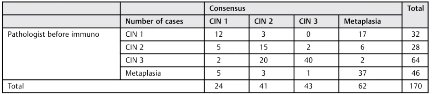
The agreement rate before and after immunohistochem-istry was low. The pathologist diagnoses before and after immunohistochemistry remained the same in 56% of cases. The kappa agreement rate was 0.39 (95% CI 0.29 - 0.49, p<0.01).
The agreement rate between the pathologist and the con-sensus increased from 61% (kappa¼0.47, 95% CI 0.38 - 0.56, p<0.01) to 77% (kappa¼0.68, 95% CI 0.60 - 0.77,p<0.01)
with the aid of immunohistochemistry (►Tables 2and3).
Discussion
Our study confirmed that the reproducibility of traditional pathological examination is not satisfactory for determining the grade of cervical intraepithelial neoplasms. Both the
agreement among three pathologists and the agreement between the original and consensus diagnoses ranged from moderate to fair (kappa¼0.44 and 0.33, respectively). The rate of agreement among each reviewing pathologist and the consensus was moderate (kappa¼0.60) and higher than other comparisons. This result may be explained by the use of the same pathologists for individual and consensus diagnoses.
The largest study of the reproducibility of cervical intra-epithelial neoplasia diagnoses reviewed 6272 cases that were diagnosed by non-specialist pathologists.14Each case was reviewed by one of three teaching gynecopathologists. The agreement rate for that study (0.46) synthesizes the agreement among non-specialists and more experienced pathologists from reference centers in the absence of biases that could affect the results.
Our results confirm the literature data that showed re-producibility rates at the lower limit of moderate agreement. Many studies show that the primary difficulty is the diagnostic reproducibility in intermediate cases; indeed, the agreement rates for CIN 2 cases are the lowest and negatively affect all statistical parameters.3,15
In our study, we found a slightly improved kappa value of 0.33 for the agreement between the original diagnosis and the consensus diagnosis when the cases were divided into two categories rather than into four. When we used CIN 2 as
Table 1 Potential immunohistochemical markers for a CIN 3 diagnosis
Marker
Qualitative p16 (%) Quantitative p16 (%) Quantitative Ki-67 (67%)
Sensitivity 97.4 79.5 53.8
Specificity 60.5 84.4 87.7
Positive predictive value 45.8 63.3 60.0
Negative predictive value 98.6 92.3 84.7
Accuracy 70.0 83.0 79.0
The cases with a high degree of staining (grade 3) were used to calculate the quantitative p16 and Ki-67 data.
the cut-off, the kappa value increased to 0.41, and when we used CIN 3 as the cut-off, the kappa value increased to 0.37. Similarly, the mean kappa value between the pathologist’s and the consensus diagnoses was 0.60 for four categories. The kappa value increased to 0.78 with CIN 2 as the cut-off and 0.67 with CIN 3 as the cut-off.
The immunohistochemical analysis revealed an expected staining pattern. The staining percentage of each CIN grade was comparable with the values found in the literature. However, a simple comparison between values should be performed with caution because the criteria used to define positive cases vary greatly among studies. Some studies use the presence of any staining as a positivity criterion, even if the staining is focal and limited.16Other studies characterize a positive case by continuous staining throughout the epi-thelium.17Genovés et al18and Nishio et al19considered both moderate and diffuse staining as a positive marker. Ki-67
values vary greatly in the literature and exhibit the same methodological issues in the definition of positivity, which hinders the ability to directly compare values.
The analysis of immunohistochemical markers has a certain degree of subjectivity; thus, this method does not provide completely objective observations. The level of agreement among observers differs when a specific degree of p16 staining is defined as positive (positive is defined as a strong and diffuse staining in most studies). Galgano et al5 reported an agreement rate of 0.87 among observers when defining strong and diffuse p16 staining as positive.
Our study shows that p16 and Ki-67 expressions in
cervical intraepithelial neoplasms are more common in high-grade lesions. These immunohistochemical markers
do not exhibit adequate accuracy as independent diag-nostic markers. However, the negative predictive value of p16 was a useful tool for the identification of cases that required more attention. The kappa agreement rate be-tween the pathologist and the consensus increased from 0.47 to a strong agreement value of 0.68. Our study confirms that the level of reproducibility of the conven-tional diagnosis of cervical intraepithelial neoplasms is fair; however, the diagnosis can be improved with the use of immunohistochemistry.
Conflict of Interest
The authors declare no conflict of interest in conducting this study.
Acknowledgments
The authors acknowledge theCoordenação de Aperfeiçoa-mento de Pessoal de Nível Superior- Capes.
Referências
1 -. Brasil. Ministério da Saúde. Instituto Nacional de Câncer José
Alencar Gomes da Silva. Estimativa 2014: incidência de câncer no Brasil. Rio de Janeiro: INCA; 2014
2 Arbyn M, Anttila A, Jordan J, et al. European guidelines for quality
assurance in cervical cancer screening. Ann Oncol 2010;21(3):
448–458
3 Stoler MH, Schiffman M; Atypical Squamous Cells of Undetermined
Significance-Low-grade Squamous Intraepithelial Lesion Triage
Study (ALTS) Group. Interobserver reproducibility of cervical
Table 3 The agreement between pathologists using immunohistochemistry for p16 and Ki-67
Consensus Total
Number of cases CIN 1 CIN 2 CIN 3 Metaplasia
Pathologist after immuno CIN 1 19 0 1 8 28
CIN 2 1 21 3 3 28
CIN 3 0 14 35 3 52
Metaplasia 0 2 0 43 45
Total 20 37 39 57 153
Kappa¼0.68 (95% CI 0.60 - 0.77),p<0.01.
Table 2 The agreement between pathologists without the use of immunohistochemistry
Consensus Total
Number of cases CIN 1 CIN 2 CIN 3 Metaplasia
Pathologist before immuno CIN 1 12 3 0 17 32
CIN 2 5 15 2 6 28
CIN 3 2 20 40 2 64
Metaplasia 5 3 1 37 46
Total 24 41 43 62 170
cytologic and histologic interpretations: realistic estimates from
the ASCUS-LSIL Triage Study. JAMA 2001;285(11):1500–1505
4 Mittal S, Ghosh I, Banerjee D, et al. Reproducibility of cervical
intraepithelial neoplasia diagnosis on histological review of cer-vical punch biopsies from a visual inspection with acetic acid and HPV detection-based screening program. Int J Gynaecol Obstet
2014;126(3):227–231
5 Galgano MT, Castle PE, Atkins KA, Brix WK, Nassau SR, Stoler MH.
Using biomarkers as objective standards in the diagnosis of
cervical biopsies. Am J Surg Pathol 2010;34(8):1077–1087
6 Darragh TM, Colgan TJ, Cox JT, et al; Members of LAST Project
Work Groups. The lower anogenital squamous terminology stan-dardization project for HPV-associated lesions: background and consensus recommendations from the College of American Path-ologists and the American Society for Colposcopy and Cervical
Pathology. Arch Pathol Lab Med 2012;136(10):1266–1297
7 - Dabbs DJ. Diagnostic immunohistochemistry: theranostic and
genomic applications, 2nd ed. Philadelphia: Elsevier; 2006
8 Lesnikova I, Lidang M, Hamilton-Dutoit S, Koch J. p16 as a
diagnostic marker of cervical neoplasia: a tissue microarray study of 796 archival specimens. Diagn Pathol 2009;4:22
9 Cardoso FA, Campaner AB, Silva MA. Prognostic value of p16
(INK4a) as a marker of clinical evolution in patients with cervical intraepithelial neoplasia grade 3 (CIN 3) treated by cervical
conization. APMIS 2014;122(3):192–199
10 Reuschenbach M, Seiz M, von Knebel Doeberitz C, et al. Evaluation
of cervical cone biopsies for coexpression of p16INK4a and Ki-67
in epithelial cells. Int J Cancer 2012;130(2):388–394
11 Carozzi F, Gillio-Tos A, Confortini M, et al; NTCC working group.
Risk of high-grade cervical intraepithelial neoplasia during fol-low-up in HPV-positive women according to baseline p16-INK4A results: a prospective analysis of a nested substudy of the NTCC
randomised controlled trial. Lancet Oncol 2013;14(2):168–176
12 Malpica A, Matisic JP, Niekirk DV, et al. Kappa statistics to measure
interrater and intrarater agreement for 1790 cervical biopsy specimens among twelve pathologists: qualitative histopatholog-ic analysis and methodologhistopatholog-ic issues. Gynecol Oncol 2005;99(3,
Suppl 1):S38–S52
13 Nam EJ, Kim JW, Hong JW, et al. Expression of the p16 and Ki-67 in
relation to the grade of cervical intraepithelial neoplasia and high-risk human papillomavirus infection. J Gynecol Oncol 2008;
19(3):162–168
14 Stoler MH, Ronnett BM, Joste NE, Hunt WC, Cuzick J, Wheeler CM;
New Mexico HPV Pap Registry Steering Committee. The interpre-tive variability of cervical biopsies and its relationship to HPV
status. Am J Surg Pathol 2015;39(6):729–736
15 Cai B, Ronnett BM, Stoler M, et al. Longitudinal evaluation of
interobserver and intraobserver agreement of cervical intraepi-thelial neoplasia diagnosis among an experienced panel of
gyne-cologic pathologists. Am J Surg Pathol 2007;31(12):1854–1860
16 Reuschenbach M, Wentzensen N, Dijkstra MG, von Knebel
Doeberitz M, Arbyn M. p16INK4a immunohistochemistry in cervical biopsy specimens: A systematic review and meta-analysis of the interobserver agreement. Am J Clin Pathol
2014;142(6):767–772
17 Liao GD, Sellors JW, Sun HK, et al. p16INK4A
immunohistochemi-cal staining and predictive value for progression of cerviimmunohistochemi-cal intraepithelial neoplasia grade 1: a prospective study in China.
Int J Cancer 2014;134(7):1715–1724
18 Genovés J, Alameda F, Mancebo G, et al. Human papillomavirus
detection and p16INK4a expression in cervical lesions: a
compar-ative study. Hum Pathol 2014;45(4):826–833
19 Nishio S, Fujii T, Nishio H, et al. p16(INK4a)
immunohistochemis-try is a promising biomarker to predict the outcome of low grade cervical intraepithelial neoplasia: comparison study with HPV