Sensitivity of caloric test and video head impulse as
screening test for chronic vestibular complaints
Raquel Mezzalira,I,II,* Roseli Saraiva Moreira Bittar,IMarcia Maria do Carmo Bile´cki-Stipsky,I,II Cibele Brugnera,ISigne Schuster GraselI
IDepartamento de Otorrinolaringologia, Faculdade de Medicina FMUSP, Universidade de Sao Paulo, Sao Paulo, SP, BR.IIClı´nica de Otorrinolaringologia do Instituto Penido Burnier, Campinas, SP, BR.
OBJECTIVE: This study compared the results of the caloric test with those of the video head impulse test obtained during the same session and evaluated whether the former can be used to screen for non-acute vestibular dysfunction.
METHODS:A total of 157 participants complaining of dizziness with vestibular characteristics of varying dura-tions and clinical courses completed the caloric test and video head impulse test.
RESULTS:Significantly more caloric test results than video head impulse test results were abnormal.
CONCLUSIONS:The results of the caloric test and video head impulse test are distinct but complement each other. Within our sample, the caloric test was more sensitive for vestibular dysfunction. Therefore, the video head impulse test is not a suitable screening tool of the vestibular system in patients with chronic complaints.
KEYWORDS: Caloric Test; Video Head Impulse Test; Vestibular Ocular Reflex; Vertigo.
Mezzalira R, Bittar RS, Bile´cki-Stipsky MM, Brugnera C, Grasel SS. Sensitivity of caloric test and video head impulse as screening test for chronic vestibular complaints. Clinics. 2017;72(8):469-473
Received for publication onJanuary 18, 2017;First review completed onMarch 5, 2017;Accepted for publication onApril 7, 2017 *Corresponding author. E-mail: [email protected]
’ INTRODUCTION
The caloric test (CT) is the most accepted method of eval-uating peripheral vestibular function. However, its limitation is well known: it only stimulates the lateral semicircular canal at low frequencies (1), whereas everyday head movements occur at higher frequencies and along all three planes (2).
The video head impulse test (vHIT) is the computerized version of the head impulse test (HIT) described by Halmagyi and Curthoys in 1988 (3). The HIT uses small-amplitude and high-acceleration movements around the vertical axis while the patient fixates on a stable target.
Gain is the magnitude of the eye movement that results from the head impulse. When the vestibular ocular reflex (VOR) is normal, the patient is able to keep the eyes fixed on the target, and the eye movement shows the same angular velocity in the same plane and opposite direction as the head movement. In this case, the gain is 1. When the compensat-ing ocular movement is insufficient, a central command will launch a corrective saccade to bring the eye back to the target. When the corrective saccade occurs after the head movement has stopped, it is called anovertsaccade. An overt
saccade indicates a VOR alteration due to an abnormality of the stimulated canal. During vestibular compensation over time, this saccade will be substituted by a saccade that occurs during the head movement called a covertsaccade. Covert saccades are likely a part of the central compensation that anticipates the final eye position according to the expected head movement. It has been proposed that cervical proprio-ceptors deliver this information or that the function of the peripheral organ is suppressed during the head impulse (4,5,6). The HIT is most important for cases of acute dizziness and when abnormal, is highly suggestive of a peripheral vestibular lesion. Along with spontaneous nystagmus and skew eye deviation, the HIT has higher sensitivity than magnetic resonance imaging for detecting stroke within the first 48 hours (7). The vHIT is more sensitive to saccades, particularly covert saccades (8), than the HIT, and the VOR measurement and registration are more reliable and can be used during patient follow up. Currently, this test is considered as a fundamental tool for the physical examina-tion of patients with acute dizziness.
Because the vHIT is important for the evaluation of dizziness, the question arises whether vHIT can substitute the bithermal CT for the diagnosis of vestibular disorders. Several studies have compared vHIT and CT results among patients with vestibular complaints. According to the literature, the vHIT cannot serve as a substitute for the CT as a screening tool for patients with vestibular dysfunction because these tests measure different aspects of the VOR (2,9,10).
DOI:10.6061/clinics/2017(08)03
Copyright&2017CLINICS–This is an Open Access article distributed under the terms of the Creative Commons License (http://creativecommons.org/licenses/by/ 4.0/) which permits unrestricted use, distribution, and reproduction in any medium or format, provided the original work is properly cited.
One explanation for the discrepant results between these tests is the anatomic and physiologic background of the VOR. The receptor of the angular VOR is the crista ampullaris, which contains type I and type II cells. This structure receives regular and irregular neural afferent discharges. Type I cells are in the middle of the crista and decode high frequency and fast acceleration head movements. These cells are connected to irregular afferent fibers. Type II cells are found in the peri-phery of the crista and decode low frequency and low accele-ration head movements. They are connected to regular afferent fibers.
Thus, regular vestibular afferent fibers show a relative higher gain at low frequencies, whereas irregular fibers demonstrate higher gain at high frequencies (11). Both vHIT and the CT are used to evaluate unilateral VOR but at dif-ferent frequencies: the vHIT evaluates frequencies above 5 Hz using rapid and short head impulses, whereas caloric irrigation activates lower frequency bands (0.003 Hz) (12).
Nevertheless, the vHIT and CT differ not only in frequen-cies but also in the way the stimulation occurs. During the vHIT, a rapid head impulse generates a physiologic endo-lymphatic flow. In contrast, the caloric stimulus induces an endolymphatic flow via a temperature gradient. The CT stimu-lates the inner ear independent of gravity (13).
The aims of the current study are as follows:
To compare the results of the CT and vHIT obtained during the same session and
To evaluate the viability of the vHIT as a screening tool for non-acute vestibular dysfunction.
’ MATERIALS AND METHODS
This multicenter cross-sectional cohort study was con-ducted in accordance with the standards of the ethical com-mittee (CAPPesq) of the Hospital das Clinicas, University of Sao Paulo Medical School (approved on March 2nd, 2016, number 1433854) and complies with the Helsinki Declaration of 1975.
Study population
This casuistic study included 157 participants (88 women and 69 men) attending the Neurotology Outpatient Clinic of Hospital das Clinicas, University of Sao Paulo Medical School and the Otolaryngologic Clinic Penido Burnier, Cam-pinas, SP. The mean age of the participants was 49±16.7
years. The common clinical complaints included vestibular dizziness of a variable duration and clinical course. All of the patients received standard clinical testing including medi-cal history, otolaryngologic and cranial nerve examinations, static and dynamic balance tests (Romberg and Fukuda), coordination tests (diadochokinesis with alternating prona-tion, forearm supination and index finger–nose tests) and
complete videonystagmography. After patient selection, the CT and vHIT were applied to the sample.
Protocol
We analyzed and compared the CT and vHIT results of patients with dizziness complaints.
Caloric stimulations were performed with water at 30o
C and 44o
C or with air at 24o
C and 50o
C. Responses to caloric stimuli are similar using air at these temperatures (14). Both ears were stimulated separately with a 5-minute interval between tests. We used an Interacoustics VN415 (Denmark) and ICS Chartr 200 Otometrics (Denmark) for these tests. The
Jongkees formula was applied to calculate the unilateral weakness (UW) and gain asymmetry (GA) based on the angular velocity values of the slow phase obtained via the warm and cold stimulations of the right and left ears.
UW¼ðRWþRCÞðLWþLCÞ=RWþRCþLWþLC
GA¼ðRWþLCÞðLWþRCÞ=RWþRCþLWþLC
Jongkees Formula: RW=right warm, RC=right cold, LW=left warm, LC=left cold
UW and GA values above 20% were considered abnormal. The angular velocity values of the slow component were also analyzed. We considered values under 5 degrees/second to represent hypofunction and values above 50 degrees/ second to represent hyperfunction (3,14,15). A morphologic or qualitative analysis of the CT recording was performed.
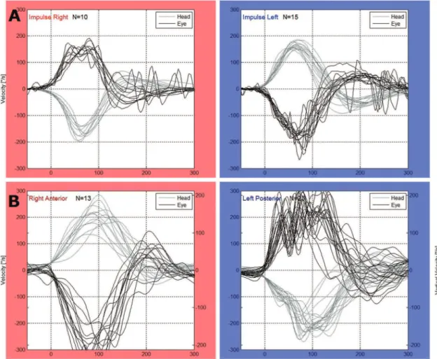
For the vHIT, we used the Eye See Cam vHIT Interacou-stics and ICS Impulse Otometrics. The horizontal VOR was evaluated. Therefore, unpredictable manual head impulses were applied at approximately 20 degrees with a mean velocity of 150 degrees/second and a mean acceleration of 1,000 to 2,500 degrees/second2in the horizontal plane with the patient fixing his or her eyes on a target placed 1 m in front of them. At least 20 adequate impulses were applied to the right and left sides for each test. The test was considered as abnormal when the VOR gain was lower than 0.8 (2,8,16). The presence of overt or covert saccades was also considered (Figure 1).
Statistical analysis
The participants’ mean (standard deviation) age was calculated. Fisher’s exact test was used to analyze the clini-cal data. The results are shown in 22 contingency tables that applied a two-tailed test. Significance was set ata=0.05.
’ RESULTS
Our sample was composed of 157 participants (88 women and 69 men) aged 49±16.7 years.
A total of 113 abnormal CT and 41 abnormal vHIT tests were found among the sample (Table 1). The absolute value of the angular velocity of the slow phase (AVSP), the UW and GA of the CT, and the gain of the vHIT was considered abnormal. The CT identified significantly more abnormal results than the vHIT (Fisher’s exact test, p=0.008). When we compared the results between the tests, the proportion was 3:1. Therefore, we had 3 abnormal CT results for every abnormal vHIT result.
The vHIT revealed abnormal UW values in 19 participants and normal values in 53 normal participants (p=1). A more detailed analysis of the UW values showed that in the UW range between 20 and 40%, eight patients exhibited an abnormal and 33 a normal vHIT test. Regarding UW values
440%, we found that 11 patients showed abnormal results,
and 20 had normal vHIT results (p=0.18). No significant difference was found in either case. No significant difference in vHIT gain was observed among normal and abnormal CT results.
found 24 patients with hypofunction and 16 abnormal vHIT results. Of the 133 patients with normal function, 25 exhi-bited an abnormal vHIT test. The CT was more sensitive than the vHIT (p=0.0001). These data are shown in Table 2.
The comparison between the angular velocity value of the slow phase below 5 degrees (hypofunction) and the correc-tive (overt and covert) saccades was statistically significant (p=0.0001). Conversely, when the angular velocity values of
the slow phase exceeded 50 degrees, they were not related with saccades (p=0.5; Tables 3 and 4).
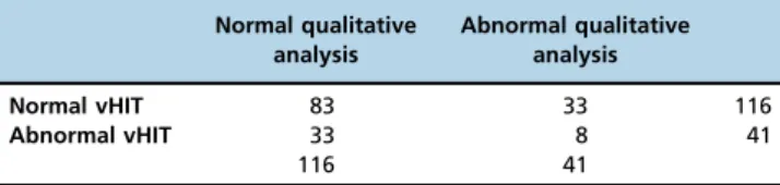
The qualitative analysis of the CT (post caloric rhythm, morphologic nystagmus alterations and fixation index absence) revealed no significant correlation with the vHIT gain (p=0.30) or vHIT saccades (p=0.57).The vHIT did not show abnormalities when the CT demonstrated qualitative alterations. These data are shown in Tables 5 and 6.
Table 2-CT hypofunction compared with abnormal gain in the vHIT.
Presence of caloric hypofunction
Absence of caloric hypofunction
Abnormal vHIT 16 25 41
Normal vHIT 8 108 116
24 133
Table 3-Hypofunction (AVSPo5 degrees) and the presence of
overt and covert saccades.
Overt Covert
AVSP45 10 34 44
AVSPo5 34 20 54
44 54
Figure 1 -vHIT chart. A: normal test, B: velocity decay, low gain and corrective saccades on the left side (see Cam vHIT Interacoustics).
Table 1-CT and vHIT test results.
Normal CT results Abnormal CT results
Normal vHIT 39 77 116
Abnormal vHIT 5 36 41
44 113
Table 4-Hyperfunction (AVSP450 degrees) and the presence of
overt and covert saccades.
Overt Covert
AVSP450 0 2 2
AVSPo50 44 52 96
’ DISCUSSION
Undoubtedly, the vHIT plays an important role during the bedside physical examination of patients with acute dizzi-ness. It is a relevant diagnostic tool for acute vertigo and is currently considered essential in emergency room units in which dizzy patients are treated. Alterations in VOR gain are key to differentiating between stroke and peripheral lesions. The current study focused on elements that can be evaluated in vestibular diseases.
The CT is the most commonly used vestibular function test in clinical practice, and our major objective was to compare these VOR gain results with those measured using the vHIT in patients with vestibular disorders. We questioned whether the vHIT could be used instead of the CT as a screening tool for vestibular dysfunction. If the results of these tests are corre-lated, then the vHIT can be used as a screening tool for patients with balance disorders to identify those who require a CT. The ability of the vHIT to document semicircular canal function over a brief time period would be a clear advantage over the CT.
Although other studies have described a higher prevalence of vHIT gain alterations during the first five symptomatic days (8), we did not consider symptom duration in this study, nor did we describe the vestibular diagnosis of our patients because we believed that our sample was not large enough.
Overall, we found 113 abnormal CT results and 41 abnor-mal vHIT results, indicating 3 abnorabnor-mal CT results for every abnormal vHIT result. Thus, the probability of diagnosing a vestibular dysfunction via CT was 72%, whereas that via vHIT was 26%. In our sample, the CT was ultimately more sensitive for diagnosing vestibular diseases.
When we analyzed the relationship between CT abnor-malities and vHIT gain, we observed that abnormal post caloric responses were significantly more frequent than vHIT gain alterations (o0.0085). Only 5 participants with normal
CT results presented with an abnormal vHIT result, whereas 77 showed abnormal CT results but normal vHIT results. These data are consistent with the findings of Bell et al. (2) who compared both tests and did not find any correlation between them.
Some authors have reported a linear correlation between CT asymmetry and vHIT gain. To observe an abnormal vHIT result, the asymmetry should be near 40% (8,10). When we consider the UW values between 20 and 40% and those
440% compared to the vHIT gain, we did still not obtain a
correlation between the measures (p=0.1779). Therefore, our results suggest that lateral VOR gain does not predict the degree of lateral semicircular canal paresis. Likewise, we did not observe any correlation between UW, regardless of its value, and vHIT gain alteration.
When we analyzed the relationship between CT hypofun-ction and abnormal vHIT gain, we found a significant cor-relation (po0.0001) between low vHIT gain and lack of
function of the lateral semicircular canal during the CT (2). A significant relationship was also found between overt and covert saccades and AVSP values below 5 degrees. In our sample, the likelihood that the vHIT would diagnose vestibular hypofunction was 66%.
However, when we analyzed AVSP values greater than 50% as well as overt and covert saccades, no significant relationship was found. We suggest that the inappropriate standardization of the upper reference limit of the vHIT gain explains this finding (2).
In addition to the quantitative analysis, we performed a qualitative analysis of the CT. The most frequent morpho-logic abnormality was dysrhythmia, showing major varia-tions of amplitude and frequency without AVSP variation. Because the cerebellum controls the nystagmus amplitude (17), these alterations suggest that this structure is involved. The vHIT findings did not indicate the presence of rhythm abnormalities (p=0.1458). This result was not surprising because vHIT gain is related to semicircular canal function, whereas post-caloric rhythm variations reveal the involve-ment of the central nervous system. We did not find refe-rences in the literature regarding this approach; however, these findings suggest that the vHIT is unable to diagnose the morphologic abnormalities observed with the CT.
The differences between the vHIT and the CT are based on the alterations documented by the VOR. Two pathways are involved in VOR activation: fast-and-direct (type I neuron) and slow-and-indirect (type II neuron) pathways. The latter is responsible for storage velocity. Storage velocity describes the characteristic of the vestibular system that maintains the response from the peripheral organ even after hair cell stimulation has ceased. This ability results from the joint activation of the primary and secondary neurons that show different characteristics of activation and depolarization. The direct pathway consists of three neurons and transmits the signal from the semicircular canals directly to the ocular effector muscles without modulation.
The indirect pathway also receives information from the semicircular canals but exhibits an elevated time constant for charge and discharge. In this way, the neurons of the indirect pathway ‘‘store’’the energy acquired by the peri-pheral organ stimulation and continue to discharge even after the stimulation has ceased (17). The caloric stimulus is a perfect example of storage velocity. It produces a nystag-mus response that begins approximately 20 seconds after the beginning of the stimulation, peaks at approximately 40 seconds and decreases until it disappears at approximately 3 minutes (type II neurons). However, the vHIT response depends on the direct pathway of three type I neurons (18). These tests demonstrate different responses to crista ampul-laris stimulation.
As in cochlear disease, the involvement of the semicircular canals can lead to specific frequency dysfunctions: whereas the vHIT represents stimuli at approximately 5 Hz, the CT stimulus is approximately 0.003 Hz. Because these tests
Table 6-Comparison between the qualitative alterations of the CT and vHIT saccades.
Normal qualitative analysis
Abnormal qualitative analysis
Absent saccades 69 27 96
Present saccades 47 14 61
116 41
Table 5-Comparison between the qualitative alterations of the CT and vHIT gain.
Normal qualitative analysis
Abnormal qualitative analysis
Normal vHIT 83 33 116
Abnormal vHIT 33 8 41
evaluate different segments of the semicircular canals, both are necessary for a comprehensive vestibular evaluation (2,8,9,10,11,16,18,19).
The vHIT and CT results are diverse and complement each other. They can be considered tests that describe the tono-topy of the crista ampullaris depending on the stimulation frequency. In our sample, the CT was more sensitive to vesti-bular dysfunction. Therefore, we do not believe that vHIT is an adequate screening tool of the vestibular system for patients with chronic complaints.
’ AUTHOR CONTRIBUTIONS
Mezzalira R and Bittar RS were responsible for the literature review, patient selection, interpretation of the tests and preparation of the manuscript. Bilécki-Stipsky MM was responsible for the literature review and interpretation of the tests. Brugnera C was responsible for the tests executions. Grasel SS was responsible for the statistical analysis and text structuring.
’ REFERENCES
1. Perez N, Rama-Lopez J. Head-impulse and caloric tests in patients with dizziness. Otol Neurotol. 2003;24(6):913–7, http://dx.doi.org/10.1097/
00129492-200311000-00016.
2. Bell LS, Barker F, Heselton H, MacKenzie E, Dewhurst D, Sanderson A. A study of the relationship between the video head impulse test and air calorics. Eur Arch Otorhinolaringol. 2015;272(5):1287-94, http://dx.doi. org/10.1007/s00405-014-3397-4.
3. Halmagyi GM, Curthoys IS. A clinical sign of canal paresis. Arch Neurol. 1988;45(7):737–9, http://dx.doi.org/10.1001/archneur.1988.0052
0310043015.
4. MacDougall HG, Curthoys I. Plasticity during Vestibular Compensation: The Role of Saccades. Front Neurol. 2012;3:21, http://dx.doi.org/10.3389/ fneur.2012.00021.
5. Manzari L, Burgess AM, MacDougall HG, Curthoys IS. Vestibular func-tion after vestibular neuritis. Int J Audiol. 2013;52(10):713–8, http://dx.
doi.org/10.3109/14992027.2013.809485.
6. Tjernstrom F, Nystrom A, Magnusson M. How to uncover the covert saccade during the head impulse test. Otol Neurotol. 2012;33(9):1583–5,
http://dx.doi.org/10.1097/MAO.0b013e318268d32f.
7. Kattah JC, Talkad AV, Wang DZ, Hsieh YH, Newman-Toker DE. HINTS to diagnose stroke in the acute vestibular syndrome: three-step bed-side oculomotor examination more sensitive than early MRI diffusion-weighted imaging. Stroke. 2009;40(11):3504-10, http://dx.doi.org/10.1161/ STROKEAHA.109.551234.
8. Mahringer A, Rambold HA. Caloric test and video-head-impulse: a study of vertigo/dizziness patients in a community hospital. Eur Arch Otorhinolar-yngol. 2014;271(3):463–72, http://dx.doi.org/10.1007/s00405-013-2376-5.
9. Zellhuber S, Mahringer A, Rambold HA. Relation of video-head-impulse test and caloric irrigation: a study on the recovery in unilateral vestibular neuritis. Eur Arch Otorhinolaryngol. 2014;271(9):2375–83, http://dx.doi.
org/10.1007/s00405-013-2723-6.
10. McCaslin DL, Jacobson GP, Bennett ML, Gruenwald JM, Green AP. Pre-dictive Properties of the Video Head Impulse Test: Measures of Caloric Symmetry and Self-Report Dizziness Handicap. Ear Hear. 2014;35(5): e185-91, http://dx.doi.org/10.1097/AUD.0000000000000047.
11. McCaslin DL, Rivas A, Jacobson GP, Bennett ML. The dissociation of Video Head Impulse Test (vHIT) and bithermal caloric test results provide topological localization of vestibular system impairment in patients with
‘‘Definite’’Ménière’s Disease. Am J Audiol. 2015;24(1):1-10, http://dx.doi. org/10.1044/2014_AJA-14-0040.
12. Halmagyi GM, Curthoys IS, Cremer PD, Henderson CJ, Todd MJ, Staples MJ, et al. The human horizontal vestibulo-ocular reflex in response to high-acceleration stimulation before and after unilateral vestibular neur-ectomy. Exp Brain Res. 1990;81(3):479–90, http://dx.doi.org/10.1007/
BF02423496.
13. Scherer H, Clarke AH. The caloric vestibular reaction in space. Physio-logical considerations. Acta Otolaryngol. 1985;100(5–6):328–36, http://dx.
doi.org/10.3109/00016488509126556.
14. Albertino S, Bittar RS, Bottino MA, Gananc¸a MM, Gonc¸alves DU, Greters
ME, et al. Air caloric test references values. Braz J Otorhinolaryngol. 2012;78(3):2, http://dx.doi.org/10.1590/S1808-86942012000300001. 15. Gonc¸alves DU, Felipe L, Lima TM. Interpretac¸ão e utilidade da prova
calórica. Rev Bras Otorrinolaringol. 2008;74(3):440-6, http://dx.doi.org/ 10.1590/S0034-72992008000300020.
16. Rambold HA. Economic management of vertigo/dizziness disease in a county hospital: video-head-impulse test vs. caloric irrigation. Eur Arch Otorhinolaryngol. 2015;272(10):2621–8,
http://dx.doi.org/10.1007/s00405-014-3205-1.
17. Mezzalira R, Bittar RS, Albertino S, Gonc¸alves DU, Gananc¸a FF, Bottino MA,
et al. editors. Otoneurologia clínica. 1st ed. Rio de Janeiro: Revinter; 2014. 18. Halmagyi GM, Aw ST, Cremer PD, Curthoys IS, Todd MJ. Impulsive
Testing of Individual Semicircular Canal Function. Ann N Y Acad Sci. 2001;942:192-200, http://dx.doi.org/10.1111/j.1749-6632.2001.tb03745.x. 19. Alhabid SF, Saliba I. Video head impulse test: a review of the literature.