Correspondence to: Suad Jaganjac, Department of Radiology, Schön Klinik Hamburg Eilbek, Dehnhaide 120, 22 081 Hamburg, Germany. Phone.: 0049 49 209 21 812,.E-mail:[email protected]
O R I G I N A L A R T I C L E UDC: 616.61-006-08
DOI: 10.2298/VSP140502122J
Palliative embolization of renal tumors
Palijativna embolizacija tumora bubrega
Suad Jaganjac, Lennart Schefe
Department of Radiology, Schön Klinik Hamburg Eilbek, Hamburg, Germany
Abstract
Background/Aim. Palliative embolization of renal tumors is the method of choice in the treatment of advanced inoperable renal cell carcinoma in patients with hematuria and pain. Patients with small tumors in the remaining solitary kidney who refuse surgery are suitable for this type of therapy as well as patients with centrally located inoperable tumors. The prerequisite for successful capillary embolization is the closure of the main arte-rial trunk with metal spirals. Methods. In the period from 2000 to 2010 we conducted 42 palliative embolizations. The average age of the patients was 75 years, including 26 men and 16 women. In 8 of the patients the intervention was repeated and in one with severe AV shunts embolization was performed 4 times. Embolization was performed with alcohol, Ivalon 150–250 μm and with metal coils. Results. No serious complications were observed during and after the intervention. Fourteen patient were still alive then and among the deceased patients the average survival time was 13.5 ± 10.8 months with the range of 1 to 56 months. The minimal survival time was 1 month with a maxi-mum survival time of 56 months. Conclusion. Our results are consistent with data in the literature. Survival in patients without metastases was longer than in those with metastases, as con-firmed by the 14 of the patients from the study. An additional therapeutic safety in the treatment of small cancers is provided with a combination therapy of embolization and radiofrequency thermoablation.
Key words:
kidney neoplasms; carcinoma, renal cell; hematuria; palliative care; embolization, therapeutic; radiography, interventional.
Apstrakt
Uvod/Cilj. Palijativna embolizacija tumora bubrega je metoda izbora u lečenju uznapredovalih inoperabilnih karcinoma bub-rega kod bolesnika sa hematurijom i bolovima. Bolesnici sa ma-lim tumorom na preostalom bubregu su pogodni za ovaj vid terapije, ukoliko ti bolesnici odbijaju operativni zahvat ili ukoli-ko tumor leži centralno, taukoli-ko da operativna enukleacija nije moguća. Preduslov za uspešnu embolizaciju je kapilarna embo-lizacija, a glavno arterijsko stablo se zatvori metalnim spiralama.
Metode. U periodu 2000–2010 godine kod 42 bolesnika ura-đena je palijativna embolizacija. Prosečna starost bolesnika iz-nosila je 75 godina. U studiju je bilo uključeno 16 žena i 26 mu-škaraca. Kod 8 bolesnika intervencija je ponovljena, a kod jed-nog, sa izraženim AV-šantovima, embolizacija je urađena četiri puta. Embolizacija je rađena alkoholom, Ivalonom 150–250 μm i metalnim spiralama. Rezultati. U toku intervencije i na-kon intervencije nije bilo teških komplikacija. Ukupno 14 bole-snika još su živi, a kod umrlih prosečno preživljavanje iznosilo je 13,5 ± 10,8 meseci. Minimalno preživljavanje iznosilo je je-dan mesec, a maksimalno 56 meseci. Zaključak. Naši rezultati podudaraju se sa podacima iz literature. Preživljavanje kod bo-lesnika bez metastaza duže je nego kod bobo-lesnika sa metasta-zama, što potvrđuje i 14 bolesnika iz naše studije. Dodatnu si-gurnost u terapiji malih karcinoma pruža kombinovana terapija: embolizacija i radiofrekventna termoablacija.
Ključne reči:
bubreg, neoplazme; hipernefrom; hematurija; lečenje, palijativno; embolizacija, terapijska; radiografija, interventna.
Introduction
Renal cell carcinoma is a rare tumor which accounts for 1.2– 3% of all newly diagnosed cancers. Men are afflicted twice as of-ten as women. It usually occurs between the 5th and the 7th deca-de of life. Renal cell carcinoma is the third most common cancer in the field of urology following bladder cancer and prostate
can-cer. The therapy of choice is nephrectomy or enucleation surgery of smaller tumors if feasible, depending on tumor location.
Percutaneous transarterial embolization of the kidney is a minimally invasive therapeutic procedure by which com-plete or partial radiological nephrectomy can be performed.
changed, but also the diagnosis of renal cell carcinoma was re-volutionized by the introduction of computed tomography (CT) and magnetic resonance imaging (MRI). Palliative embo-lization is the treatment of choice for patients with advanced kidney cancer in whom surgery is technically unfeasible or if they have a high surgical risk. The main indication for the im-plementation of embolization in these patients is hematuria, which can be treated successfully with this method regarding aspects of technique and clinical outcome. Furthermore these patients often have severe pain because of the infiltration of the organ as well as the surrounding anatomical structures, in which case embolization is also an adequate paintherapy. Me-tastases are not a contraindication to implementing this proce-dure regarding the two above mentioned indications.
In asymptomatic patients without distant metastases the complete necrosis of the tumor is achieved by capillary em-bolization so that a portion of these patients are converted from the inoperable to the operabable status.
Another indication for palliative embolization is a small tumor in the remaining solitary kidney in which partial resec-tion of the kidney is not technically possible or in patients who refuse surgery on the remaining kidney.
Patients with advanced metastases and small tumors of the kidney are also indications for embolization. Embolizati-on is performed Embolizati-on the capillary level. For capillary emboli-zation alcohol (96%), Ethibloc and polyvinyl alcohol foam – Ivalon (150–250 μm) are used. Alcohol is the cheapest and most efficient embolization agent. The disadvantage of using alcohol as the only agent to embolize is that it must be appli-ed to a blockappli-ed blood vessel in order to prevent reflux or otherwise must be made visible so as to be able to make sure, that reflux can be seen.
After capillary embolization is completed, the occlusion of the central main stem of the renal artery is performed by using spirals.
Methods
This study included 42 patients in whom palliative em-bolizations of renal tumors were performed in the period from 2000 to 2010. The average age of patients was 75 years, including 26 men and 16 women. Indications for em-bolization were as follows: inoperability demonstrated by CT or MRI, surgery contraindicated due to poor general health status, surgery refusal, tumors in both kidneys, and tumor in the remaining kidney after unilateral nephrectomy.
All the patients were diagnosed by CT scan or MRI. CT examinations were biphasic with a portal venous phase after 65 sec and an urographic phase after 10 min. As contrast agent we used Xenetix 350. The contrast medium was appli-ed by using an injector with a flow rate of 3.5 mL /sec.
MRI was performed with T2 and T1 sequences without contrast agent and after the contrast agent (Dotarem®) had been injected manually in a quantity according to the patients weight (0.1 mg/kg). After administration of the contrast
me-After selective angiography has been performed, the embolization procedure follows. Embolization was done at the first capillary level. The embolic materials used in this study at the capillary level were alcohol or Ivalon® (150–250 μm particles).
For the intervention 96% alcohol was used. In the begin-ning of the study the alcohol embolization was done by balloon occlusion using latex balloons. In the second half of the study 1/3 of lipiodol was added (to alcohol so as to make it visible), as occluding catheters were not availably anymore. Some patients underwent combined embolization with alcohol and Ivalon® – the so-called “sandwich” technique. The advantage of using Ivalon® is that the supplying blood vessel need not to be bloc-ked. Upon completion of capillary embolization, control angiography was performed. If renal tumor is sufficiently em-bolized, central embolization of the major supplying vessel (in-let vessel) should be done using metal coils. In the study, Torna-do spirals provided by Cook Medical were used.
After having embolized the tumor on the capillary and mo-re central level, aortography was performed to confirm the com-plete embolization and to show the state of the surrounding non-targeted blood vessels.
Results
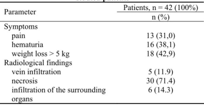
The major symptoms that lead the patients to their physicians are shown in Table 1.
Table 1 The major symptoms and radiological findings in the
studied patients
Patients, n = 42 (100%) Parameter
n (%)
Symptoms
pain 13 (31,0)
hematuria 16 (38,1)
weight loss > 5 kg 18 (42,9) Radiological findings
vein infiltration 5 (11.9)
necrosis 30 (71.4)
infiltration of the surrounding organs
6 (14.3)
One of the patients was diagnosed with bilateral tu-mors, so unilateral nephrectomy was done and superselective embolization was performed on the remaining kidney.
In 7 of the patients after nephrectomy tumor was dis-covered in the remaining kidney. Some of these patients re-fused surgery or the tumor had the central location, making partial nephrectomy impossible.
Angiographic findings of small residual tumor on the right kidney before and after embolization as well as CT scan after a year are shown in Figure 1.
Fig. 1 – Small residual tumor on the right kidney: a) and b) – angiographic findings; c) angiographic finding after embolization; d) computed tomography scan after a year showing scar tissue in the kidney.
Fig. 2 – Metastatic tumor of the left kidney: a) and b) – computed tomography scan of lung showing metastases; c) an-giographic finding before, and d) anan-giographic finding after embolization.
Lymph node metastases were present in 10 patients and 11 patients had distant metastases. Two of the patients had infiltration of the hollow system.
In 8 of the patients embolization was repeated, and in
(73.8%). A total of 27 (64.3%) patients had postinterventional pain. In 15 (35.7%) patients nausea or vomiting was reported.
Figure 3 shows survival in months in patients receiving palliative embolization. The median survival time was 13.5 ± 10.8 months, with the shortest survival being 1 month and the maximal one 56 months. Fourteen patients were still
ali-tion could not be performed. These patients underwent ther-moablation.
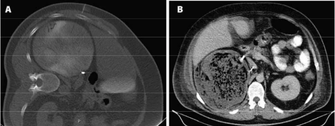
Figure 4 shows CT scan in a patient with a small tumor on the left kidney and negative angiographic findings.
Figure 5 shows renal tumor immediately after embolizati-on and a few days after embolizatiembolizati-on with air development.
Survival in months
Fig. 3 – The survival time in patients receiving palliative embolization of renal tumors.
Fig. 4 – a) Computed tomography scan in a patient with small tumor on the left kidney; b) negative angiographic finding in the same patient.
Discussion
Palliative embolization is a feasible treatment for pati-ents with inoperable kidney cancer, especially in patipati-ents with hematuria or severe pain. These palliative, therapeutic goals are achieved in our patients, which is consistent with the literature 3, 4.
Capillary embolization in the case of inoperability in asymptomatic patients without distant metastases can induce complete tumor necrosis, showing that embolization is equivalent to nephrectomy. In patients who undertook nephrectomy and were diagnosed with the second tumor on the remaining kidney embolization is one of the therapeutic options. According to our experience patients having a solitary kidney (only one remaining kidney) very often refu-se total nephrectomy of. It should be noted that in our hospi-tal surgery is the method of choice if enucleation of the tu-mor is possible. In case of unfavorable location of the tutu-mor, when operational enucleation is impossible, superselective embolization is offered as therapeutic option in which healthy tissue of the remaining kidney is preserved, and pati-ents do not have to undergo hemodialysis.
Advanced inoperable tumors often develop AV shunts which may be disastrous for the patient, as blood flow thro-ugh these shunts is very large and often leads to right heart failure. Intervention in these patients is not simple and often needs to be repeated at least once. The literature on the sur-vival rate of patients undergoing palliative embolization is controversial. According to Hansmann et al. 5 in 9 patients that underwent palliative embolization the survival was 3 years in 3 patients with metastases and 6 years and 4 months in 6 patients without metastases.
In the Onishi et al. 4 study the average survival time of patients in the group that underwent palliative embolization with alcohol was extended to 229 days, compared to the me-an survival time of 116 days in the control group without embolization.
Kauffmann et al. 6 report on the extension of lifespan in 6 patients with metastases by the mean value of 3 years. The embolizations were performed with capillary Ethibloc.
In another study Demirci et al. 7 report that a group of patients with kidney tumors and metastases that was treated with an adjuvant therapy using interferon-alpha and 5-fluorouracil in combination with nephrectomy, had approximately the same survival rate as a group of patients treated only with embolization.
Hallscheidt et al. 8 states that in a group of 7 patients with metastases, there was no significant prolongation of life when treated with palliative tumor embolization. In 6 pati-ents without metastases median survival was 2.3 years. The same study reports that other studies have shown the average survival time of 4 to 11 months after palliative embolization.
In their publication from 2007 Maxwell et al. 9 report the median survival time of 6 months in their group of 19 pa-tients. These authors did not distinguish between patients with or without metastases. This group of patients was very heterogeneous, also including patients with local recurrence after nephrectomy.
Vaicekavicus and Pranculis 10 state that one patient with lung metastases out of the group of 8 patients undergoing palliative embolization lived for 4 years.
Munro et al. 11 report in a retrospective study including 25 patients, that restaging was assessed in a period from 27 to 39 months in 14 patients. The results were as follows: in a patients the tumor size increased, in 7 patients the tumor remained un-changed, and in 5 patients there was a decrease of the tumor size. In patients without metastases there were no cases of newly appearing metastases during the follow-up time. In two patients with metastases a regression of the metastases occurred. All embolizations in this group were performed with absolute alcohol and spirals.
Superselective embolization is performed in cases of small tumors in the remaining kidney (< 4 cm) when surgery is not possible. Additional security for those patients is offe-red by ablative therapeutic methods such as radiofrequency ablation. We prefer superselective embolization if abnormal blood vessels can be found at angiography. Following embo-lization, thermoablation is to be performed after 24 hours under CT supervision. In our opinion this approach is a lot more convenient for the patient and for the interventional ra-diologists. Because renal cell carcinoma is extremely well vascularized tumor, there is a certain risk of bleedings occur-ring when punctuoccur-ring the tumor, which must be treated surgically. We exclude this complication by previous embo-lization. It is interesting to note that in two patients out of our group of patients with small renal tumors the size of 2 cm we did not have positive angiographic findings. In both patients only thermoablation was performed, which passed without complications. One of the above-mentioned two patients had multiple metastases to the spine that were surgically treated, and histopathology confirmed that the metastases originated from a clear-cell renal tumor.
At the beginning of the era of embolization a complica-tion rate of about 4% was described in the literature 12. Du-ring the beginnings of embolization a group of authors in Germany applied alcohol without using occlusion catheter, leading to more serious complications. Löhr and Ross 13 de-scribe death during embolization of tumors of the right kidney with alcohol, probably due to massive pulmonary embolism. In this patient an AV malformation was identifi-ed, so the authors assume a direct crossing of alcohol into the venous circulation, which led to pulmonary embolism. Seve-ral authors 14–16 report bowel infarction as a complication du-ring embolization. Besides, Laarmann et al. 17 and other aut-hors 18 also report gonadal damage during kidney emboliza-tion. Individual descriptions of paraplegia as a result of renal embolization exist in literature 19, 20.
me-the release of oxygen which can not be dissolved in me-the regi-on of the coliquatiregi-on necrosis. At the beginning of the era of embolization the exact cause of air bubbles in embolized tu-mors was unknown. Bacteria that produces gases was assu-med to be responsible for this situation 23. In later works it is stated that the sterile products of tumor cells decomposition are responsible for the development of gas bubbles 24.
It is well known from embolizing other organs that ab-scess formation can occur after embolization. Sometimes it is difficult to discern whether symptoms like temperature and leukocytosis develop due to a postembolization syndrome including sterile gas formation or due to abscess formation. The answer to this question is given by follow-up CT scans proving the considerable colliquation of the abscess close to
lization. Sometimes general condition of patient’s is so bad that drainage must be performed after nephrectomy.
With our embolization technique we had no serious complications in terms of reflux and unwanted embolization into other vascular regions including the venous system.
Conclusion
Our results are consistent with data in the literature. Survival in patients without metastases was longer than in those with metastases, as confirmed by 14 of the patients from the study. An additional therapeutic safety in the treat-ment of small cancers is provided with a combination therapy of embolization and radiofrequency thermoablation.
R E F E R E N C E S
1. Lalli AF, Peterson N, Bookstein JJ. Roentgen-guided infarctions of kidneys and lungs. A potential therapeutic technic. Radiol-ogy. 1969; 93(2): 434–5.
2. Almgård LE, Fernström I, Haverling M, Ljungqvist A. Treatment of renal adenocarcinoma by embolic occlusion of the renal cir-culation. Br J Urol 1973; 45(5): 474−9.
3. Marx FJ, Chaussy C, Moser E. Limitations and hazards of pallia-tive renal tumor embolization. Urologe A 1982; 21(4): 206−10. 4. Onishi T, Oishi Y, Yanada S, Abe K, Hasegawa T, Maeda S. Prog-nostic implications of histological features in patients with chromophobe cell renal carcinoma. BJU Int 2002; 90(6): 529−32.
5. Hansmann HJ, Hallscheidt P, Aretz K, Kauffmann GW, Richter GM.
Renal tumor embolization. Radiologe 1999; 39(9): 783−9. 6. Kauffmann GW, Richter GM, Roeren TK. Renal tumor
emboliza-tion. Radiologe 1992; 32(3): 127−31.
7. Demirci D, Tatlişen A, Ekmekçioğlu O, Ozcan N, Kaya R. Does radical nephrectomy with immunochemotherapy have any su-periority over embolization alone in metastatic renal cell carci-noma? A preliminary report. Urol Int 2004; 73(1): 54−8. 8. Hallscheidt P, Besharati S, Noeldge G, Haferkamp A, Lopez R,
Kauffmann G. Präoperative und palliative Embolisation des Nierenzellkarzinoms: Nachsorge von 49 Patienten. RöFo 2006; 178(4): 391−9.
9. Maxwell NJ, Saleem AN, Rogers E, Kiely D, Sweeney P, Brady AP.
Renal artery embolisation in the palliative treatment of renal carcinoma. Br J Radiol 2007; 80(950): 96−102.
10.Vaicekavicius E, Pranculis A. Transcatheter renal arterial emboli-zation in malignant renal neoplasms: clinical results and indica-tions for use of the method in multi-profile hospitals. Medi-cina (Kaunas) 2002; 38(9): 888−91.
11.Munro NP, Woodhams S, Nawrocki JD, Fletcher MS, Thomas PJ. The role of transarterial embolization in the treatment of renal cell carcinoma. BJU Int 2003; 92(3): 240−4.
12.Kauffmann GW, Rohrbach R, Richter G, Rassweiler J, Sommerkamp H. Kidney tumor embolization. Progress, experiences and complications. Urologe A 1984; 23(2): 109−16.
13.Löhr E, Ross S. Embolisations-Therapie von Nierentumoren-Erfahrungen an einem Krankengut von 60 Patienten. Ra-diologe 1985; 25: 354−8.
14.Cox GG, Lee KR, Price HI, Gunter K, Noble MJ, Mebust WK. Co-lonic infarction following ethanol embolization of renal-cell carcinoma. Radiology 1982; 145(2): 343−5.
15.Teertstra HJ, Winter WA, Frensdorf EL. Ethanol embolization of a renal tumor, complicated by colonic infarction. Diagn Imag-ing Clin Med 1984; 53: 250−4.
16.Sutherland PD, Howard PR, Marshall VR. Colonic infarction fol-lowing ethanol embolisation of the kidney. Br J Urol 1986; 58(2−4): 337.
17.Laarmann S, Straube W, Timmermann J. Toxic alcohol-induced gonadal damage caused by alcohol embolization of kidney tu-mors. Urologe A 1987; 26(2): 94−5.
18.Siniluoto TM, Hellström PA, Päivänsalo MJ, Leinonen AS. Testicu-lar infarction following ethanol embolization of a renal neo-plasm. Cardiovasc Intervent Radiol 1988; 11(3): 162−4. 19.Gang DL, Dole KB, Adelman LS. Spinal cord infarction
follow-ing therapeutic renal artery embolization. JAMA 1977; 237(26): 2841–2.
20.Lang EK, Sullivan J, DeKernion JB. Work in progress: transcathe-ter embolization of renal cell carcinoma with radioactive in-farct particles. Radiology 1983; 147(2): 413−8.
21.Schwartz MJ, Smith EB, Trost DW, Vaughan EDJr.. Renal artery embolization: clinical indications and experience from over 100 cases. Br J Urol 2007; 99(4): 881−6.
22.Cofan F, Real M, Vilardell J, Montanya X, Blasco J, Martin P, et al. Percutaneous renal artery embolisation of non-functioning re-nal allografts with clinical intolerance. Transpl Int 2002; 15(4): 149−55.
23.Basche S. Gas formation following renal tumor embolization. RöFo 1987; 147(4): 459−60.
24.Weckermann D, Schlotmann R, Tietze W, Häckel T. Gas formation after renal artery embolisation: genesis and clinical relevance. Urol Int 1992; 49(4): 211−4.