Critical Role of Bcr1-Dependent
Adhesins in
C. albicans
Biofilm
Formation In Vitro and In Vivo
Clarissa J. Nobile1,2, David R. Andes3, Jeniel E. Nett3, Frank J. Smith, Jr.1, Fu Yue4, Quynh-Trang Phan4, John E. Edwards, Jr.4,5, Scott G. Filler4,5, Aaron P. Mitchell1*
1Department of Microbiology, Columbia University, New York, New York, United States of America,2Biological Sciences Program, Department of Biological Sciences, Columbia University, New York, New York, United States of America,3Department of Medicine, Section of Infectious Diseases, University of Wisconsin, Madison, Wisconsin, United States of America,4Los Angeles Biomedical Research Institute at Harbor-UCLA Medical Center, Torrance, California, United States of America,5The David Geffen School of Medicine, University of California Los Angeles, Los Angeles, California, United States of America
The fungal pathogenCandida albicansis frequently associated with catheter-based infections because of its ability to form resilient biofilms. Prior studies have shown that the transcription factor Bcr1 governs biofilm formation in an in vitro catheter model. However, the mechanistic role of the Bcr1 pathway and its relationship to biofilm formation in vivo are unknown. Our studies of biofilm formation in vitro indicate that the surface protein Als3, a known adhesin, is a key target under Bcr1 control. We show that an als3/als3 mutant is biofilm-defective in vitro, and that ALS3
overexpression rescues the biofilm defect of thebcr1/bcr1mutant. We extend these findings with an in vivo venous catheter model. Thebcr1/bcr1mutant is unable to populate the catheter surface, though its virulence suggests that it has no growth defect in vivo.ALS3overexpression rescues thebcr1/bcr1biofilm defect in vivo, thus arguing that Als3 is a pivotal Bcr1 target in this setting. Surprisingly, theals3/als3mutant forms a biofilm in vivo, and we suggest that additional Bcr1 targets compensate for the Als3 defect in vivo. Indeed, overexpression of Bcr1 targetsALS1, ECE1,and
HWP1 partially restores biofilm formation in a bcr1/bcr1 mutant background in vitro, though these genes are not required for biofilm formation in vitro. Our findings demonstrate that the Bcr1 pathway functions in vivo to promote biofilm formation, and that Als3-mediated adherence is a fundamental property under Bcr1 control. Known adhesins Als1 and Hwp1 also contribute to biofilm formation, as does the novel protein Ece1.
Citation: Nobile CJ, Andes DR, Nett JE, Smith FJ Jr, Yue F, et al. (2006) Critical role of Bcr1-dependent adhesins inC. albicansbiofilm formation in vitro and in vivo. PLoS Pathog 2(7): e63. DOI: 10.1371/journal.ppat.0020063
Introduction
Biofilms are microbial communities that are associated with solid surfaces. Most bacteria and fungi exist predom-inantly in such communities in nature, and they form the basis for numerous interactions that affect human health. Cells in a biofilm display phenotypes that are distinct from their free-living counterparts, including extreme resistance to many antimicrobial agents [1–4]. Their health impact is reflected in the fact that implanted medical devices, such as intravascular catheters, are major risk factors for blood-stream and deep tissue infection [5, 6]. These devices serve as substrates for biofilm development; the mass and intrinsic drug resistance of the biofilm limits efficacy of host defenses and antimicrobial therapy. These biofilm-based infections are estimated to cause about 50% of all nosocomial infections [5, 7].
The fungal pathogen Candida albicans is a major cause of device-associated infections [5, 8, 9]. It produces adherent biofilms on a variety of different surfaces in vitro [3, 4, 10, 11]. Biofilm formation begins with surface adherence of yeast-form cells, which grow to yield a basal layer. Basal layer cells include some hyphae, or long tubular chains of cells, which extend to yield an upper layer that is almost exclusively hyphae. As the biofilm matures, it produces an extracellular matrix containing predominantly carbohydrate and protein [1, 12, 13].
C. albicansBcr1, a C2H2zinc finger protein, has a significant role in biofilm formation: bcr1/bcr1 insertion and deletion mutants form only rudimentary biofilms on silicone catheter material in vitro [14]. Bcr1 is required for expression of several cell wall protein genes, and we have proposed that Bcr1 is a positive regulator of adherence. Many Bcr1 target genes had been identified initially as hyphal-specific genes, and BCR1 RNA accumulation depends upon the hyphal developmental activator Tec1 [14]. Bcr1 is not required for hyphal morphogenesis, and we believe that it acts down-stream of Tec1 to activate the acquisition of hyphal adherence properties.
Biofilms are considerably more complex in vivo than in vitro. For example, in vivo, biofilms form on intravascular catheters under conditions of vascular flow, and are exposed to and incorporate many plasma constituents. The
complex-Editor:Alexander Johnson, University of California San Francisco, United States of America
ReceivedJanuary 3, 2006;AcceptedMay 12, 2006;PublishedJuly 7, 2006
DOI:10.1371/journal.ppat.0020063
Copyright:Ó2006 Nobile et al. This is an open-access article distributed under the terms of the Creative Commons Attribution License, which permits unrestricted use, distribution, and reproduction in any medium, provided the original author and source are credited.
Abbreviations:CSLM, confocal scanning laser microscopy
ity involved in forming a biofilm in vivo underscores the question of whether the same mechanisms are required for biofilm formation in vitro as in vivo. Indeed, several fungal and bacterial mutants have medium-dependent biofilm defects in vitro [15, 16]. Thus, the functions of key regulators must be appraised in vivo in order to connect questions in developmental biology to answers in antimicrobial therapy.
Recently developed animal models permit analysis of C. albicans biofilm formation in vivo. Central venous catheter infection models have been described for both rabbits [17] and rats [18]. These catheter surfaces are substrates for extensive biofilm formation, and biofilm cells on these substrates exhibit reduced antifungal susceptibility. These models further reflect the circumstances of human infection, in that the biofilm cells can lead to seeding and infection of organs [18].
In this report, we test the roles of Bcr1 target genes in biofilm formation in vitro. Our findings substantiate the proposal that Bcr1 is a regulator of adherence. We extend this analysis to an in vivo model, where our findings argue that adherence is a fundamental property under Bcr1 control that promotes biofilm formation and that the adhesin Als3 is a pivotal functional target of Bcr1 both in vitro and in vivo. Our findings highlight the complexity of in vivo biofilm formation, yet reveal a convergence of in vitro and in vivo studies to define a significant biofilm regulatory mechanism.
Results
Bcr1 Promotes Adherence In Vitro
We proposed that Bcr1 acts in the hyphal development pathway to promote adherence through stimulation of expression of several cell surface protein genes. This hypothesis predicts that overexpression of BCR1 in yeast form cells may stimulate adherence. We tested this prediction by examining the effects of BCR1 expression in a tec1/tec1 mutant (Figures 1 and 2), which is defective in producing hyphae in vitro [19]. The tec1/tec1 mutant is defective in biofilm formation [14], and the mutant cells fail to adhere to silicone catheter material (Figures 1A, 1C, and 2, Strain Set A). Introduction of a TEF1-BCR1 overexpression construct restored biofilm formation ability partially to the tec1/tec1 mutant (Figures 1F and 2, Strain Set A; p¼0.018 for the comparison of biomass determinations). Overexpression of
BCR1 in the tec1/tec1 mutant does not restore hyphal formation ability (Figure 1G–1I). We believe that the partial suppression by TEF1-BCR1 reflects the failure to restore hyphal formation to thetec1/tec1 mutant. In any case, these findings support the idea that Bcr1 is a positive regulator of adherence, but not of hyphal formation.
To understand the mechanism of Bcr1-promoted adher-ence, we compared expression of Bcr1-dependent genes in strains with or without the TEF1-BCR1construct (Figure 3). The genesHYR1, HWP1, CHT2, ECE1, RBT5, ALS1,andALS3 were expressed at much lower levels in abcr1/bcr1mutant than in the wild-type reference strain (Figure 3, samples 3 and 7). The presence of the TEF1-BCR1 construct restored expres-sion of these genes and biofilm formation in the bcr1/bcr1 mutant, thus verifying the function of the construct (Figure 2; Figure 3, samples 2 and 7). The surface protein geneECM331 was expressed at higher levels in thebcr1/bcr1mutant than in the wild-type reference strain, and this elevated expression was also reversed by the TEF1-BCR1 construct (Figure 3, samples 2, 3, and 7). We note that TEF1-BCR1 did not substantially increase expression of Bcr1-dependent genes in the otherwise wild-type reference strain background (Figure 3, samples 5 and 3). Among the Bcr1-dependent genes, we found that HYR1, HWP1, CHT2, ECE1, and ALS3 were expressed at reduced levels in the tec1/tec1 strain compared to the reference strain (Figure 3, samples 3 and 6). Introduction of theTEF1-BCR1 construct increased expres-sion of these genes in thetec1/tec1strain (Figure 3, samples 4 and 6). These findings suggest that Bcr1 acts downstream of Tec1 to activate expression of target genes HYR1, HWP1, CHT2, ECE1,and ALS3. Furthermore, the augmented adher-ence during biofilm formation of the tec1/tec1 TEF1-BCR1 strain highlights this particular group of Bcr1-dependent genes as candidates for mediators of Bcr1-dependent adherence.
Key Role of Bcr1 Target GeneALS3in Biofilm Formation In Vitro
To test the roles of Bcr1 target genes in biofilm formation, we carried out biofilm formation assays with mutants defective in each gene. We observed no significant biofilm defect inhyr1/hyr1(p¼0.463), ece1/ece1(p¼0.850), cht2/cht2(p¼
0.909), or rbt5/rbt5 (p ¼ 0.323) mutant strains versus the reference strain (Figure 2, Strain Set B; Figure 4). Theals1/als1 and hwp1/hwp1 mutants also produced substantial biofilms (Figure 4), although the biofilms often sloughed off the substrate. Biofilm biomass determinations further indicated that the hwp1/hwp1 mutant has a partial biofilm defect compared to the reference strain (Figure 2, Strain Set B, p
¼0.022). In contrast to theals1/als1andhwp1/hwp1strains, the als3/als3 mutant displayed a severe defect in biofilm for-mation compared to the reference strain (Figure 2, Strain Set B; Figure 4;p¼0.005), and introduction of a single wild-type ALS3allele rescued the defect substantially (Figure 2, Strain Set B;p¼0.005). Confocal scanning laser microscopy (CSLM) imaging revealed that the als3/als3 mutant formed a rudi-mentary biofilm of 20lm in depth, while the wild-type and als3/als3þpALS3complemented strains produced biofilms of over 200lm in depth (Figure 5). CSLM depth images showed that the rudimentaryals3/als3mutant biofilm was comprised mainly of yeast cells, with few hyphae, whereas the biofilms of the wild-type and als3/als3 þpALS3 complemented strains CriticalC. albicansBiofilm Adhesins
Synopsis
The formation of biofilms (surface-attached microbial communities) on implanted medical devices such as catheters is a major cause of fungal and bacterial infections. Prior studies of the fungal pathogen
included abundant hyphae (Figure 5). It should be noted that theals3/als3mutant is not defective in hyphal formation as it forms normal hyphae when assayed under hyphal inducing conditions (Figure 5). Hyphae are also apparent among the cells in the surrounding medium of an als3/als3 mutant biofilm (unpublished data). These findings argue that Als3 has a major role in biofilm formation and suggest that reduced expression ofALS3in thebcr1/bcr1mutant may account for its biofilm defect.
If reduced expression ofALS3is the cause of thebcr1/bcr1 mutant biofilm defect, then increased expression ofALS3in a bcr1/bcr1 mutant background should promote biofilm for-mation. To test this prediction, we introduced the TEF1 promoter adjacent to the nativeALS3coding region to create a TEF1-ALS3 allele, permitting Bcr1-independent ALS3 expression. RT-PCR measurement of ALS3 RNA levels confirmed that the TEF1-ALS3 allele permits expression of ALS3in bothBCR1/BCR1 andbcr1/bcr1backgrounds (Figure 6). In the wild-type reference strain background,TEF1-ALS3 had no obvious effect on biofilm formation (Figure 6, top row). In the bcr1/bcr1 mutant background, TEF1-ALS3 im-proved biofilm formation substantially (Figure 2; Figure 6, top row; p ¼ 0.002). These observations indicate that
increasedALS3expression in thebcr1/bcr1mutant promotes significant biofilm formation ability.
We used CSLM imaging to examine the structure of biofilms that resulted from increased ALS3 expression. The TEF1-ALS3 allele did not alter biofilm structure in the otherwise wild-type background (Figure 6, CSLM depth and side views): biofilm depth was about 400 lm; little staining occurred in the basal region; and hyphal staining was prominent. Thebcr1/bcr1strain produced a thin rudimentary biofilm comprised largely of yeast form cells, as expected [14]. Thebcr1/bcr1 TEF1-ALS3strain produced a substantial biofilm that included a basal poorly stained region (Figure 6), similar in appearance to those of the wild-type strain (Figure 6) and complemented bcr1/bcr1 mutant [14]. Thus, increased ALS3 expression permits at least partial rescue of the bcr1/bcr1 mutant defect in biofilm formation.
Bcr1 Function in Biofilm Formation In Vivo
In order to determine whether Bcr1 may have a role in biofilm formation in vivo, we turned to a rat venous catheter model [18]. Implanted catheters were allowed to stabilize for 24 h and were then inoculated with wild-type, bcr1/bcr1 mutant, orbcr1/bcr1þpBCR1complemented strains. Biofilm formation was visualized after 12, 24, and 48 h by scanning Figure 1.Effect of IncreasedBCR1Expression on Adherence and Hyphal Morphogenesis
Strains were grown under in vitro biofilm assay conditions for 60 h and photographed (A–F) or grown in Spider suspension cultures and examined by phase contrast microscopy at3400 magnification (G–I). For the biofilms assays, turbid medium with all cells free-floating in the medium rather than attached to the silicone substrate indicates a biofilm-negative phenotype; clear medium with the silicone substrate completely covered with cells indicates a biofilm-positive phenotype. Relevant genotypes are given above each panel for strains CJN1015 (reference strainþTEF1) (A, G), CJN1060 (bcr1/bcr1þTEF1) (B), CJN1052 (tec1/tec1þTEF1) (C, H), CJN1039 (reference strainþTEF1-BCR1) (D), CJN1011 (bcr1/bcr1þTEF1-BCR1) (E), and CJN1035 (tec1/tec1þTEF1-BCR1) (F, I).
electron microscopy of the intraluminal catheter surface (Figure 7). The wild-type and bcr1/bcr1 þ pBCR1 comple-mented strains initiated biofilm formation by 12 h and yielded extensive adherent populations by 24 h (Figure 7A, 7B, 7G, and 7H). Both strains produced mature biofilms by 48 h that included abundant matrix material (Figure 7C and 7I), as previously reported for strain K1 [18]. In contrast, thebcr1/ bcr1mutant yielded few adherent cells at 12 and 24 h (Figure 7D and 7E), and the catheter surface was devoid of biofilm material after 48 h (Figure 7F). Despite the dramatic differ-ences in biofilm formation ability, the three strains grew comparably in a mouse disseminated infection model; median mouse survival time was 13 d after inoculation with the wild-type strain and 10 d after inoculation with either the bcr1/bcr1mutant orbcr1/bcr1þpBCR1complemented strains. Based on this evidence, Bcr1 is not required for growth in vivo under non–biofilm-forming conditions but is required for biofilm formation in vivo.
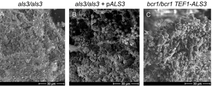
Als3 Function in Biofilm Formation In Vivo
Our observations above indicate that Als3 is a key mediator of Bcr1-dependent biofilm formation in vitro. To verify that these findings extend to in vivo biofilm formation, we compared als3/als3 mutant and als3/als3 þ pALS3 comple-mented strains in the rat venous catheter model. Both strains formed extensive biofilms within 24 h (Figure 8A and 8B). Therefore, Als3 is not absolutely required for biofilm formation in vivo.
To determine whether Als3 may contribute to biofilm formation in vivo, we tested the ability of the TEF1-ALS3 expression construct to rescue thebcr1/bcr1 mutant biofilm defect. Thebcr1/bcr1 TEF1-ALS3strain produced an extensive
biofilm containing both cells and matrix material (Figure 8C). This biofilm, formed after 24 h, was similar in overall appearance to that formed by theBCR1/BCR1control strains (Figures 7B and 8B). TEF1-ALS3 expression thus rescues biofilm formation in abcr1/bcr1background (compare Figures 7E and 8C). These findings support the model thatALS3is a critical Bcr1 target gene that functions in biofilm formation in vivo.
Overexpression Assays of Bcr1 Target Gene Function In Vitro
Our in vivo assays suggest that Als3 may be one of several Bcr1 targets that contribute to biofilm formation. The analysis of insertion and deletion mutant strains above pointed toward Als1 and Hwp1 as additional candidate functional targets, although their biofilm defects were mild: biofilm biomass was reduced only slightly (Figure 2), and CSLM visualization revealed no qualitative defects (unpub-lished data). Thus, we turned to an alternative functional analysis strategy, gene overexpression, which has recently been applied with considerable success on a genome-wide scale in Saccharomyces cerevisiae [20]. Gene overexpression is particularly useful in identifying functions among partially redundant genes, the situation that we postulate to exist here. To determine if increased expression of Bcr1-activated target genes, other thanALS3, may rescue the biofilm defect of the bcr1/bcr1 mutant, we created genomic fusions of the TEF1promoter to theCHT2,HYR1,RBT5,ALS1,HWP1, and ECE1coding regions. TheTEF1-ALS1,TEF1-HWP1, and TEF1-ECE1alleles improved biofilm formation ability considerably (p,0.004 for all comparisons tobcr1/bcr1), although not to the extent ofTEF1-ALS3(Figure 2, Strain Set C;p,0.006 for Figure 2.Biofilm Dry Mass Determinations
Biofilm dry mass determinations were made in quadruplicate after 60 h growth under standard biofilm conditions, as detailed in Materials and Methods. Reference strains DAY185 (shown) and CAI4-URA3 (not shown) gave similar results. Strains are grouped for convenience of comparison. Strain Set A contains CJN896 (tec1/tec1), CJN1052 (tec1/tec1þTEF1), CJN1035 (tec1/tec1þTEF1-BCR1), CJN1023 (tec1/tec1þpTEC1), CJN702 (bcr1/bcr1), CJN1060 (bcr1/bcr1þTEF1), CJN1011 (bcr1/bcr1þTEF1-BCR1), CJN698 (bcr1/bcr1þpBCR1), respectively. Strain Set B contains FJS2 (hyr1/hyr1), FJS6 (ece1/ece1), FJS5 (cht2/cht2), FJS8 (rbt5/rbt5), CAYC2YF1U (als1/als1), CAH7-1A1E2 (hwp1/hwp1), CAYF178U (als3/als3), CAQTP178U (als3/als3þpALS3), respectively. Strain Set C contains CJN1153 (bcr1/bcr1þTEF1-ALS3), CJN1144 (bcr1/bcr1þTEF1-ALS1), CJN1288 (bcr1/bcr1þTEF1-ECE1), CJN1222 (bcr1/bcr1þ TEF1-HWP1), CJN1281 (bcr1/bcr1þTEF1-CHT2), CJN1259 (bcr1/bcr1þTEF1-HYR1), CJN1276 (bcr1/bcr1þTEF1-RBT5), respectively.
DOI: 10.1371/journal.ppat.0020063.g002
all comparisons to bcr1/bcr1 TEF1-ALS3). These same TEF1 promoter fusion alleles did not augment biofilm formation in theBCR1/BCR1background (unpublished data). These results indicate that Als1, Hwp1, and Ece1 may act in addition to Als3 to contribute to biofilm formation.
Discussion
We have recently taken a genetic approach to elucidate the mechanistic basis ofC. albicansbiofilm formation [14, 15]. A central issue is how in vitro biofilm models are related to biofilm growth in vivo and, thus, to disease. Here we have shown that the transcription factor Bcr1 is required in vivo, as it is in vitro, for biofilm formation. One key target gene under Bcr1 control isALS3, as demonstrated by the rescue of biofilm formation through increasedALS3expression in vitro and in vivo. These results argue that Als3-mediated adher-ence is a key factor in formation of biofilms in vitro and in vivo. However, absence of Als3 causes a biofilm defect only in vitro and not in vivo. One implication from this result is that Bcr1 activates additional biofilm adhesin genes. In support of this model, we find that overexpression of three additional Bcr1 target genes partially restores biofilm formation ability in vitro to abcr1/bcr1mutant. Our findings are summarized in
Figure 9. Clearly, the interplay of in vitro and in vivo analyses holds great promise for defining biofilm regulatory mecha-nisms.
Relationship of Bcr1 and Hyphal Gene Expression Our studies here solidify the concept that Bcr1 relays a signal within the hyphal developmental program because an increase inBCR1expression leads to increased expression of the hyphal-specific genesHYR1, HWP1,andALS3in a hyphal-defectivetec1/tec1mutant. However, we find that some Bcr1-dependent genes are expressed substantially in the tec1/tec1 strain, includingRBT5, ECE1,andALS1, despite the reduced expression ofBCR1.Two simple explanations can account for this apparent paradox. One possibility is that the 4-fold reduced level of Bcr1 in the tec1/tec1mutant is sufficient to activate a subset of target genes. These genes may have the highest-affinity Bcr1 binding sites, or their promoter regions may include binding sites for additional transcription factors that interact cooperatively with Bcr1. A second possibility is that some Bcr1 target genes are subject to a compensatory regulatory mechanism in thetec1/tec1background. The latter explanation seems particularly plausible for RBT5, which responds to numerous genetic and environmental regulatory Figure 3.Effect of IncreasedBCR1Expression on Target Gene RNA Levels
RNA prepared from mid-log phase Spider medium cultures was used to prepare Northern blots or in RT-PCR assays, as indicated. Northern blots were probed for the transcripts indicated along the left side, and PhosphorImager exposures are shown. RT-PCR assays forALS1, ALS3,andTEF1were conducted on serial 2-fold dilutions of cDNA preparations and fractionated on agarose gels; only the last two dilutions are shown.TEF1transcript levels were used as an expression control. Strains included DAY185 (reference strain) (sample 1), CJN1011 (bcr1/bcr1þTEF1-BCR1) (sample 2), CJN1015 (reference strainþTEF1) (sample 3), CJN1035 (tec1/tec1þTEF1-BCR1) (sample 4), CJN1039 (reference strainþTEF1-BCR1) (sample 5), CJN1052 (tec1/tec1þ
signals [21–23]. Identification of the Bcr1 binding site will help to distinguish between these explanations.
One unexpected observation is that CHT2 expression is both Bcr1 and Tec1 dependent.CHT2,which specifies a cell wall chitinase homolog, is expressed at higher levels in yeast cells than hyphal cells under many growth conditions [24, 25]. However,CHT2is not exclusively a yeast phase-specific gene. For example, it was found to be coregulated with numerous hyphal-specific genes in a study of pH-regulated gene expression [26], and theCHT2transcript has been detected previously in cells induced to form hyphae in Spider medium or serum [21]. Our results indicate that Bcr1 and Tec1 target genes are not restricted to hyphal-specific genes.
Control of Adherence by Bcr1 during Biofilm Formation In Vitro
The proposal that Bcr1 is a positive regulator of biofilm adherence stems from two prior observations. First, Bcr1 is required for biofilm formation, a process that depends upon both cell-cell and cell-substrate adherence. Second, numerous Bcr1-dependent genes encode proteins that contribute to cell wall or cell surface structure. The in vitro studies reported
here include three lines of evidence in support of this proposal. First, expression of Bcr1 in a tec1/tec1 mutant promotes substantial adherence to a silicone substrate. Second, a deletion of one Bcr1-dependent adhesin gene, ALS3,causes a biofilm formation defect similar to that of the bcr1/bcr1mutant. Third, thebcr1/bcr1biofilm formation defect is fully rescued through increased expression of Als3 and partially rescued through increased expression of two other known adhesins, Als1 and Hwp1. Our results thus indicate that the adhesin expression defect is a major cause of thebcr1/ bcr1mutant biofilm formation defect.
The tec1/tec1mutant has a severe biofilm defect: it grows under our in vitro cultivation conditions as a suspension of yeast cells. Introduction of TEF1-BCR1 alters that mutant phenotype by promoting growth primarily on the surface of the silicone substrate. The biofilm so formed is unstable in that it disperses into clumps of cells during manipulation, and its biomass is 3-fold less than that of the wild-type and complemented mutant strains. Thus, expression of Bcr1 is not sufficient to promote extensive biofilm formation by yeast cells. However, increased adherence of the tec1/tec1 Figure 4.Biofilm Formation In Vitro by Bcr1 Target Gene Mutants
Strains were grown in our standard biofilm assay and photographed after 60 h. Relevant genotypes are given above each panel and include DAY286 (reference strain), CJN459 (bcr1/bcr1), FJS2 (hyr1/hyr1), CAH7-1A1E2 (hwp1/hwp1), FJS5 (cht2/cht2), FJS6 (ece1/ece1), FJS8 (rbt5/rbt5), FJS10 (ecm331/
ecm331), CAYF178U (als3/als3), and CAYC2YF1U (als1/als1). Turbid medium with all cells free-floating in the medium rather than attached to the silicone substrate indicates a biofilm-negative phenotype; clear medium with the silicone substrate completely covered with cells indicates a biofilm-positive phenotype. An uninoculated control is shown in the panel labeled‘‘Blank.’’
DOI: 10.1371/journal.ppat.0020063.g004
TEF1-BCR1 strain, compared to atec1/tec1 strain, is readily apparent, thus connecting Bcr1 function to adherence.
The finding that the Bcr1-dependent adhesin Als3 is required for biofilm formation strengthens this connection. Als3 belongs to a large C. albicans protein family with structural features similar to those of theamating agglutinin ofS. cerevisiae[27, 28]. Direct assays have demonstrated roles for C. albicans Als1, Als3, Als5, and Als6 in adherence to diverse substrates [29], and mutational analysis indicates that Als2 and Als4 are also adhesins [30]. The two Bcr1-dependent family members, Als1 and Als3, have highly related sequences throughout their N-terminal domains, the region implicated in substrate binding. This close relationship is reflected by their similar substrate binding properties [29]. Our studies here also support a close functional relationship between Als1 and Als3, because overexpression of either adhesin in a bcr1/bcr1 background restores biofilm formation to a
meas-urable extent. There are some functional distinctions between Als1 and Als3, because Als3 is required for biofilm formation under our in vitro assay conditions, while Als1 is not. Similarly,TEF1-ALS3is more efficient thanTEF1-ALS1in suppression of the bcr1/bcr1biofilm defect. Nonetheless, the clear connection between Bcr1, Als1, and Als3 argues that a major functional role of Bcr1 is to promote adherence.
One unexpected conclusion from our findings is that Hwp1 contributes to biofilm formation in vitro. Hwp1 is a well-characterized hyphal adhesin that serves as a substrate for mammalian transglutaminases, thus mediating covalent at-tachment of C. albicans to host cells [28, 31]. It has not previously been shown to mediate interactions between C. albicanscells, and the transglutaminases that modify Hwp1 are of mammalian origin [31]. Our observations suggest that Hwp1 can mediateC. albicanscell-cell interactions, and that it does so in the absence of transglutaminase activity. An interesting implication is that Hwp1 may contribute to adherence between mating partners, thus explaining its up-regulation by mating factor [32, 33].
Our findings also implicate Ece1 in adhesion, thus providing the first functional insight into this protein.ECE1 was discovered as a hyphal-induced gene and was among the firstC. albicansgenes disrupted with the Ura-blaster method [34, 35]. However, the ece1/ece1 mutant has no apparent phenotypic defect [35]. The idea that Ece1 functions in adhesion is suggested by our observation that its over-expression restores biofilm formation to a bcr1/bcr1mutant. ECE1, like HWP1, is induced by mating pheromone [33], another possible connection between Ece1 and adherence. Ece1 does not resemble an adhesin: it is comprised of novel 34-residue repeats that surround a possible transmembrane domain [35]. Although its mechanism of action is uncertain, an interesting possibility is that Ece1 promotes surface exposure of adhesins.
Genetic Control of Biofilm Formation In Vivo
Biofilm formation in vivo is considerably more complex than in vitro and involves dynamic interactions with many host proteins, cells, and environmental factors. These differ-ences raise the question of whether the major genetic factors operative in vitro play a commensurate role in vivo. We have addressed this issue for two gene products: Bcr1 and Als3. Although the experimental outcomes were different in detail, they argue that both proteins have significant roles in vivo.
The significance of Bcr1 is clearest: it is required in vivo for biofilm formation but not for growth. The fact that the mutant leaves catheter surfaces essentially clear of material suggests that there is a defect in early events of biofilm formation in vivo, much as observed in vitro. The defects under the two circumstances, however, are slightly different: a thin layer of bcr1/bcr1 mutant cells is associated with the substrate transiently in vivo but stably in vitro. It is possible that the few substrate-bound cells that appear early in vivo may be destroyed later by host defenses. An alternative possibility is that larger cell masses are dislodged efficiently by blood flow if their adherence is compromised by thebcr1 defect. In either case, it is clear that Bcr1 governs a mechanism that contributes to biofilm formation in vivo.
The potency ofTEF1-ALS3as a suppressor of thebcr1/bcr1 defect argues that Als3 also has a critical role in biofilm formation in vivo. How can that observation be reconciled Figure 5.In Vitro Filamentation and Biofilm Formation by theals3/als3
Mutant
Cells were grown in free-living (planktonic) cultures in Spider medium; filamentation was examined by phase contrast microscopy at3400 magnification (top panels). Biofilms were grown under standard conditions in Spider medium, and stained with concanavalin A conjugate for CSLM visualization. Artificially colored CSLM depth views, in which blue color represents cells closest to the silicone and red color represents cells farthest from the silicone, are shown in middle panels. For the depth views of reference strain CAI4-URA3 (ALS3/ALS3), blue¼0lm and red¼ 800lm; CAYF178U (als3/als3), blue¼0lm, red¼80lm; CAQTP178U (als3/als3þpALS3), blue¼0lm, red¼600 lm. CSLM side views are shown in lower panels. For the side views, the scale bars represent 50lm for CAI4-URA3 (ALS3/ALS3) and CAQTP178U (als3/als3þpALS3); and 20 lm for CAYF178U (als3/als3).
with the fact that an als3/als3 null mutant has no biofilm defect in vivo? One simple model is that additional adhesins can partially compensate for the absence of Als3 in vivo but not in vitro. Our overexpression studies implicate Als1 and Hwp1 as candidate compensatory adhesins, in keeping with this model. The distinction between the in vivo and in vitro situations may reflect a higher level of expression of the compensatory adhesins in vivo than in vitro. A second possibility is that host constituents, for example serum components, may contribute to adherence. Thus, the same low level of surface adhesin activity may support biofilm formation in vivo but not in vitro.
The restoration of biofilm formation through ALS3 over-expression in thebcr1/bcr1mutant both in vitro and in vivo indicates that Bcr1 governs one main function relevant for biofilm formation: adherence. Although transcription factor mutants are useful for definition of functionally related genes, the mechanistic basis for their phenotypic defects can be complex because of the extent of their gene expression defects. Moreover, functional overlap among targets can obscure loss-of-function target gene mutant phenotypes. Our results here illustrate the utility of gene overexpression for identification of critical target genes that govern a complex process.
Figure 6.Overexpression ofALS3in thebcr1/bcr1Mutant Restores Substantial Biofilm Formation In Vitro
Biofilms were grown under standard conditions and stained with concanavalin A conjugate for CSLM visualization. The top panels show the visual appearance. The next set of panels show depth views, in which blue color represents cells closest to the silicone and red color represents cells farthest from the silicone. The next set of panels show side views. For the depth views of reference strain DAY185 (BCR1/BCR1), blue¼0lm and red¼600lm; CJN1149 (BCR1/BCR1þTEF1-ALS3), blue¼0lm and red¼500lm; CJN702 (bcr1/bcr1), blue¼0lm and red¼80lm; CJN1153 (bcr1/bcr1þTEF1-ALS3), blue¼0lm and red¼180lm. For the side views, the scale bars represent 50lm for DAY185 (BCR1/BCR1), CJN1149 (BCR1/BCR1þTEF1-ALS3), and CJN1153 (bcr1/bcr1þTEF1-ALS3); and 20lm for CJN702 (bcr1/bcr1). The next set of panels show RT-PCR analysis ofALS3expression of the indicated strains with successive 2-fold dilutions of cDNA from left to right. The bottom panels show RT-PCR of controlTEF1transcript levels.
DOI: 10.1371/journal.ppat.0020063.g006
Materials and Methods
Media.C. albicansstrains were grown at 308C in either YPD (2% Bacto Peptone, 2% dextrose,1% yeast extract) for Uraþstrains or in YPDþuri (2% Bacto Peptone, 2% dextrose,1% yeast extract, and 80 lg/ml uridine) for Ura strains. C. albicans transformants were selected for on synthetic medium (2% dextrose, 6.7% YNB with ammonium sulfate, and auxotrophic supplements) or on YPDþ clon-NAT (2% Bacto Peptone, 2% dextrose,1% yeast extract, and 400lg/ ml clonNAT [WERNER BioAgents, Jena, Germany]) for Natþstrains. For biofilm growth, strains were grown at 378C in Spider medium [36]. Assays for hyphal induction of thetec1/tec1 mutant (þvector) (CJN1052), thetec1/tec1mutant overexpressingBCR1(CJN1035), the reference strain (þvector) (CJN1015), the reference strain (DAY185), theals3/als3mutant (CAYF178U), and theals3/als3þpALS3
comple-mented strain (CAQTP178U) were also done at 37 8C in Spider medium.
Plasmid andC. albicansstrain construction.All strains used in this
study are listed in Table 1. All strains are derived from BWP17 (ura3D::kimm434/ura3D::kimm434 arg4::hisG/arg4::hisG his1::hisG/his1::
hisG) [37] except for the following CAI4 derivatives [34]: CAI4-URA3 [38], CAYC2YF1U, theals1/als1mutant strain [39], and CAH7-1A1E2 [28], thehwp1/hwp1mutant strain. Construction of thebcr1/bcr1
insertion mutant strain, CJN459; thetec1/tec1insertion mutant strain, CJN308; the bcr1/bcr1 deletion mutant strain, CJN702, and its complemented strain, CJN698, was described previously [14].
For construction of the insertion mutant strains for Bcr1 target genes, we took advantage of aTn7-UAU1plasmid insertion mutant library containing our genes of interest, made by The Institute for Genome Research (TIGR). Each TIGR plasmid containing the
orf::Tn7-Figure 7.Bcr1 Requirement for Biofilm Formation In Vivo
Central venous catheters were introduced into rats, inoculated withC. albicansstrain DAY185 (BCR1/BCR1) (A–C), CJN 702 (bcr1/bcr1) (D–F), or CJN698 (bcr1/bcr1þpBCR1) (G–I) and then flushed and incubated [18]. Catheters were the removed and their contents visualized by scanning electron microscopy after 12 h (A, D, G), 24 h (B, E, H), and 48 h (C, F, I).
UAU1segment fororf19.4975 (HYR1), orf19.3895(CHT2), orf19.3374
(ECE1),orf19.5636(RBT5), andorf19.4255(ECM331) was released by NotI digestion and then transformed into strain BWP17 using standard C. albicans transformation protocols described previously
[40], except thatC. albicanscells were heat shocked at 448C for 20 min, which increased efficiency, instead of the standard 428C for 1 h. The Argþ heterozygous transformants were then used to obtain Argþ Uraþhomozygous insertion mutant strains FJS2 (hyr1/hyr1), FJS5 (cht2/
cht2), FJS6 (ece1/ece1), FJS8 (rbt5/rbt5), and FJS10 (ecm331/ecm331) using methods described previously [40]. These homozygous insertion mutants were then screened by colony PCR to ensure absence of the wild-type allele. We used strain DAY286 (ArgþUraþHis) [40] as a reference strain for these mutants.
For construction of the TEF1-BCR1 overexpression plasmid pCJN491, PCR was done using primers OE723-ATG (59-ATGTCAGG
GACATCACAAGTACTTCA-39) and OE723-908 (59-AATAA
TAGTTTCCCAATTGAAAAAAGAGAGGAC-39) to generate a
2,723-bp fragment beginning from the ATG of the BCR1 ORF (orf19.8342) to 500 bp downstream of the stop codon. This fragment was inserted into the pGEMT-Easy vector (Promega, Madison, Wisconsin, United States) and then digested with EcoRI and SpeI (releasing a 1,650-bp fragment containing the larger portion of the
BCR1ORF including the start codon and 1,650 bp downstream of the start codon), and cloned into an EcoRI- and SpeI-linearized vector pTEF1 [15], to yield plasmid pCJN491 in the correct orientation. pTEF1 [15] is a vector that harbors the constitutively activeTEF1
promoter that is derived from pDDB78, aHIS1vector [41]. A unique SbfI site lying within the 1,650-bp portion ofBCR1was used to direct integration of the plasmid to the natural BCR1 locus via SbfI digestion. TheTEF1-BCR1overexpressionC. albicansstrains CJN1011, CJN1035, and CJN1039 were constructed by transforming CJN459 (a Hisbcr1/bcr1insertion mutant), CJN308 (a Histec1/tec1 insertion mutant), and DAY286 (a Hisreference strain), respectively, with SbfI-linearized pCJN491 to generate Hisþ strains overexpressing
BCR1. TheTEF1vector aloneC. albicansstrains CJN1060, CJN1052, and CJN1015 were constructed by transforming CJN459, CJN308, and DAY286, respectively, with NruI-linearized pTEF1 to generate Hisþ strains with the vector alone.
TheNAT1-TEF1overexpression plasmid pCJN498 was generated as
follows. PCR was done using primers AgNat1F (59-AT
CAAGCTTGCCTCGTCC-39) and AgNat1R (59-GCGTTAGTATC
GAATCGACAG-39) with the template plasmid pJK799 [42] to generate a 1,220-bp fragment amplifying the Ashbya gossypii TEF1
promoter next to theC. albicans NAT1ORF and followed by theA. gossypii TEF1terminator. The use ofA. gossypiisequences instead ofC. albicans sequences in pJK799 surrounding theNAT1 ORF prevents misintegration of the construct [42]. This fragment was inserted into the pGEMT-Easy vector (Promega) in the correct orientation to create plasmid pCJN495. PCR was done using primers TEF1-SpeIF
(59-AAACTAGTGCATCTAAACATCAATTGAC-39) and TEF1-Nde1R
(59-GATTGATCATATGTATATAAAATGTATACTTAG-39) to
gener-ate an 800-bp product containing theC. albicans TEF1promoter with Figure 8.Role of Als3 in Biofilm Formation In Vivo
Central venous catheters were introduced into rats, inoculated withC. albicansstrains CAYF178U (als3/als3) (A), CAQTP178U (als3/als3þpALS3) (B), or CJN1153 (bcr1/bcr1þTEF1-ALS3) (C), and then flushed and incubated for 24 h [18]. Catheters were subsequently removed and their contents visualized by scanning electron microscopy.
DOI: 10.1371/journal.ppat.0020063.g008
Figure 9.Role of Bcr1 Target Genes in Biofilm Formation
Bcr1 is required for full expression of adhesins Als3, Als1, and Hwp1 and of novel protein Ece1. Gene mutation and overexpression analyses together prove that Als3 is necessary and sufficient among Bcr1 targets for biofilm formation in vitro. Overexpression analysis indicates that Als1, Hwp1, and Ece1 can also restore biofilm formation in the absence of Bcr1 in vitro. The fact that overexpression suppressors Als3, Als1, and Hwp1 are all known adhesins indicates that adherence is the property through which Bcr1 governs biofilm formation. Bcr1 is required for biofilm formation in vivo, and overexpression of Als3 permits biofilm formation in the absence of Bcr1 in vivo. Thus, Bcr1-dependent adherence is critical for biofilm formation in vivo and in vitro.
DOI: 10.1371/journal.ppat.0020063.g009
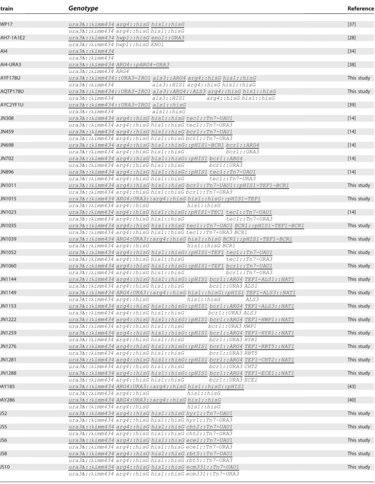
Table 1.C. albicansStrains Used in This Study
Strain Genotype Reference
BWP17 ura3D::kimm434 arg4::hisG his1::hisG [37]
ura3D::kimm434 arg4::hisG his1::hisG
CAH7-1A1E2 ura3D::kimm434 hwp1::hisG eno1::URA3 [28]
ura3D::kimm434 hwp1::hisG ENO1
CAI4 ura3D::kimm434 [34]
ura3D::kimm434
CAI4-URA3 ura3D::kimm434 ARG4::pARG4-URA3 [38]
ura3D::kimm434 ARG4
CAYF178U ura3D::kimm434::URA3-IRO1 als3::ARG4 arg4::hisG his1::hisG This study ura3D::kimm434 als3::HIS1 arg4::hisG his1::hisG
CAQTP178U ura3D::kimm434::URA3-IRO1 als3::ARG4::ALS3 arg4::hisG his1::hisG This study
ura3D::kimm434 als3::HIS1 arg4::hisG his1::hisG
CAYC2YF1U ura3D::kimm434::URA3-IRO1 als1::hisG [39]
ura3D::kimm434 als1::hisG
CJN308 ura3D::kimm434 arg4::hisG his1::hisG tec1::Tn7-UAU1 [14]
ura3D::kimm434 arg4::hisG his1::hisG tec1::Tn7-URA3
CJN459 ura3D::kimm434 arg4::hisG his1::hisG bcr1::Tn7-UAU1 [14]
ura3D::kimm434 arg4::hisG his1::hisG bcr1::Tn7-URA3
CJN698 ura3D::kimm434 arg4::hisG his1::hisG::pHIS1-BCR1 bcr1::ARG4 [14] ura3D::kimm434 arg4::hisG his1::hisG bcr1::URA3
CJN702 ura3D::kimm434 arg4::hisG his1::hisG::pHIS1 bcr1::ARG4 [14]
ura3D::kimm434 arg4::hisG his1::hisG bcr1::URA3
CJN896 ura3D::kimm434 arg4::hisG his1::hisG::pHIS1 tec1::Tn7-UAU1 [14] ura3D::kimm434 arg4::hisG his1::hisG tec1::Tn7-URA3
CJN1011 ura3D::kimm434 arg4::hisG his1::hisG bcr1::Tn7-UAU1::pHIS1-TEF1-BCR1 This study ura3D::kimm434 arg4::hisG his1::hisG bcr1::Tn7-URA3
CJN1015 ura3D::kimm434 ARG4:URA3::arg4::hisG his1::hisG::pHIS1-TEF1 This study ura3D::kimm434 arg4::hisG his1::hisG
CJN1023 ura3D::kimm434 arg4::hisG his1::hisG::pHIS1-TEC1 tec1::Tn7-UAU1 [14] ura3D::kimm434 arg4::hisG his1::hisG tec1::Tn7-URA3
CJN1035 ura3D::kimm434 arg4::hisG his1::hisG tec1::Tn7-UAU1 BCR1::pHIS1-TEF1-BCR1 This study ura3D::kimm434 arg4::hisG his1::hisG tec1::Tn7-URA3 BCR1
CJN1039 ura3D::kimm434 ARG4:URA3::arg4::hisG his1::hisG BCR1::pHIS1-TEF1-BCR1 This study ura3D::kimm434 arg4::hisG his1::hisG BCR1
CJN1052 ura3D::kimm434 arg4::hisG his1::hisG::pHIS1-TEF1 tec1::Tn7-UAU1 This study ura3D::kimm434 arg4::hisG his1::hisG tec1::Tn7-URA3
CJN1060 ura3D::kimm434 arg4::hisG his1::hisG::pHIS1-TEF1 bcr1::Tn7-UAU1 This study ura3D::kimm434 arg4::hisG his1::hisG bcr1::Tn7-URA3
CJN1144 ura3D::kimm434 arg4::hisG his1::hisG::pHIS1 bcr1::ARG4 TEF1-ALS1::NAT1 This study ura3D::kimm434 arg4::hisG his1::hisG bcr1::URA3 ALS1
CJN1149 ura3D::kimm434 ARG4:URA3::arg4::hisG his1::hisG::pHIS1 TEF1-ALS3::NAT1 This study
ura3D::kimm434 arg4::hisG his1::hisG ALS3
CJN1153 ura3D::kimm434 arg4::hisG his1::hisG::pHIS1 bcr1::ARG4 TEF1-ALS3::NAT1 This study ura3D::kimm434 arg4::hisG his1::hisG bcr1::URA3 ALS3
CJN1222 ura3D::kimm434 arg4::hisG his1::hisG::pHIS1 bcr1::ARG4 TEF1-HWP1::NAT1 This study ura3D::kimm434 arg4::hisG his1::hisG bcr1::URA3 HWP1
CJN1259 ura3D::kimm434 arg4::hisG his1::hisG::pHIS1 bcr1::ARG4 TEF1-HYR1::NAT1 This study ura3D::kimm434 arg4::hisG his1::hisG bcr1::URA3 HYR1
CJN1276 ura3D::kimm434 arg4::hisG his1::hisG::pHIS1 bcr1::ARG4 TEF1-RBT5::NAT1 This study ura3D::kimm434 arg4::hisG his1::hisG bcr1::URA3 RBT5
CJN1281 ura3D::kimm434 arg4::hisG his1::hisG::pHIS1 bcr1::ARG4 TEF1-CHT2::NAT1 This study ura3D::kimm434 arg4::hisG his1::hisG bcr1::URA3 CHT2
CJN1288 ura3D::kimm434 arg4::hisG his1::hisG::pHIS1 bcr1::ARG4 TEF1-ECE1::NAT1 This study ura3D::kimm434 arg4::hisG his1::hisG bcr1::URA3 ECE1
DAY185 ura3D::kimm434 ARG4:URA3::arg4::hisG his1::hisG::pHIS1 [43]
ura3D::kimm434 arg4::hisG his1::hisG
DAY286 ura3D::kimm434 ARG4:URA3::arg4::hisG his1::hisG [40]
ura3D::kimm434 arg4::hisG his1::hisG
FJS2 ura3D::kimm434 arg4::hisG his1::hisG hyr1::Tn7-UAU1 This study
ura3D::kimm434 arg4::hisG his1::hisG hyr1::Tn7-URA3
FJS5 ura3D::kimm434 arg4::hisG his1::hisG cht2::Tn7-UAU1 This study
ura3D::kimm434 arg4::hisG his1::hisG cht2::Tn7-URA3
FJS6 ura3D::kimm434 arg4::hisG his1::hisG ece1::Tn7-UAU1 This study
ura3D::kimm434 arg4::hisG his1::hisG ece1::Tn7-URA3
FJS8 ura3D::kimm434 arg4::hisG his1::hisG rbt5::Tn7-UAU1 This study
ura3D::kimm434 arg4::hisG his1::hisG rbt5::Tn7-URA3
FJS10 ura3D::kimm434 arg4::hisG his1::hisG ecm331::Tn7-UAU1 This study
ura3D::kimm434 arg4::hisG his1::hisG ecm331::Tn7-URA3
added NdeI and SpeI restriction sites upstream and downstream of the promoter, respectively. This PCR fragment was digested with NdeI and SpeI and ligated into NdeI- and SpeI-digested plasmid pCJN495 to create pCJN498 containing theA. gossypii TEF1promoter next to theC. albicans NAT1ORF, followed by theA. gossypii TEF1
terminator, followed by theC. albicans TEF1promoter in the correct orientation.
The TEF1-ALS3 overexpression C. albicans strains CJN1149 and CJN1153 were constructed by transforming DAY185 (a Hisþ reference strain) [43] and CJN702 (a Hisþbcr1/bcr1deletion mutant), respectively, using PCR products from template plasmid pCJN498
and primers ALS3-F-OE-Ag-NAT-Ag-TEF1p (59-AGCCAAA
CAATCCGAAGCAACGTAAAGTACGATATCAAAGAATCATAACT TTGCTTTCTATTTGATAACCCGCCTCAAATCAAGATTGGGAGG
TTAACAATCAAGCTTGCCTCGTCCCC-39) and
ALS3-R-OE-Ag-NAT-Ag-TEF1p (59-TAGACCAAGTCAATGAATTAAAACTGTT
GAAAACACCAGTGATTGTCTTTGCAGTCGCAACCGACAAATA TATGAGTAACAATGTATATTGTTGTAGCATTATAAAATGTAT ACTTAGAA-39). These primers amplify the entire A. gossypii TEF1
promoter, theC. albicans NAT1ORF, theA. gossypii TEF1terminator, and theC. albicans TEF1promoter with 100 bp of hanging homology to 500 bp upstream into the promoter of ALS3 for the forward primer and 100 bp of hanging homology from exactly the start codon of the ALS3 ORF. The homology in these primers allows for homologous recombination of the entire cassette directly upstream of theALS3natural locus so thatALS3can be overexpressed with the
TEF1promoter instead of its natural promoter. The transformation into C. albicans strains was done as described above except an additional 5-h recovery step in YPD at 308C was done after the cells were heat shocked at 448C for 20 min in order to allow forNAT1
expression. The cells were then plated onto YPDþ400lg/ml clonNat plates for 2 d at 30 8C to select for Natþ transformants, and transformants were checked by colony PCR. We used strain DAY185 (ArgþUraþHisþ) [43] as a reference strain for these strains.
Theals3D/als3Dmutant, CAYF178U, was constructed from strain BWP17. The two alleles ofALS3 were serially disrupted using the markersHIS1 and ARG4. The disruption cassettes were amplified with the following primers: ALS3-5DR (59-CCTCATTACACCAAC CATACAACTTTGTGGTCTACAACTTGGGTTATTGAAACAAAAA
CAGT TTTCC CAGTC ACGACG TT-39) a nd ALS3-3DR (59
-GGTTGATTCAGCAGTAGTAGTAACAGTAGTAGTTTCATCAGC
ACTAGAAGAAATGATAGGTGTGGAATTGTGAGCGGATA-39).
The disruption ofALS3was verified by PCR using the following
primers: 3Confirm-1 (59-ATGACACCATGTCAAGTTCAGA-39)
and 3Confirm-2 (59-GTTGGTTGTTCAATGACACTGG-39). To
complement the als3D/als3D mutant with a wild-type copy of
ALS3,a full–length version of ALS3was digested from pGEMT with PvuI and SphI [29], and then subcloned into pDS10 at the SphI site [44]. The construct was linearized with Bsp1407I and integrated into the ALS3 locus of the als3D/als3D Urastrain, selecting Uraþ. Excision of the URA3-dpl200 marker was then selected by plating on 5-FOA medium. ALS3 complementation
was confirmed by PCR using primers 59-TGAAGCAGCCTT
TAGTGGCCT-39and 59-AGAAGTGGAAGCAGCTGTGGA-39.
URA3and the adjacentIRO1locus was restored in theals1D/als1D strain [39],als3D/als3D, andals3D/als3D::ALS3strains as follows. Ura derivatives of these mutants were selected by plating on synthetic media containing 5-FOA and uridine. A 3.9-kbURA3-IRO1fragment was released from pBSK-URA3 by NotI/PstI digestion and used to transform the Urastrains [44]. The restoration ofURA3to its native l o c u s w a s c o n fi r m e d b y P C R u s i n g t h e p r i m e r s 59
-TGCTGGTTGGAAT GCTTATTTG-39 and 59-TGCAAATTCTGC
TACTGGAGTT-39.
In vitro biofilm growth conditions. For in vitro biofilm growth assays, strains were grown in YPD overnight at 308C, diluted to an
OD600 ¼ 0.5 in 2 ml of Spider medium (with auxotrophic
supplements), and added to a sterile 12-well plate with a prepared silicone square (1.531.5 cm cut from Cardiovascular Instrument silicone sheets [Wakefiled, Massachusetts, United States]). The silicone square was previously treated with bovine serum (B-9433; Sigma, St. Louis, Missouri, United States) overnight and washed with PBS in order to prepare it for the biofilm assay. The inoculated plate was incubated at 378C for 90 min at 150 rpm agitation for initial adhesion of cells. To remove unadhered cells, the squares were washed with 2 ml of PBS, and the squares were moved to a fresh 12-well plate containing 2 ml of fresh Spider medium. This plate was incubated at 37 8C for an additional 60 h at 150-rpm agitation to allow for biofilm formation.
Microscopic visualization of in vitro biofilms. For the in vitro experiments, biofilms were observed visually and by CSLM. For in
vitro CSLM imaging, biofilms were stained with 25 lg/ml con-canavalin A Alexa Fluor 594 conjugate (C-11253; Molecular Probes, Eugene, Oregon, United States) for 1 h in the dark at 378C with 150 rpm agitation. CSLM was performed with an upright Zeiss Axioskop2 FS MOT LSM 510 multiphoton microscope using a Zeiss Achroplan340/0.8W objective. In order to visualize concanavalin A conjugate staining, a HeNe1 laser with 543-nm excitation wave-length was used. All in vitro CSLM images were assembled into side and depth views using the Zeiss LSM Image Browser (version 3.2.0.115) software. For all side views, the silicone is located at the top of the image. Depth views are artificially colored images indicating cell depth using a color gradient, where blue represents cells closest to and red represents cells farthest from the silicone substrate.
RNA isolation and expression analysis. Overnight cultures were inoculated in 5 ml of YPD at 308C. The next day, 100 ml of Spider medium was inoculated with the YPD overnight culture to obtain an OD600¼0.05, and was grown at 378C for 12 h (OD600¼’8). Cells were immediately harvested by vacuum filtration. RNA extraction and Northern analysis were performed as previously described [40]. For RT-PCR analysis for detection ofALS1andALS3, 10lg of total RNA was DNase treated at 378C for 1 h, ethanol precipitated, and resuspended in 100 ll of DEPC water. cDNA was synthesized and RT-PCR was done as previously described forALS1andALS3[45] with reverse transcriptase and without reverse transcriptase (as a control).
In vivo biofilm model. A rat central venous catheter infection model [18] was selected for in vivo biofilm studies. The catheter diameter was chosen in an attempt to permit blood flow around the extraluminal catheter surface. To mimic material used in patients, polyethylene tubing (inner diameter0.76 mm, outer diameter 1.52 mm) was chosen. Specific-pathogen-free Sprague-Dawley rats weighing 400 g were used (Harlan Sprague-Dawley, Indianapolis, Indiana, United States). A heparinized (100 U/ml) catheter was surgically inserted into the external jugular vein and advanced to a site above the right atrium (2 cm length). The catheter was secured to the vein and the proximal end tunneled subcutaneously to the midscapular space and externalized through the skin. The catheters were placed 24 h prior to infection to allow a conditioning period for deposition of host protein on the catheter surface. Infection was achieved by intraluminal instillation of 500ll ofC. albicanscells (106 cells/ml). After a dwelling period of 4 h, the catheter volume was withdrawn and the catheter was flushed with heparinized 0.15 M NaCl.
Catheters from two animals were removed at three time points (12, 24, and 48 h) after C. albicans infection to determine biofilm development on the internal surface of the intravascular devices. The distal 2 cm of the catheter was cut from the entire catheter length, and biofilms were imaged using both CSLM and scanning electron microscopy. Scanning electron microscopy was used for architectural investigation of the biofilm process. Catheter segments were washed with 0.1 M phosphate buffer (pH 7.2) and placed in fixative (1% glutaraldehyde and 4% formaldehyde). The samples were then washed with buffer for 5 min and placed in 1% osmium tetroxide for 30 min. The samples were then dehydrated in a series of 10-min ethanol washes (30%, 50%, 70%, 85%, 95%, and 100%). Final desiccation was accomplished by critical point drying (Tousimis, Rockville, Maryland, United States). Specimens were mounted on aluminum stubs and sputter-coated with gold. Samples were imaged in a scanning electron microscope (Hitachi S-5700 or JEOL JSM-6100) in the high-vacuum mode at 10 kV. The images were processed for display using Adobe Photoshop 7.0.1.
Disseminated murine candidiasis models.Groups of ten 20-g male Balb/C mice were inoculated via the lateral tail vein with 53105 blastospores with each strain ofC. albicans. The mice were monitored three times daily for survival.
Biofilm dry mass measurements.For dry mass measurements, each silicone square was weighed prior to inoculation with the strain of interest. Biofilms were grown for 60 h on the silicone square (as described above). The silicone squares containing their respective biofilms were then removed from the wells, dried overnight in a fume hood, and weighed the following day. Total biomass of each biofilm was calculated by subtracting the weight of the silicone prior to biofilm growth from the weight of the silicone after biofilm growth. The average total biomass for each strain was calculated from four independent samples after subtracting the mass of a blank silicone square with no cells added. Statistical significance (p-values) was calculated with the Student’s two-tailedt-test function in Microsoft Excel.
Supporting Information
Figure S1.Mouse Survival Data
Disseminated murine candidiasis assays. Groups of ten 20-g male Balb/C mice were inoculated via the lateral tail vein with 53105 blastospores with each strain ofC. albicans. The mice were monitored three times daily for survival.
Found at DOI: 10.1371/journal.ppat.0020063.sg001 (44 KB PDF).
Accession Numbers
Information for the followingC. albicansgenes can be found at the
Candida Genome Database (CGD) Web site (http://www. candidagenome.org): BCR1 (orf19.723), TEC1 (orf19.5908), HWP1
(orf19.1321),ALS3(orf19.1816),ALS1(orf19.5741),HYR1(orf19.4975),
CHT2(orf19.3895),ECE1(orf19.3374),RBT5 (orf19.5636),andECM331
(orf19.4255).
Acknowledgments
We are grateful for the online availability of theCandida genome database (CGD) and the CandidaDB Web Server. We thank all
members of the Mitchell laboratory for insightful discussions and comments on this manuscript. We also acknowledge our manuscript reviewers, who suggested that we broaden our gene overexpression analysis. We are grateful to Allison Fay for her help in constructing plasmid pCJN498, Paula Sundstrom for generously providing us with herhwp1/hwp1mutant strain, Julia Kohler for sharing her plasmid pJK799, and Bill Nierman and his colleagues at The Institute for Genome Research (TIGR) for constructing a plasmid insertion mutant library from which severalC. albicansinsertion mutants were made. We are indebted to Theresa Swayne, Sudhindra Swamy, and Sharmilee Ramcharan for CSLM advice.
Author contributions.CJN, DRA, JEN, SGF, and APM conceived and designed the experiments. CJN, DRA, JEN, and QTP performed the experiments. CJN, DRA, JEN, JEE, SGF, and APM analyzed the data. FJS, FY, QTP, JEE, and SGF contributed reagents/materials/ analysis tools. CJN, DRA, JEE, SGF, and APM wrote the paper.
Funding.This study was supported by National Institutes of Health (NIH) grants R01 AI057804 and R01 AI067703 (APM), NIH K08 AI01767 (DRA), NIH T32 HL07899 (JEN), RO1 AI19990 (JEE), and R01 AI054928 (SGF).
Competing interests.The authors have declared that no competing interests exist.
References
1. Douglas LJ (2003) Candida biofilms and their role in infection. Trends Microbiol 11: 30–36.
2. Fux CA, Costerton JW, Stewart PS, Stoodley P (2005) Survival strategies of infectious biofilms. Trends Microbiol 13: 34–40.
3. Kumamoto CA (2002) Candida biofilms. Curr Opin Microbiol 5: 608–611. 4. Ramage G, Saville SP, Thomas DP, Lopez-Ribot JL (2005) Candida biofilms:
An update. Eukaryot Cell 4: 633–638.
5. Kojic EM, Darouiche RO (2004) Candida infections of medical devices. Clin Microbiol Rev 17: 255–267.
6. von Eiff C, Jansen B, Kohnen W, Becker K (2005) Infections associated with medical devices: Pathogenesis, management and prophylaxis. Drugs 65: 179–214.
7. Bertagnolio S, de Gaetano Donati K, Tacconelli E, Scoppettuolo G, Posteraro B, et al. (2004) Hospital-acquired candidemia in HIV-infected patients. Incidence, risk factors and predictors of outcome. J Chemother 16: 172–178.
8. Beck-Sague C, Jarvis WR (1993) Secular trends in the epidemiology of nosocomial fungal infections in the United States, 1980–1990. National Nosocomial Infections Surveillance System. J Infect Dis 167: 1247–1251. 9. Maki DG, Tambyah PA (2001) Engineering out the risk for infection with
urinary catheters. Emerg Infect Dis 7: 342–347.
10. Chandra J, Patel JD, Li J, Zhou G, Mukherjee PK, et al. (2005) Modification of surface properties of biomaterials influences the ability of Candida albicansto form biofilms. Appl Environ Microbiol 71: 8795–8801. 11. Kuhn DM, Chandra J, Mukherjee PK, Ghannoum MA (2002) Comparison of
biofilms formed byCandida albicansandCandida parapsilosison bioprosthetic surfaces. Infect Immun 70: 878–888.
12. Baillie GS, Douglas LJ (2000) Matrix polymers of Candida biofilms and their possible role in biofilm resistance to antifungal agents. J Antimicrob Chemother 46: 397–403.
13. Chandra J, Kuhn DM, Mukherjee PK, Hoyer LL, McCormick T, et al. (2001) Biofilm formation by the fungal pathogen Candida albicans: Development, architecture, and drug resistance. J Bacteriol 183: 5385–5394.
14. Nobile CJ, Mitchell AP (2005) Regulation of cell-surface genes and biofilm formation by theC. albicanstranscription factor Bcr1p. Curr Biol 15: 1150– 1155.
15. Richard ML, Nobile CJ, Bruno VM, Mitchell AP (2005)Candida albicans
biofilm-defective mutants. Eukaryot Cell 4: 1493–1502.
16. Chagneau C, Saier MH Jr (2004) Biofilm-defective mutants ofBacillus subtilis. J Mol Microbiol Biotechnol 8: 177–188.
17. Schinabeck MK, Long LA, Hossain MA, Chandra J, Mukherjee PK, et al. (2004) Rabbit model of Candida albicans biofilm infection: Liposomal amphotericin B antifungal lock therapy. Antimicrob Agents Chemother 48: 1727–1732.
18. Andes D, Nett J, Oschel P, Albrecht R, Marchillo K, et al. (2004) Development and characterization of an in vivo central venous catheter
Candida albicansbiofilm model. Infect Immun 72: 6023–6031.
19. Schweizer A, Rupp S, Taylor BN, Rollinghoff M, Schroppel K (2000) The TEA/ATTS transcription factor CaTec1p regulates hyphal development and virulence in Candida albicans. Mol Microbiol 38: 435–445.
20. Sopko R, Huang D, Preston N, Chua G, Papp B, et al. (2006) Mapping pathways and phenotypes by systematic gene overexpression. Mol Cell 21: 319–330.
21. Braun BR, Johnson AD (2000) TUP1, CPH1 and EFG1 make independent contributions to filamentation inCandida albicans. Genetics 155: 57–67. 22. Liu TT, Lee RE, Barker KS, Lee RE, Wei L, et al. (2005) Genome-wide
expression profiling of the response to azole, polyene, echinocandin, and pyrimidine antifungal agents in Candida albicans. Antimicrob Agents Chemother 49: 2226–2236.
23. Weissman Z, Kornitzer D (2004) A family of Candida cell surface haem-binding proteins involved in haemin and haemoglobin-iron utilization. Mol Microbiol 53: 1209–1220.
24. McCreath KJ, Specht CA, Robbins PW (1995) Molecular cloning and characterization of chitinase genes fromCandida albicans. Proc Natl Acad Sci U S A 92: 2544–2548.
25. Cao YY, Cao YB, Xu Z, Ying K, Li Y, et al. (2005) cDNA microarray analysis of differential gene expression in Candida albicans biofilm exposed to farnesol. Antimicrob Agents Chemother 49: 584–589.
26. Bensen ES, Martin SJ, Li M, Berman J, Davis DA (2004) Transcriptional profiling inCandida albicansreveals new adaptive responses to extracellular pH and functions for Rim101p. Mol Microbiol 54: 1335–1351.
27. Hoyer LL (2001) The ALS gene family ofCandida albicans. Trends Microbiol 9: 176–180.
28. Sundstrom P (2002) Adhesion inCandidaspp. Cell Microbiol 4: 461–469. 29. Sheppard DC, Yeaman MR, Welch WH, Phan QT, Fu Y, et al. (2004)
Functional and structural diversity in the Als protein family ofCandida albicans. J Biol Chem 279: 30480–30489.
30. Zhao X, Oh SH, Yeater KM, Hoyer LL (2005) Analysis of theCandida albicans
Als2p and Als4p adhesins suggests the potential for compensatory function within the Als family. Microbiology 151: 1619–1630.
31. Staab JF, Bradway SD, Fidel PL, Sundstrom P (1999) Adhesive and mammalian transglutaminase substrate properties of Candida albicans
Hwp1. Science 283: 1535–1538.
32. Zhao R, Daniels KJ, Lockhart SR, Yeater KM, Hoyer LL, et al. (2005) Unique aspects of gene expression duringCandida albicansmating and possible G(1) dependency. Eukaryot Cell 4: 1175–1190.
33. Bennett RJ, Uhl MA, Miller MG, Johnson AD (2003) Identification and characterization of aCandida albicansmating pheromone. Mol Cell Biol 23: 8189–8201.
34. Fonzi WA, Irwin MY (1993) Isogenic strain construction and gene mapping inCandida albicans. Genetics 134: 717–728.
35. Birse CE, Irwin MY, Fonzi WA, Sypherd PS (1993) Cloning and character-ization of ECE1, a gene expressed in association with cell elongation of the dimorphic pathogenCandida albicans. Infect Immun 61: 3648–3655. 36. Liu H, Kohler J, Fink GR (1994) Suppression of hyphal formation inCandida
albicansby mutation of a STE12 homolog. Science 266: 1723–1726. 37. Wilson RB, Davis D, Mitchell AP (1999) Rapid hypothesis testing with
Candida albicansthrough gene disruption with short homology regions. J Bacteriol 181: 1868–1874.
38. Sanchez AA, Johnston DA, Myers C, Edwards JE Jr, Mitchell AP, et al. (2004) Relationship between Candida albicans virulence during experimental hematogenously disseminated infection and endothelial cell damage in vitro. Infect Immun 72: 598–601.
39. Fu Y, Ibrahim AS, Sheppard DC, Chen YC, French SW, et al. (2002)Candida albicans Als1p: An adhesin that is a downstream effector of the EFG1 filamentation pathway. Mol Microbiol 44: 61–72.
40. Davis DA, Bruno VM, Loza L, Filler SG, Mitchell AP (2002)Candida albicans
Mds3p, a conserved regulator of pH responses and virulence identified through insertional mutagenesis. Genetics 162: 1573–1581.
41. Spreghini E, Davis DA, Subaran R, Kim M, Mitchell AP (2003) Roles of
Candida albicansDfg5p and Dcw1p cell surface proteins in growth and hypha formation. Eukaryot Cell 2: 746–755.
selectable marker for transformation of Candida albicans and other pathogenicCandidaspecies. Infect Immun 73: 1239–1242.
43. Davis D, Wilson RB, Mitchell AP (2000) RIM101-dependent and-independ-ent pathways govern pH responses inCandida albicans. Mol Cell Biol 20: 971–978.
44. Park H, Myers CL, Sheppard DC, Phan QT, Sanchez AA, et al. (2005) Role of
the fungal Ras-protein kinase A pathway in governing epithelial cell interactions during oropharyngeal candidiasis. Cell Microbiol 7: 499–510. 45. Green CB, Cheng G, Chandra J, Mukherjee P, Ghannoum MA, et al. (2004) RT-PCR detection of Candida albicans ALS gene expression in the reconstituted human epithelium (RHE) model of oral candidiasis and in model biofilms. Microbiology 150: 267–275.