J of Evolution of Med and Dent Sci/ eISSN- 2278-4802, pISSN- 2278-4748/ Vol. 4/ Issue 39/ May 14, 2015 Page 6841
SURGICALLY INDUCED ASTIGMATISM AFTER 20G VS 23G PARS PLANA
VITRECTOMY
J. M. Lokabhi Reddy1, G. Suneetha 2
HOW TO CITE THIS ARTICLE:
J. M. Lokabhi Reddy, G. Suneetha. Surgically Induced Astigmatism after 20g VS 23g Pars Plana Vitrectomy . Journal of Evolution of Medical and Dental Sciences 2015; Vol. 4, Issue 39, May 14; Page: 6841-6848,
DOI: 10.14260/jemds/2015/992
ABSTRACT: Pars Plana Vitrectomy is done to clear the Vitreous cavity of the Eye. Trans conjunctival Sutureless Vitrectomy with 23G & 25G has become more popular over the Conventional 20G Vitrectomy in recent times. It has many advantages. Less amount of Surgically Induced Astigmatism is one of the Advantages with Sutureless Vitrectomy, which will have the Advantage of Early Visual rehabilitation with better Vision. An interventional comparative study was done between 20G & 23G Pars Plana Vitrectomy in 2 Groups of 30 patients each to assess the amount of Post-Operative Astigmatism. The cases were followed up for 6 months to assess the long term effects. There was a significant difference in immediate Post-Operative Astigmatism. But after some time the difference is much less showing that the main advantage on Astigmatism with Trans conjunctival Sutureless Vitrectomy is noted mainly during the first few weeks after the Surgery.
KEYWORDS: Astigmatism, Conventional 20G Pars plana Vitrectomy, Trans conjunctival Sutureless Vitrectomy.
INTRODUCTION: AIM: The purpose of the study was to Compare the Preoperative and Post-operative Corneal Astigmatism and assess corneal stability in 20G Sutured versus 23G Suture less vitrectomy. Most of the studies on 20G /23G/25G Vitrectomies are from outside India, and there are very few studies on 23G Vitrectomy. This work shows the clinical experience with 23G Pars Plana Vitrectomy and Conventional 20G Pars Plana Vitrectomy comparing the changes in Astigmatism (Keratometry readings).
MATERIALS & METHODS: The study was done at Sarojini Devi eye Hospital, Hyderabad. 60 cases were studied for the change in preoperative and post-operative astigmatism after Pars Plana Vitrectomy. The cases were divided into 2 groups. 30 cases for Standard 20 G vitrectomy & 30 cases for 23G Trans conjunctival Suture less PPV and were done by a single surgeon on Alcon Accurus Vitrectomy machine and instruments. The indications for the PPV were Primary Vitrectomies for non-resolving Vitreous Hemorrhages, ERM, PDR etc. Eyes which underwent earlier surgeries and Corneal Pathologies were excluded. All cases which required sutures in 23G PPV were excluded. All cases underwent detailed Slit lamp examination, Fundus Examination, Keratometry (Bausch & Lomb Keratometer), I.O.P., BCVA. FFA, OCT & B scan were done wherever necessary. Follow up examination done pre-operatively and Post operatively (Day1, end of 1 week, end of 1 month and end of 6 months).
J of Evolution of Med and Dent Sci/ eISSN- 2278-4802, pISSN- 2278-4748/ Vol. 4/ Issue 39/ May 14, 2015 Page 6842 Variables including age, gender, presenting complaints, comorbid risk factors were recorded. BCVA, IOP were recorded at base line and at each follow up visit. Keratometric readings recorded at each follow up visit.
GENDER & AGE: The study included 19 males and 11 females in 23G PPV group and 18 males and 12 females in 20G PPV. The mean Age of the patients in 23G PPV was 44.6±12.9 yrs. The mean age of patients in 20G PPV was 47.1±10.5 years, Most of the patients belonging to age group of 40-59 years.
THE DISTRIBUTION OF CASES: In 23G PPV group there were 14 cases of VH with Proliferative diabetic retinopathy, 6 cases of traumatic VH, 3 cases of full thickness macular hole, 5 cases of Tractional retinal detachment, 2 cases of Rhegmatogenous retinal detachment.
In 20G PPV group, there were 13 cases of Proliferative diabetic retinopathy with vitreous hemorrhage, 5 cases of traumatic VH, 4 cases of full thickness macular hole, 5 cases of Diabetic TRD, 3 cases of Rhegmatogenous RD.
INTRA OCULAR PRESSURE:
The Average Preoperative IOP in 23G PPV was 14.9±2.14 mm Hg. The Average pre op IOP in 20G PPV was 15.5±2.27mmHg.
There was no statistically significant difference observed in the mean baseline IOP in both the groups. (p=0.28).
Graph 1: 23G PPV - Case distribution
J of Evolution of Med and Dent Sci/ eISSN- 2278-4802, pISSN- 2278-4748/ Vol. 4/ Issue 39/ May 14, 2015 Page 6843 The difference between mean IOP on all follow up visits, compared to mean baseline IOP remained statistically insignificant in both 20G and 23G PPV groups (p>0.05).
VISUAL ACUITY: The mean base line Log Mar BCVA in 23G PPV was 1.68±0.31. The mean base line Log Mar BCVA in 20G PPV was 1.59±0.28.
In 23G Group, The mean Log Mar Visual acuity compared to the pre op levels changed significantly from 1 week and remained significant on all subsequent follow up visits.
In 20G group, the mean Log Mar BCVA changed significantly from 1 week and remained statistically significant on all follow up visits compared to the pre-operative mean BCVA. (p<0.05).
There was no difference observed in the change in BCVA on all follow up visits in both the groups. The difference remained statistically insignificant (p>0.05).
ASTIGMATISM CHANGE: The mean baseline astigmatism in 23G group 0.59±0.40. The mean baseline astigmatism in 20G group 0.80±0.73.
Astigmatism in 23G group on 1st post-operative day ranged from 0.25D to 0.75D with mean of 0.6±0.17, on 7th post-operative day with mean of 0.56±0.11, after 1 month with mean of 0.55±0.12 and 0.55±0.15 after 6 months.
In 23G group the mean astigmatism change is insignificant from Day1 and remained as such on all follow up visits, compared to Pre-operative mean astigmatism (p>0.05).
23 G PPV - Astigmatism Change
Mean+/ -SD P Value
Pre Op 0.59±0.40
Day 1 0.60±0.17 0.91
1 Week 0.56±0.11 0.71
1 Month 0.55±0.12 0.69
6 Months 0.55±0.15 0.63
J of Evolution of Med and Dent Sci/ eISSN- 2278-4802, pISSN- 2278-4748/ Vol. 4/ Issue 39/ May 14, 2015 Page 6844 Astigmatism in 20G group on 1st day had ranged from 2.5D to 3.5D with a mean of 3.00±0.51 which reduced to a mean of 1.95±0.58 on the 7th post-operative day, 1.38±0.38 after 1 month, 1.00± 0.21 after 6 months.
In 20G group the mean astigmatism change is significant from 1 day and remained as such on all follow up visits, compared to pre-operative mean astigmatism (p<0.05).
20 G PPV - Astigmatism Change
Mean±SD p Value
Pre Op 0.80±0.73
Day 1 3.00±0.51 0.0001
1 Week 1.95±0.58 0.0001
1 Month 1.38±0.38 0.0017
6 Months 1.00±0.21 0.0258
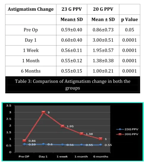
J of Evolution of Med and Dent Sci/ eISSN- 2278-4802, pISSN- 2278-4748/ Vol. 4/ Issue 39/ May 14, 2015 Page 6845 The corneal induced astigmatism was lower 1 week post operatively in the 23G group as compared with the 20G group. 1 month post operatively, the surgically induced corneal astigmatism was still lower in the 23G group.
There was statistically significant difference observed in the astigmatism change on day 1 and on all follow up visits in both the groups. The difference remained statistically significant (p<0.05).
Astigmatism Change 23 G PPV 20 G PPV
Mean± SD Mean ± SD p Value
Pre Op 0.59±0.40 0.86±0.73 0.05
Day 1 0.60±0.40 3.00±0.51 0.0001
1 Week 0.56±0.11 1.95±0.57 0.0001
1 Month 0.55±0.12 1.38±0.38 0.0001
6 Months 0.55±0.15 1.00±0.21 0.0001
Table 3: Comparison of Astigmatism change in both the groups
There was no major complication in either group pre operatively and post operatively, No choroidal detachment or Bacterial endophthalmitis developed in either group.
DISCUSSION: 23 gauge pars plana vitrectomy has more advantages compared to 20 gauge pars plana vitrectomy.
One of the obvious advantages of using 23-gauge vitrectomy is the Shortening of operation time during creation and closure of Sclerotomies. The wound opening and closure time was significantly shorter for the 23-gauge system, when compared with the conventional 20-gauge system.
J of Evolution of Med and Dent Sci/ eISSN- 2278-4802, pISSN- 2278-4748/ Vol. 4/ Issue 39/ May 14, 2015 Page 6846 Compared with conventional 20-gauge vitrectomy, there is less surgically induced Astigmatism following 23-gauge vitrectomy.[2] This allows for faster stabilization of postoperative refraction and faster visual rehabilitation of the patients.
Although much work has been done on 25 gauge pars plana vitrectomy and its role in surgically induced astigmatism, only limited studies are available regarding surgically induced astigmatism in 20 gauge pars plana vitrectomy and especially 23 gauge pars plana vitrectomy.
The sclerotomy size in 23-gauge Transconjunctival sutureless vitrectomy (23GTSV) is 0.7mm and has a greater potential for causing surgically induced astigmatism than 25-gauge sclerotomy does (0.5 mm). However, most of the reports on postoperative astigmatic changes after transconjunctival sutureless vitrectomy have evaluated the 25-gauge system, so no data is available to determine whether surgically induced astigmatism is significant after 23G transconjunctival sutureless vitrectomy.
Low astigmatic changes in 23 gauge pars plana vitrectomy are thought to be related to the absence of sutures.[3] In 20 gauge pars plana vitrectomy astigmatic effect might have been induced by sclerotomy wound suturing.
Domniz and associates described that the induced astigmatism in patients undergoing 20-guage vitrectomy, usually transient, may be attributed to suturing at the entry ports.[4]
Bergmann et al reported that scleral cautery near the incisions changed corneal curvature by causing thermal contracture of the treated tissue and immediate central steepening.[5]
In our study the mean astigmatism on day one in 23G PPV group was 0.60±0.17. The mean astigmatism on day one in 20G PPV group was 3.30±0.51.
Astigmatism in 23G group on 1st post-operative day ranged from 0.25D to 0.75D with mean of 0.6±0.17, on 7th post-operative day with mean of 0.56±0.11, after 1 month with mean of 0.55± 0.12 and 0.55± 0.15 after 6 months.
Astigmatism in 20G group on 1st day had ranged from 2.5D to 3.5D with a mean 3.00±0.51 which reduced to a mean of 1.95±0.58 on the 7th post-operative day, 1.38±0.38 after 1 month, 1.00± 0.21 after 6 months.
In 23G group the mean astigmatism change is insignificant from 1 day and remained as such on all follow up visits, compared to pre-operative mean astigmatism (p>0.05).
In 20G group the mean astigmatism change is significant from 1 day and remained as such on all follow up visits, compared to pre-operative mean astigmatism (p<0.05).
The p-value between both the groups was statistically significant (p<0.05). The p- value for astigmatism change on all subsequent follow up visits was statistically significant between both the groups, showing less surgically induced astigmatism was seen in 23G PPV group compared to 20G PPV group.
Syed Raza Ali Shah, et al, study, showed that surgically induced corneal astigmatism was lower at one week postoperatively in the 23 gauge group (p=006) compared with the 20 gauge group (p=.001). One month postoperatively, the surgically induced corneal astigmatism was still lower in the 23 gauge group.[6]
In our study, 1(3.3%) 1 out of 30 cases 23G PPV group had hypotony, which normalized within 3days. In a study conducted by Raza Ali Shah et al, reported 1(5%) out of 20 cases in 23G PPV group had hypotony which normalized in 3 days.[6]
J of Evolution of Med and Dent Sci/ eISSN- 2278-4802, pISSN- 2278-4748/ Vol. 4/ Issue 39/ May 14, 2015 Page 6847 inspection of the sclerotomy for leakage at the end of the operation can be made difficult by the overlying blood. In patients taking Aspirin or Anti- coagulants, withholding the medications before the operation might help to decrease the risk of this complication.
Hypotony due to Intra operative sclerotomy leakage is another complication which needs to be seen at the end of surgery. The risk factors of intraoperative sclerotomy leakage requiring suture placement after 23-gauge transconjunctival sutureless vitrectomy are prior vitrectomy, young age at operation, and vitreous base dissection. Caution should be exercised to ensure the detection of sclerotomy leakage and hypotony in cases with these risk factors.[7]
Endophthalmitis or intraocular inflammation of the eye is one of the most serious complications of intraocular surgery. In a large retrospective case series, the incidence of Endophthalmitis after 25-gauge vitrectomy has been shown to be 12 times higher than conventional 20-gauge vitrectomy.[8]
The suture less nature of the scleral wound poses a definite risk for this devastating complication. However, subsequent series using 25- and 23-gauge Transconjunctival vitrectomy have shown that there were no obvious increased incidence of Endophthalmitis compared with 20-gauge vitrectomy. Wound leakage and postoperative Hypotony should be avoided in order to minimize the chance of bacterial entry and hence the risk of Endophthalmitis.[7,8]
There were no complications like endophthalmitis and choroidal detachments in both the groups in our study. In a study by Raza Ali Shah, et al reported no complications in both the groups.[6]
CONCLUSION AND SUMMARY: Our study shows that in the early post-operative period 23 gauge Pars plana vitrectomy resulted in less corneal astigmatic changes as compared to conventional 20 gauge Pars plana vitrectomy. During subsequent visits there is no significant difference in surgically induced astigmatism between both the groups. There was 1 case of Hypotony in 23 gauge pars plana vitrectomy which is normalized in 3 days. There was no statistical difference in the mean post-operative day1 IOP compared to mean pre-post-operative IOP in both the groups. The BCVA improved in both the groups and there was no statistical difference between the two groups.
Further prospective, case control studies with large number of patients should be considered to compare surgically induced astigmatism between patients undergoing 23 gauge Trans conjunctival sutureless vitrectomy and those undergoing 20 gauge conventional vitrectomy.
REFERENCES:
1. Wimpissinge B, Kellner L, Brannath, et al. 23-gauge versus 20-gauge system for pars planar
vitrectomy: a prospective randomized clinical trial. Br J Ophthalmol 2008; 92: 1483-1487. 2. Park DH, Shin JP, Kim SY. Surgically induced astigmatism in combined phacoemulsification and
vitrectomy; 23-gauge transconjunctival sutureless vitrectomy versus 20-gauge standard vitrectomy. Graedes Arch Clin Exp Ophthalmol 2009; 10: 1331-1337.
3. Okamoto F, Okamoto C, Sakata N, Hiratsuka K, Yamane N, Hiraoka T et al. Changes in corneal
topography after 25-gauge transconjunctival sutureless vitrectomy versus after 20-gauge standard vitrectomy. Ophthalmology 2007; 114 (12): 2138–2141.
4. Domniz YY, Cahana M, Ayni I. Corneal surface changes after pars plana vitrectomy and scleral
buckling surgery. J Cataract Refract Surg. 2001; 27: 868- 72.
5. Bergmann MT, Koch DD, Zeiter JH. The effect of scleral cautery on corneal astigmatism in
J of Evolution of Med and Dent Sci/ eISSN- 2278-4802, pISSN- 2278-4748/ Vol. 4/ Issue 39/ May 14, 2015 Page 6848 6. Syed Raza Ali Shah, Tehseen Mehmood Mahju, Qasim Lateef Chaudry, Asad Aslam Khan,
Chaudry Nasir Ahmad, Zoya Raza, Surgically Induced Corneal Astigmatism in Conventional 20 – Gauge Versus Trans-Conjunctival Sutureless 23 – Gauge Vitrectomy, Pak J Ophthalmol 2013, Vol. 29 No. 4.
7. Woo SJ, Park KH, Hwang JM, et al. Risk factors associated with sclerotomy leakage and
postoperative hypotony after 23-gauge transconjunctival sutureless vitrectomy. Retina 2009; 29: 456-463.
8. Kunimoto DY, Kaiser RS, Wills Eye Retina Service. Incidence of endophthalmitis after 20- and
25-gauge vitrectomy. Ophthalmology 2007; 114: 2133-2137.
AUTHORS:
1. J. M. Lokabhi Reddy 2. G. Suneetha
PARTICULARS OF CONTRIBUTORS:
1. Associate Professor, Department of
Retinal Services, Sarojini Devi Eye Hospital, Hyderabad.
2. Post Graduate, Department of Retinal
Services, Sarojini Devi Eye Hospital, Hyderabad.
FINANCIAL OR OTHER
COMPETING INTERESTS: None
NAME ADDRESS EMAIL ID OF THE CORRESPONDING AUTHOR:
Dr. J. M. Lokabhi Reddy, Associate Professor, Sarojini Devi Eye Hospital, Mehadipatnam,
Hyderabad-500028. E-mail: [email protected]