www.rbo.org.br
0102-3616/$–see front matter © 2013 Sociedade Brasileira de Ortopedia e Traumatologia. Publicado pela Elsevier Editora Ltda. Todos os direitos reservados Work performed in the Medical Investigation Laboratory for the Musculoskeletal System (LIM41), Department of Orthopedics and Traumatology, Hospital das Clínicas, School of Medicine, Universidade de São Paulo, São Paulo, SP, Brazil
*Corresponding author at: Rua Louis Pasteur, 75, casa 25, Parque Alto Taquaral, Campinas, SP, Brazil. CEP: 13087-773 E-mail: [email protected] (G.C. Campos).
Original Article
Randomized prospective study evaluating addition of
corticoid to viscosupplementation: three months of
follow-up
Gustavo Constantino de Campos,
a,* Márcia Uchôa de Rezende,
bAlexandre Felício Pailo,
cRenato Frucchi,
cThiago Pasqualim,
cand Olavo Pires de Camargo
daPostgraduate Student in the Discipline of Orthopedics and Traumatology, School of Medicine, Universidade de São Paulo, São Paulo, SP,
Brazil
bMSc and PhD in Orthopedics and Traumatology from the School of Medicine, Universidade de São Paulo; Head of the Osteometabolic
Diseases Group, Institute of Orthopedics and Traumatology, Hospital das Clínicas, School of Medicine, Universidade de São Paulo, São Paulo, SP, Brazil
cOrthopedist in the Department of Orthopedics and Traumatology, Hospital das Clínicas, School of Medicine, Universidade de São Paulo,
São Paulo, SP, Brazil
dTitular Professor of the Department of Orthopedics and Traumatology, Hospital das Clínicas, School of Medicine, Universidade de São
Paulo, São Paulo, SP, Brazil
doi:
a b s t r a c t
Objective: To assess if the initial results of viscosupplementation are improved by the addition of corticosteroid. Design: We evaluated 104 patients receiving usual care for knee osteoarthritis at the Universidade de São Paulo Medical Center. Patients were randomized to receive either a single intra-articular injection of 6 mL of Hylan GF-20 (Group 1) or a single intra-articular injection of 6 mL of Hylan GF-20 plus 1 mL (20 mg) of Triamcinolone Hexacetonide (Group 2). VAS, WOMAC and Lequesne questionnaires were applied at weeks zero (prior the injection), and after one, four, and 12 weeks. Results: The baseline measurements of the two groups with 52 patients each were not statistically different. At week one, WOMAC and VAS showed significantly better results for Group 2 compared to Group 1 (p < 0,05). At week four the scores did not show a statistically significant differences. The groups showed similar results at week 12. Conclusion: The addition of Triamcinolone Hexacetonide improves the short term symptom/functional scores of viscosupplementation.
© 2013 Sociedade Brasileira de Ortopedia e Traumatologia. Published by Elsevier Editora Ltda. All rights reserved. A RT I C L E I N F O
Article history:
Received on June 28, 2012 Accepted on August 3, 2012
Keywords:
Adrenal cortex hormones Clinical trial
Controlled clinical trial Double-blind method Hyaluronic acid
Palavras-chave:
Hormônios do córtex adrenal Ensaio clínico
Ensaio clínico controlado Método duplo-cego Ácido hialurônico Injeções, intra-articular Osteoartrite do joelho Escala de dor Tratamento de saída Viscossuplementação
r e s u m o
Objetivo: Avaliar se há melhora dos resultados iniciais da viscossuplementação com a adição de corticosteroide. Métodos: Foram avaliados 104 pacientes em tratamento para osteoartrite do joelho no Instituto de Ortopedia e Traumatologia do Hospital das Clínicas da Faculdade de Medicina da Universidade de São Paulo. Os pacientes foram distribuídos aleatoriamente para receber uma única injeção intra-articular de 6 mL de Hylan GF-20 (Grupo 1) ou uma injeção intra-articular de 6 mL de Hylan GF-20, mais 1 mL (20 mg) de hexacetonido de triancinolona (Grupo 2). Foram aplicados a escala visual analógica de dor (VAS) e os questionários de WOMAC e Lequesne antes da infiltração e após uma, quatro e 12 semanas. Resultados: As medidas basais dos dois grupos com 52 pacientes cada não apresentaram diferença estatística. Após uma semana, o WOMAC e a VAS mostraram resultados significativamente melhores para o Grupo 2 em relação ao Grupo 1 (p < 0,05). Com quatro semanas os resultados não apresentaram diferenças estatisticamente significativas entre os grupos. Os grupos apresentaram resultados semelhantes na 12ª. semana. Conclusão: A adição de hexacetonido de triancinolona melhora os resultados de curto prazo da viscossuplementação.
© 2013 Sociedade Brasileira de Ortopedia e Traumatologia. Publicado pela Elsevier Editora Ltda. Todos os direitos reservados.
Estudo prospectivo e randomizado que avalia a adição de corticoide à viscossuplementação: três meses de seguimento
Introduction
Osteoarthritis is the commonest form of joint disease1 and is a pathological condition of multifactorial origin that leads to destruction of the joint cartilage and inflammatory alterations throughout the joint.2 With the progressive increase in life expectancy, this pathological condition is further increasing in importance, not only with regard to health, but also with regard to the costs generated by the disease. In Brazil, a projection from the Brazilian Institute for Geography and Statistics (IBGE) has indicated that in 2050, the proportion of individuals over the age of 65 years, which in 2000 was only 5% of the population, will have increased to approximately 18%.3
There are more than 50 methods for treating knee osteoarthritis.4 The main treatment options include non-pharmacological management, non-pharmacological management,5 use of intra-articular injections and surgical treatments.6
Intra-articular injections have been used for many years to treat painful joint disorders, especially by means of injecting crystalline suspensions of long-duration corticosteroids.7 Viscosupplementation, which is a relatively new intervention, consists of injection of exogenous hyaluronic acid into diarthrodial joints in order to treat osteoarthritis.8
Hyaluronic acid is a polysaccharide that is produced naturally by type B cells of the synovial membrane. Its molecules, of high molecular weight, form a high-viscosity solution that serves both as a lubricant and as a shock absorber.9 Viscosupplementation has short-term efficacy due to its pain-relief effect, but it is also considered to be a drug that modifies the course of osteoarthritis, with benefits within a period lasting for between six months and a few years.10 It is believed that the long-term results from hyaluronic acid are due to its modulating mechanism of action, especially through its interaction with the CD44 receptors of synoviocytes.11
Most placebo-controlled studies demonstrate clinical improvement within two to five weeks after the
intra-articular injection of hyaluronic acid.12 In comparing viscosupplementation with intra-articular injection of corticosteroids, the most recent data suggest that over the first four weeks, intra-articular corticosteroids seem to be relatively more effective against pain. From the fourth week onwards, the two approaches have equal efficacy, but after the eighth week, hyaluronic acid has greater efficacy.13 The late start, combined with reports of synovitis as a reaction to the procedure, may discourage physicians and patients in relation to using viscosupplementation.
Objective
To assess whether the initial clinical results from viscosupplementation could be improved through addition of corticosteroid.
Methods
T h i s s t u dy wa s c o n d u c t e d i n t h e D ep a r t m e n t o f Orthopedics and Traumatology, Institute of Orthopedics and Traumatology, Hospital das Clínicas, School of Medicine of the University of São Paulo (DOT-IOT-HCFMUSP), in accordance with the CONSORT guidance (Consolidated Standards of Reporting Trials). It was approved by the Ethics Committee for Analysis of Research Projects (CAPPesq), under no. 0073/10. It was registered on the website clinicaltrials.org. This study was fully funded from the Research Support Foundation of the State of São Paulo (Fapesp) (2010/11450-9).
group of IOT-HCFMUSP. Our usual treatment consisted of education through classes, class notes, audiovisual material and guidance from orthopedists, nutritionists, psychologists, occupational therapists, physical educators and social assistants. All the patients, except for those with contraindications, were making use of analgesics as required (paracetamol and codeine). According to their knee alignment, use of shoe insoles would also be indicated. None of the patients routinely used non-steroidal anti-inflammatory drugs (NSAIDs), and their use was discouraged throughout the study, until seven days after the procedure.
Inclusion criteria
- fulfilling the diagnostic criteria for osteoarthritis of the American College of Rheumatology;14
- understanding, agreeing with and signing the free and informed consent statement;
- absence of any history of previous fracture in the knee to be assessed;
- absence of any history of previous surgery in the knee to be assessed;
- absence of any history of allergy to any of the substances used;
- not having undergone any infiltration in the knee to be assessed over the last six months;
- being under treatment with our group for at least six months; - not having used NSAIDs over the last seven days.
Exclusion criteria
- undergoing surgery on the knee assessed, during the follow-up period;
- need for new infiltration in the knee assessed, during the follow-up period;
- severe reaction to the procedure;
- development of active infection during the study; - use of NSAIDs at any time.
One week before the infiltration, the patients were asked to fill out the free and informed consent statement, the visual analogue pain scale (VAS),15 and the WOMAC16 and Lequesne questionnaires.17 The visual scale and the questionnaires were also answered one week after the infiltration (week 1)), four weeks afterwards (week 4) and 12 weeks afterwards (week 12). Anteroposterior and lateral radiographs with loading on the affected knee were evaluated, and the radiological classification was made by three observers using the system of Kellgren and Lawrence.18
The patient sample size was estimated by calculating an n that would enable statistical power of 80% and a significance level of 5%. The patients were randomized into two groups of 52 patients each (groups 1 and 2), by means of simple randomization generated by a computer program. The investigator who performed the randomization did not know the patients and did not participate in any intervention. The three investigators who performed the infiltrations did not have any contact with the patients at any other time. The investigators who applied the questionnaires, both before the
procedure and at the return visits in weeks 1, 4 and 12, did not know which group the patients belonged to. The patients were not allowed to know which group they belonged to, or to see what was being infiltrated.
The patients in group 1 underwent a viscosupplementation procedure in the arthritic knee consisting of infiltration of 6 mL of hylan GF-20. The patients in group 2 underwent a viscosupplementation procedure in the arthritic knee consisting of infiltration of 6 mL of hylan GF-20 and 1 mL (20 mg) of triamcinolone hexacetonide.
All the procedures were performed in an outpatient environment, using the same technique. The infiltration into the knee was done with the patient seated with the knees at 90 degrees and lower legs off the bed. The access route chosen for the joint injection was an anterolateral route.19 The procedures were performed by three investigators with experience of viscosupplementation. Immediately after the procedure, the patients were released without restrictions, with the instruction to take paracetamol (500 mg) every 6 hours for three days.
To investigate whether the groups differed in relation to nominal characteristics, we described the characteristics according to the groups using absolute and relative frequencies. We investigated whether associations existed by using the chi-square or Fisher exact test, or the likelihood ratio when the sample was insufficient to apply the chi-square test.
The quantitative characteristics were described according to the groups by using summary measurements (mean, standard deviation, median, minimum and maximum), and the values were compared between the groups by using Student’s t test.
The pain and functionality scales were described according to the groups and the times of the evaluations, by using summary measurements, and the values were compared between the groups and times by using analysis of variance with repeated measurements, with two factors that made the assumption of a first-order autoregressive correlation matrix between the times, followed by Tukey multiple comparisons in order to compare the groups and times in pairs. Figure 1 presents the flow diagram for the study.
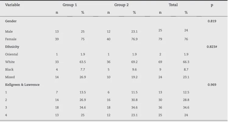
Table 1 describes the nominal characteristics of the patients according to the groups, and the results from the association tests. In this table, it can be seen that the characteristics evaluated did not present any statistically significant associations with the groups of patients (p > 0.05), i.e. the groups were homogenous in relation to the nominal characteristics evaluated.
Table 2 describes the numerical characteristics of the patients according to the groups, and the results from the association tests. In this table, it can be seen that the characteristics evaluated were on average statistically equal in the two groups of patients (p > 0.05).
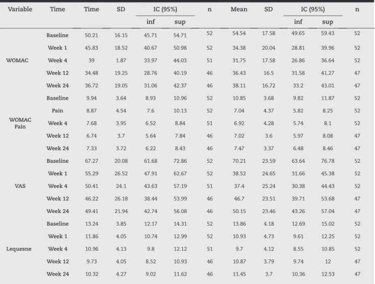
The numerical characteristics of the preoperative scores were on average statistically equal in the two groups of patients, with p > 0.05 (Table 3). Table 3 presents the results from the VAS, WOMAC, WOMAC pain and Lequesne questionnaires at all the times evaluated.
Variable Group 1 Group 2 Total p
n % n % n %
Gender 0.819
Male 13 25 12 23.1 25 24
Female 39 75 40 76.9 79 76
Ethnicity 0.823#
Oriental 1 1.9 1 1.9 2 1.9
White 33 63.5 36 69.2 69 66.3
Black 4 7.7 5 9.6 9 8.7
Mixed 14 26.9 10 19.2 24 23.1
Kellgreen & Lawrence 0.969
1 7 13.5 6 11.5 13 12.5
2 14 26.9 16 30.8 30 28.8
3 18 34.6 18 34.6 36 34.6
4 13 25 12 23.1 25 24
Result from chi-square test; # result from Fisher’s exact test..
Table 1 - Description of the nominal characteristics of the patients according to groups, and results from association tests.
Figure 1 - Flow diagram for the study.
Osteometabolic diseases outpatient clinic IOT-FMUSP
250 patients
Fulfilled the inclusion criteria
147 patients RECRUITMENT
GROUP 1 GROUP 2
RANDOMIZATION
Before infiltration
52 patients
1 patient excluded (severe reaction)
5 patients did not attend return visit
2 moved home 3 unknown causes
4 patients did not attend return visit
1 moved home 3 unknown causes +
1 patient excluded who underwent arthroscopy Before infiltration
52 patients
week 1
52 patients
week 1
52 patients
week 4
51 patients
week 4
52 patients
week 12
46 patients
week 12
presented a statistically significant mean reduction between the preoperative situation and the postoperative weeks (p < 0.05), which stabilized. The mean WOMAC value in week 1 was statistically lower in group 2 than in group 1 (p = 0.038).
Variable Group Mean SD IC
MIN
(95%) MAX
n p
Age 1 60.75 11.71 57.49 64.01
52
0.062
2 64.65 9.14 62.11 67.2 52
Weight 1 79.63 14.6 75.57 83.7 52 0.136
2 75.77 11.47 72.58 78.96 52
Height 1 1.63 0.09 1.6 1.65 52 0.773
2 1.62 0.08 1.6 1.64 52
BMI 1 30.18 5.24 28.72 31.64 52 0.157
2 28.87 4.08 27.73 30 52
Variable Time Time SD IC (95%) n Mean SD IC (95%) n
inf sup inf sup
WOMAC
Baseline 50.21 16.15 45.71 54.71 52 54.54 17.58 49.65 59.43 52
Week 1 45.83 18.52 40.67 50.98 52 34.38 20.04 28.81 39.96 52
Week 4 39 1.87 33.97 44.03 51 31.75 17.58 26.86 36.64 52
Week 12 34.48 19.25 28.76 40.19 46 36.43 16.5 31.58 41.27 47
Week 24 36.72 19.05 31.06 42.37 46 38.11 16.72 33.2 43.01 47
WOMAC Pain
Baseline 9.94 3.64 8.93 10.96 52 10.85 3.68 9.82 11.87 52
Pain 8.87 4.54 7.6 10.13 52 7.04 4.37 5.82 8.25 52
Week 4 7.68 3.95 6.52 8.84 51 6.92 4.28 5.74 8.1 52
Week 12 6.74 3.7 5.64 7.84 46 7.02 3.6 5.97 8.08 47
Week 24 7.33 3.72 6.22 8.43 46 7.47 3.37 6.48 8.46 47
VAS
Baseline 67.27 20.08 61.68 72.86 52 70.21 23.59 63.64 76.78 52
Week 1 55.29 26.52 47.91 62.67 52 38.52 24.65 31.66 45.38 52
Week 4 50.41 24.1 43.63 57.19 51 37.4 25.24 30.38 44.43 52
Week 12 46.22 26.18 38.44 53.99 46 46.7 23.51 39.71 53.68 47
Week 24 49.41 21.94 42.74 56.08 46 50.15 23.46 43.26 57.04 47
Lequesne
Baseline 13.24 3.85 12.17 14.31 52 13.86 4.18 12.69 15.02 52
Week 1 11.86 4.05 10.74 12.99 52 10.93 4.73 9.61 12.25 52
Week 4 10.96 4.13 9.8 12.12 51 9.7 4.12 8.55 10.85 52
Week 12 9.73 4.05 8.52 10.93 46 10.87 3.79 9.74 12 47
Week 24 10.32 4.27 9.02 11.62 46 11.45 3.7 10.36 12.53 47
Table 2 - Description of the numerical characteristics according to groups, and results from comparison tests.
Table 3 - Description of the scales according to groups, and the times evaluated.
scale was significantly lower in group 2 than in group 1 (p = 0.014).
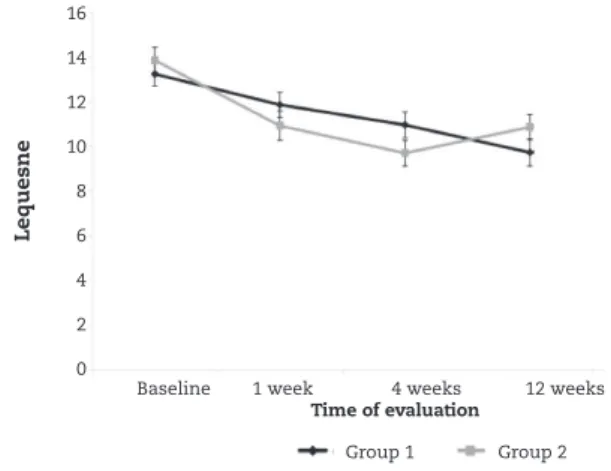
In Figure 4, it can be seen that the mean Lequesne value showed a statistically significant reduction from the preoperative situation to the times from week 4 onwards (p < 0.05) in group 1. In group 2, the mean reduction from the preoperative situation to the subsequent weeks already presented statistical significance from week 1 onwards (p < 0.05). However, in week 24, the mean Lequesne value in this group became statistically equal to the preoperative value (p = 0.076). There was no statistical difference between the groups at any time.
Figures 2 to 4 suggest that there were earlier reductions on the WOMAC and VAS scales in group 2. Figure 3 does not suggest that there was a difference on the Lequesne scale between the groups, but it seemed that the behavior of the Lequesne scale over the course of the evaluations was inconstant.
There was no statistical difference between the groups regarding occurrences of adverse effects from the viscosupplementation procedure (Table 4). Among all the patients, 17.3% reported having some type of pain or discomfort after the infiltration and 4.8% presented joint effusion. Only
one patient in group 1 presented great pain and large volumes of joint effusion, and this patient underwent puncture with drainage of the effusion and infiltration of corticosteroids to alleviate the symptoms. This patient was excluded from the remainder of the study. All the other patients with adverse effects presented mild symptoms and were treated with ice, analgesics and rest.
Discussion
Improvements were observed in all the patients, but the VAS and WOMAC values diminished to lower levels and earlier in the group in which corticosteroids were used in association with viscosupplementation (group 2). This phenomenon can be explained by the faster mechanism of action of corticoids, as observed in the literature.13,20 Figures 2 to 4 show two different curve patterns for each group over the first four weeks, probably due to the effect of the corticosteroid. Group 1 showed a gentler curve, thus denoting a mechanism involving greater modulation of the action of hyaluronic acid. After reaching the fourth week, the results were similar.
Figure 2 - Average profiles and respective standard errors for WOMAC score according to groups.
Figure 3 - Average profiles and respective standard errors for VAS score according to groups.
Figure 4 - Average profiles and respective standard errors for Lequesne score according to groups.
Adverse effects
Group 1 Group 2 Total p
n % n % n %
Pain - 44 86.5 41 78.8 86 82.7 0.3
+ 8 13.5 11 21.2 18 17.3
Effusion - 49 94.2 50 96.2 99 95.2
3 5.8 2 3.8 5 4.8 > 0,999*
Table 4 - Description of occurrences of adverse effects according to groups, and result from association tests.
60
50
40
30
20
10
0
16
14
12
10
8
6
4
2
0
80
70
60
50
40
30
20
10
0
Baseline 1 week 4 weeks 12 weeks
Time of evaluation
Baseline 1 week 4 weeks 12 weeks
Time of evaluation
Baseline 1 week 4 weeks 12 weeks
Time of evaluation
WO
M
AC
Lequesne
VA
S
Group 1
Group 1
Group 1
Group 2
Group 2
It remains uncertain how the addition of corticosteroids affects the modifying effect of hyaluronic acid, despite the similar clinical results. To evaluate the progression of the disease, objective methods such as measuring the joint space (JSW)2,21 or advanced imaging studies like magnetic resonance imaging (MRI) have not been used.22 There is also some concern regarding the chondrotoxicity of intra-articular corticosteroids, but a review of the literature has shown evidence of sufficient security regarding this subject.23 A clinical trial with two years of follow-up showed that there was no loss of joint space after intra-articular injection of triamcinolone hexacetonide at three-month intervals.24
O s t e o a r t h r i t i c c h o n d r o c y t e s a r e d e f i c i e n t i n glucocorticoid receptors25 and the weak response to circulating corticosteroids may be one of the factors involved in the higher levels of cytokines and metalloproteinases in osteoarthritic joints. Therefore, in addition to improving the initial results from viscosupplementation, it can be speculated that addition of triamcinolone hexacetonide may even affect the long-term results positively. Further studies on this subject are necessary.
The present study has some limitations. The use of analgesics or any non-pharmacological treatment was not controlled. However, we believe that viscosupplementation is a procedure that should not exclude any other type of treatment for osteoarthritis, whether pharmacological or not. We maintained the usual treatment and the patients were encouraged to continue to use any medication that they were using before the viscosupplementation, as described earlier. The present study took a pragmatic approach that focused not only on efficacy or safety, but also attempted to place viscosupplementation within the context of the real world, together with the concept of efficacy.26
As mentioned earlier, clinical scores such as WOMAC and Lequesne are not capable of individualizing one knee from the other when the patient has bilateral osteoarthritis. Therefore, in the present study, the patients with bilateral disease underwent treatment for both knees with the same drug, and only the knee that the patient reported to be “worse” was taken into consideration. IN the literature, there are better results from viscosupplementation in patients with less severe osteoarthritis,27 i.e. lower K&L levels, but our results did not show a statistical association in this regard.
The present study did not have a placebo group that received saline injection. There are several studies comparing placebo with viscosupplementation and comparing placebo injection with corticosteroids. Our aim was to improve the results from viscosupplementation, since we believe that the issue of its efficacy has already been answered in the current literature.12,20 Thus, the present study had a control group that also received treatment (viscosupplementation), which avoided the ethical problems involved in using a placebo group.
Most products containing hyaluronic acid that are available on the market have to be administered by means of three to five injections. The regimen used for the present study was a single dose of 6 mL of hylan GF-20, which is only accepted for
this product in particular.28 Regarding the type of hyaluronic acid used for viscosupplementation, there is no convincing evidence to show that one product is superior to another, in relation to molecular weight and the concentration or number of cross-bonds.12,20 We believe that the results from the present study can be extrapolated to viscosupplementation procedures in general.
Adverse effects may occur in 4.2% of the patients, such as knee effusion, pain, heat and erythema.29 The present study found a higher pain rate and a similar rate for effusion. Curiously, there was no statistical difference between the groups of adverse effects. Another unexpected result was the absence, one week after the procedure, of any statistically significant difference between the groups, according to the WOMAC pain subscale (p = 0.324), in contrast with the comparison made using the total WOMAC scale at the same time point. This may provide proof of the good analgesic effect of viscosupplementation.
Conclusion
Addition of 1 ml of triamcinolone hexacetonide significantly improved the pain and function results from viscosupplemen-tation over the short term.
Conflicts of interest
The authors declare no conflicts of interest.
R E F E R E N C E S
1. Lawrence RC, Felson DT, Helmick CG, Arnold LM, Choi H, Deyo RA, et al. Estimates of the prevalence of arthritis and other rheumatic conditions in the United States. Part II. Arthritis Rheum. 2008;58(1):26-35.
2. Martel-Pelletier J, Boileau C, Pelletier JP, Roughley PJ. Cartilage in normal and osteoarthritis conditions. Best Pract Res Clin Rheumatol. 2008;22(2):351-84.
3. Projeção da população do Brasil por sexo e idade – 1980-2050 [database on the Internet. IBGE. 2008. Available from: http:// www.ibge.gov.br/home/estatistica/populacao/projecao_da_ populacao/2008/projecao.pdf.
4. Zhang W, Nuki G, Moskowitz RW, Abramson S, Altman RD, Arden NK, et al. OARSI recommendations for the management of hip and knee osteoarthritis: part III: Changes in evidence following systematic cumulative update of research published through January 2009. Osteoarthritis Cartilage. 2010;18(4):476-99.
5. Rezende MU, Gobbi RG. Tratamento medicamentoso da osteoartrose do joelho. Rev Bras Ortop. 2009;44(1):14-9. 6. Camanho GL. Tratamento da Osteoartrose do Joelho. Rev Bras
Ortop. 2001;36(5):135-40.
7. Hollander JL. The use of intra-articular hydrocortisone, its analogs and its higher esters in arthritis. Ann N Y Acad Sci. 1955;61(2):511-6.
9. Prieto JG, Pulido MM, Zapico J, Molina AJ, Gimeno M, Coronel P, et al. Comparative study of hyaluronic derivatives:
rheological behaviour, mechanical and chemical degradation. Int J Biol Macromol. 2005;35(1-2):63-9.
10. Navarro-Sarabia F, Coronel P, Collantes E, Navarro FJ, de la Serna AR, Naranjo A, et al. A 40-month multicentre, randomised placebo-controlled study to assess the efficacy and carry-over effect of repeated intra-articular injections of hyaluronic acid in knee osteoarthritis: the AMELIA project. Ann Rheum Dis. 2011;70(11):1957-62.
11. Yasuda T. Hyaluronan inhibits prostaglandin E2 production via CD44 in U937 human macrophages. Tohoku J Exp Med. 2010;220(3):229-35.
12. Bellamy N, Campbell J, Robinson V, Gee T, Bourne R, Wells G. Viscosupplementation for the treatment of osteoarthritis of the knee. Cochrane Database Syst Rev. 2006(2):CD005321. 13. Bannuru RR, Natov NS, Obadan IE, Price LL, Schmid CH,
McAlindon TE. Therapeutic trajectory of hyaluronic acid versus corticosteroids in the treatment of knee osteoarthritis: a systematic review and meta-analysis. Arthritis Rheum. 2009;61(12):1704-11.
14. Altman R, Asch E, Bloch D, Bole G, Borenstein D, Brandt K, et al. Development of criteria for the classification and reporting of osteoarthritis. Classification of osteoarthritis of the knee. Diagnostic and Therapeutic Criteria Committee of the American Rheumatism Association. Arthritis Rheum. 1986;29(8):1039-49.
15. Carlsson AM. Assessment of chronic pain. I. Aspects of the reliability and validity of the visual analogue scale. Pain. 1983;16(1):87-101.
16. Fernandes MI. Tradução e validação do questionário de qualidade de vida específico para osteoartrose WOMAC (Western Ontario McMaster Universities) para a língua portuguesa. São Paulo: Universidade Federal de São Paulo; 2003.
17. Marx FC, Oliveira LM, Bellini CG, Ribeiro MCC. Tradução e validação cultural do questionário algo funcional de Lequesne para osteoartrite de joelhos e quadris para a língua portuguesa. Rev Bras Reumatol. 2006;46(4):253-60.
18. Kellgren JH, Lawrence JS. Radiological assessment of rheumatoid arthritis. Ann Rheum Dis. 1957;16(4):485-93.
19. Esenyel C, Demirhan M, Esenyel M, Sonmez M, Kahraman S, Senel B, et al. Comparison of four different intra-articular injection sites in the knee: a cadaver study. Knee Surg Sports Traumatol Arthrosc. 2007;15(5):573-7.
20. Bannuru RR, Natov NS, Dasi UR, Schmid CH, McAlindon TE. Therapeutic trajectory following intra-articular hyaluronic acid injection in knee osteoarthritis--meta-analysis. Osteoarthritis Cartilage. 2011;19(6):611-9.
21. Reichmann WM, Maillefert JF, Hunter DJ, Katz JN, Conaghan PG, Losina E. Responsiveness to change and reliability of measurement of radiographic joint space width in osteoarthritis of the knee: a systematic review. Osteoarthritis Cartilage. 2011;19(5):550-6.
22. Hunter DJ, Zhang W, Conaghan PG, Hirko K, Menashe L, Li L, et al. Systematic review of the concurrent and predictive validity of MRI biomarkers in OA. Osteoarthritis Cartilage. 2011;19(5):557-88.
23. Bellamy N, Campbell J, Robinson V, Gee T, Bourne R, Wells G. Intra-articular corticosteroid for treatment of osteoarthritis of the knee. Cochrane Database Syst Rev. 2006(2):CD005328. 24. Raynauld JP, Buckland-Wright C, Ward R, Choquette D,
Haraoui B, Martel-Pelletier J, et al. Safety and efficacy of long-term intra-articular steroid injections in osteoarthritis of the knee: a randomized, double-blind, placebo-controlled trial. Arthritis Rheum. 2003;48(2):370-7.
25. DiBattista JA, Martel-Pelletier J, Wosu LO, Sandor T, Antakly T, Pelletier JP. Glucocorticoid receptor mediated inhibition of interleukin-1 stimulated neutral metalloprotease synthesis in normal human chondrocytes. J Clin Endocrinol Metab. 1991;72(2):316-26.
26. Schwartz D, Lellouch J. Explanatory and pragmatic attitudes in therapeutical trials. J Clin Epidemiol. 2009;62(5):499-505. 27. Lussier A, Cividino AA, McFarlane CA, Olszynski WP,
Potashner WJ, De Medicis R. Viscosupplementation with hylan for the treatment of osteoarthritis: findings from clinical practice in Canada. J Rheumatol. 1996;23(9):1579-85. 28. Chevalier X, Jerosch J, Goupille P, van Dijk N, Luyten FP, Scott
DL, et al. Single, intra-articular treatment with 6 ml hylan G-F 20 in patients with symptomatic primary osteoarthritis of the knee: a randomised, multicentre, double-blind, placebo controlled trial. Ann Rheum Dis. 2010;69(1):113-9.