RESEARCH ARTICLE
Hepatitis B Virus Pre-S2 Mutant Induces
Aerobic Glycolysis through Mammalian
Target of Rapamycin Signal Cascade
Chiao-Fang Teng1, Wen-Chuan Hsieh1, Han-Chieh Wu1, Yih-Jyh Lin2, Hung-Wen Tsai3, Wenya Huang4, Ih-Jen Su1,2,3*
1National Institute of Infectious Diseases and Vaccinology, National Health Research Institutes, Tainan, Taiwan,2Department of Surgery, National Cheng Kung University Hospital, Tainan, Taiwan,3Department of Pathology, National Cheng Kung University Hospital, Tainan, Taiwan,4Department of Medical
Laboratory Science and Biotechnology, National Cheng Kung University College of Medicine, Tainan, Taiwan
*suihjen@nhri.org.tw
Abstract
Hepatitis B virus (HBV) pre-S2 mutant can induce hepatocellular carcinoma (HCC) via the induction of endoplasmic reticulum stress to activate mammalian target of rapamycin (MTOR) signaling. The association of metabolic syndrome with HBV-related HCC raises the possibility that pre-S2 mutant-induced MTOR activation may drive the development of metabolic disorders to promote tumorigenesis in chronic HBV infection. To address this issue, glucose metabolism and gene expression profiles were analyzed in transgenic mice livers harboring pre-S2 mutant and in anin vitroculture system. The pre-S2 mutant trans-genic HCCs showed glycogen depletion. The pre-S2 mutant initiated an MTOR-dependent glycolytic pathway, involving the eukaryotic translation initiation factor 4E binding protein 1 (EIF4EBP1), Yin Yang 1 (YY1), and myelocytomatosis oncogene (MYC) to activate the sol-ute carrier family 2 (facilitated glucose transporter), member 1 (SLC2A1), contributing to ab-errant glucose uptake and lactate production at the advanced stage of pre-S2 mutant transgenic tumorigenesis. Such a glycolysis-associated MTOR signal cascade was validat-ed in human HBV-relatvalidat-ed HCC tissues and shown to mvalidat-ediate the inhibitory effect of a model of combined resveratrol and silymarin product on tumor growth. Our results provide the mechanism of pre-S2 mutant-induced MTOR activation in the metabolic switch in HBV tumorigenesis. Chemoprevention can be designed along this line to prevent HCC develop-ment in high-risk HBV carriers.
Introduction
Epidemiological studies have provided overwhelming evidence for a causal role of chronic hep-atitis B virus (HBV) infection in the development of human hepatocellular carcinoma (HCC) [1]. Although several mechanisms have been proposed to explain HBV-related tumorigenesis
PLOS ONE | DOI:10.1371/journal.pone.0122373 April 24, 2015 1 / 17
OPEN ACCESS
Citation:Teng C-F, Hsieh W-C, Wu H-C, Lin Y-J, Tsai H-W, Huang W, et al. (2015) Hepatitis B Virus Pre-S2 Mutant Induces Aerobic Glycolysis through Mammalian Target of Rapamycin Signal Cascade. PLoS ONE 10(4): e0122373. doi:10.1371/journal. pone.0122373
Academic Editor:Diego Calvisi, University of Medicine, Greifswald, Germany, GERMANY
Received:December 23, 2014
Accepted:February 13, 2015
Published:April 24, 2015
Copyright:© 2015 Teng et al. This is an open access article distributed under the terms of the
Creative Commons Attribution License, which permits unrestricted use, distribution, and reproduction in any medium, provided the original author and source are credited.
Data Availability Statement:All relevant data are within the paper and its Supporting Information files.
Funding:The authors received a grant from National Health Research Institutes (grant number: 03A1-IVPP20-014;http://www.nhri.org.tw/NHRI_WEB/ nhriw001Action.do). The funders had no role in study design, data collection and analysis, decision to publish, or preparation of the manuscript.
[2,3], the pathogenesis of HBV carcinogenesis is still elusive. Previously, we demonstrated that HBV pre-S2 mutant identified in type II ground glass hepatocytes (GGHs) can induce endo-plasmic reticulum (ER) stress and oxidative DNA damage, as well as exhibits transforming ca-pabilities [4]. Transgenic mice harboring pre-S2 mutant can induce nodular dysplasia and HCC [5]. Moreover, subsequent studies have revealed the positive predictive value of pre-S2 mutant and type II GGHs in HCC development [6–8]. Therefore, type II GGHs represent pre-neoplastic lesions of HBV-related HCC, and pre-S2 mutant is now recognized as a potential viral oncoprotein [9,10].
Metabolic changes are common features in the development of many types of human can-cers [11]. Reports have established that cancer cells frequently display high rates of aerobic gly-colysis in comparison to their nontransformed counterparts, a phenomenon known as the
“Warburg effect”, to support the increased demand of macromolecules for cell growth and pro-liferation [12]. Recently, numerous reports have uncovered multiple metabolic changes in HCC, among which elevated glycolysis is one of the principal changes linked to highly prolifer-ative malignant phenotype [13–15]. Previous study based on HBV transgenic mice has also consistently revealed a metabolic alteration of hepatocytes from the glycogen-storage (glyco-genotic) state toward an increase of glycolysis (the glycogen-poor state) during neoplastic transformation [16]. However, the underlying mechanism of HBV in aerobic glycolysis in HCC development remains to be clarified.
The mammalian target of rapamycin (MTOR) is a highly conserved serine/threonine kinase that controls cell growth and proliferation [17]. In addition to its better-known functions in promoting protein synthesis, MTOR is now emerging as a key regulator of cellular metabolism and cancer [18]. Research has documented that MTOR activation is sufficient to stimulate spe-cific metabolic pathways, including aerobic glycolysis [19]. Previously, we have demonstrated that HBV pre-S2 mutant can activate MTOR through the induction of ER stress-dependent vascular endothelial growth factor A (VEGFA)/AKT signaling in GGHs to promote tumori-genesis [20]. The activated MTOR signal can further upregulate the Yin Yang 1 (YY1) [21], a transcription factor involved in cell proliferation and regulation of oncogenes [22]. This study was designed to investigate whether pre-S2 mutant-induced MTOR activation may regulate aerobic glycolysis through YY1 signaling cascade in HBV-related tumorigenesis.
Materials and Methods
Transgenic Mice
The transgenic mice expressing HBV pre-S2 mutant and X proteins in liver were established by Professor Ting-Fen Tsai’s laboratory as described [23]. All animal experiments were performed in male mice after sacrifice by CO2 inhalation under the approval of the institutional animal care and use committees of the National Cheng Kung University College of Medicine and the National Health Research Institutes.
Histopathology, Immunohistochemistry (IHC), and Immuofluorescence
(IF) Studies
cDNA Microarray Analysis
The microarray experiments were performed by Welgene Biotech (Taipei, Taiwan) using the Agilent Mouse Whole Genome (4×44K) Oligo Microarray Chips (Agilent Technologies, Santa Clara, CA). All values were shown as fold changes relative to the data of age-matched non-transgenic livers. The1.5-fold changes at each stage were considered as significant upregula-tion or downregulaupregula-tion.
Real-Time Polymerase Chain Reaction (PCR)
Total RNA was extracted and converted to complementary DNA as described [21]. Real-time PCR was performed with the LightCycler system and Mouse Universal ProbeLibrary system (Roche Applied Science, Indianapolis, IN). The primers and probes used are shown inS1 Table.
Plasmid, Cell Line, and Transient Transfection
The plasmids pIRES-Δ2 and pIRES-X expressing HA-tagged HBV pre-S2 mutant and X pro-teins, respectively, were established as described [5]. Rapamycin was purchased from Calbio-chem (San Diego, CA) and used at a final concentration of 100 nM. All siRNAs were obtained as ON-TARGETplus SMARTpool reagents (a mixture of 4 individual siRNAs) (Dharmacon, Lafayette, CO) and used at a final concentration of 50 nM. The human hepatoma cells (HuH-7 and HepG2) were obtained from the Health Science Research Resources Bank (JCRB0403; Osaka, Japan) and the American Type Culture Collection (ATCC HB-8065; Rockville, MD), respectively. All transfections were performed with the MicroPorator (Invitrogen Life Technol-ogies, Carlsbad, CA) following the manufacturer’s instructions. Cell proliferation was mea-sured by MTT assay using the Cell Counting Kit-8 (Sigma, St. Louis, MO) following the manufacturer’s instructions.
Western Blot Analysis
Western blot analysis was performed as described.[21] The primary antibodies used in this study were anti-pMTOR (Ser2448), anti-E2F2, anti-SLC2A2, anti-SLC2A3, and anti-SLC2A4 (Abcam, Cambridge, UK), anti-YY1, and anti-CCNA2 (Santa Cruz Biotechnology), anti-MYC, anti-CCNE1, anti-EIF4EBP1, anti-pEIF4EBP1 (Thr37/46), anti-RPS6KB1, and anti-pRPS6KB1 (Thr389) (Cell Signaling Technology, Danvers, MA), anti-SLC2A1 (Novus Biologicals, Little-ton, CO), anti-HA-tag (Zymed Laboratories, South San Francisco, CA), and anti-ACTB (actin, beta) (Chemicon, Temecula, CA). The plasma membrane proteins were prepared with the Qproteome Plasma Membrane Protein Kit (Qiagen, Valencia, CA) following the
manufacturer’s instructions.
Glycogen, Glucose, and Lactate Measurements
Glycogen levels in liver tissues were determined using the Glycogen Colorimetric/Fluorometric Assay Kit (Biovision, Mountain View, CA) following the manufacturer’s instructions. Glucose uptake and lactate production are reported as glucose utilization and lactate secretion per cell within the observed period, respectively. To that end, glucose and lactate concentrations were measured in supernatants using the Glucose (HK) Assay Kit (Sigma) and Lactate Assay Kit (Sigma) following the manufacturer’s instructions, respectively. Each experiment was per-formed in triplicate and repeated at least three times independently.
Pre-S2 Mutant Induces Aerobic Glycolysis
Human HCC Liver Tissues
Decoding freshly frozen human HBV-related HCC tissues were obtained from the Human Bio-bank, Research Center of Clinical Medicine, National Cheng Kung University Hospital, Tai-nan, Taiwan (URL:http://tissuebank.med.ncku.edu.tw), under the approval of the National Cheng Kung University Hospital Institutional Review Board (A-BR-101-133). The National Cheng Kung University Hospital Institutional Review Board waived the need for informed consent.
The model of combined resveratrol and silymarin product on HCC
growth
Resveratrol (0.875 mg Vineatrol 30/g chow) (Breko GmbH, Bremen, German) and silymarin (0.875 mg Silymarin/g chow) (Indena, Milan, Italy) were mixed with normal chow diets and fed to the transgenic mice at the age of 8 months for 6 months and then sacrificed. The trans-genic mice livers were sampled for analyzing the gene expression profiles.
Statistical Analysis
The significance of selected biomarkers in transgenic mice and human livers was determined by unpaired and paired t tests, respectively. Thein vitrodata were analyzed by one-way ANOVA with Bonferroni’s multiple-comparison post-test. APvalue of<0.05 was considered
statistically significant (
Pvalue<0.05;Pvalue<0.01;Pvalue<0.001). The data
repre-sent the mean with standard deviation error bar.
Results
Pre-S2 mutant transgenic mice exhibited glycogen depletion in HCC
tissues
In the pre-S2 mutant transgenic mice model, male mice developed HCCs at the mean age of 24.5 months with a 12% occurrence rate and expressed HBsAg in the hepatocytes around the typical central vein (Fig. 1A). The transgenic livers were sampled for histopathologic studies at 1 and 3 months (early stage), 6 months (middle stage), 12 months (advanced stage), and at tumor formation. PAS staining showed that the livers contained high glycogen contents in 3-, 6-, and 12-month-old transgenic mice, but not in 1-month-old transgenic mice, as compared with the age-matched non-transgenic mice (Fig. 1Ba,b,c,d,e). Interestingly, when the transgenic livers progressed to HCCs, the glycogen staining became slightly weaker and tended to be scat-tered and mixed with the vacuoles (Fig. 1Bf). The amount of glycogen in mice livers was further determined and showed a significant decrease in transgenic HCCs (Fig. 1C).
MTOR, YY1, MYC, and SLC2A1 signals were chronologically activated
in pre-S2 mutant transgenic livers and HCCs
The cDNA microarray data of pre-S2 mutant transgenic livers was adopted to identify the YY1-activated oncogenes.Mycwas identified as the only gene showing significant upregulation (1.5-fold) in transgenic HCCs relative to the non-transgenic livers (S1A Fig.). By further analysis of the microarray data for potential MYC-activated glycolytic genes, we found that
Pre-S2 Mutant Induces Aerobic Glycolysis
hypothesis, Western blot analysis was performed to evaluate the expression of genes involved in the proposed signaling pathway. We observed that the expression levels of the phosphorylat-ed active form of MTOR (pMTOR) and YY1 were significantly elevatphosphorylat-ed at as early as one month of age and persistently activated throughout the study period, while MYC and SLC2A1 expression was found upregulated only upon tumor formation (Fig. 2).
Pre-S2 mutant activated MTOR/YY1/MYC/SLC2A1 signaling cascade in
HuH-7 cells
To confirm that the MTOR/YY1/MYC/SLC2A1 signaling cascade was indeed induced by pre-S2 mutant, HuH-7 cells were transfected with pre-pre-S2 mutant or control plasmid and then ana-lyzed by Western blot assay. As shown inFig. 3A, the expression of all the examined signaling molecules was increased at 24 and 48 hours post-transfection in pre-S2 mutant-transfected cells, as compared to the control cells. The upregulation of these signals by pre-S2 mutant was apparently mediated by MTOR, as the effect could be abolished by the MTOR inhibitor rapa-mycin (Fig. 3B). Also, selective knockdown of YY1 and MYC by siRNAs could sequentially di-minish the elevated signals from upstream activators to downstream targets, even under the conditions of MTOR activation. Similar results were obtained from HepG2 cells (S2 Fig.). Taken together, these data suggest that pre-S2 mutant could upregulate SLC2A1 via the MTOR/YY1/MYC signaling. Considering that MYC is also a master regulator of cell cycle pro-gression,[24] we further examined the candidate MYC-regulated cell cycle progression genes. The results revealed that cyclin A2 (CCNA2), cyclin E1 (CCNE1), and E2F transcription factor 2 (E2F2) were significantly increased in the transgenic HCCs and could be upregulated by pre-S2 mutant through MYC activation in HCC cells (S3 Fig.).
Activation of MTOR signal cascade by pre-S2 mutant promoted SLC2A1
translocation, aerobic glycolysis, and growth advantages in HuH-7 cells
Since the functional site of which SLC2A1 mediated cellular glucose uptake is on the plasma membrane, we further assessed whether the level of SLC2A1 translocation to cell surface was increased by pre-S2 mutant in HuH-7 cells. As shown inFig. 3C, when compared with the con-trol cells, pre-S2 mutant-expressed cells showed significantly higher levels of SLC2A1 in the plasma membrane fraction, which could be reversed by rapamycin treatment. This MTOR-me-diated SLC2A1 translocation pattern was further confirmed by confocal microscopy and IF staining, as indicated by the peripheral expression of SLC2A1 in pre-S2 mutant-expressed cells (Fig. 3D). Considering SLC2A1 as a key rate-limiting factor for aerobic glycolysis in cancer cells,[25] we next evaluated the effect of the MTOR signal cascade on aerobic glycolysis in HuH-7 cells. As shown inFig. 3Ea and 3Eb, pre-S2 mutant transfections resulted in stimulation of glucose uptake and lactate production in comparison with the control cells, and this effect could be abrogated by inhibition of MTOR, YY1, MYC, and SLC2A1 signals. It has been pro-posed that aerobic glycolysis plays a fundamental role in supporting tumor proliferation.[12] To further characterize the role of this MTOR-induced glycolytic pathway in HCC cells, we performed functionalin vitroassays with HuH-7 cells. As shown inFig. 3Ec, pre-S2 mutant-transfected cells exhibited increased proliferation as compared to control cells. Following treat-ment of the transfected cells with inhibitors or siRNAs targeting to the signaling molecules
Fig 1. Pre-S2 mutant transgenic mice exhibited glycogen depletion in HCC tissues: (A) Gross view of representative HCC (a), histological HE staining of tumor (b), and immunohistochemical detection of HBsAg (c).(B) PAS staining for glycogen. (C) Glycogen levels in mice livers. Shown were representative results from different months (M) of pre-S2 mutant (Δ2) and non-transgenic (N) livers, nontumorous livers (NT), and tumors (T). Original magnification, ×20. Scale bar, 50μm.
Pre-S2 Mutant Induces Aerobic Glycolysis
significantly slowed the cell proliferation. Similar results were obtained with HepG2 cells (S4 Fig.). Collectively, these results suggested that pre-S2 mutant could stimulate aerobic gly-colysis, which was required for tumorigenicity of HCC cells.
Pre-S2 mutant activated the MTOR signal cascade through EIF4EBP1
A major function of MTOR is to promote protein synthesis by phosphorylating EIF4EBP1 and ribosomal protein S6 kinase, 70kDa, polypeptide 1 (RPS6KB1) [17]. In this study, the phos-phorylation of EIF4EBP1 and RPS6KB1 was examined by Western blot assay and shown to in-crease throughout the entire period in pre-S2 mutant transgenic livers (Fig. 4A; raw data in
S5A Fig.). To further clarify the effects, pre-S2 mutant-transfected HuH-7 cells were treated with rapamycin,EIF4EBP1orRPS6KB1siRNA. As shown inFig. 4B, pre-S2 mutant-expressed cells exhibited increased levels of pMTOR, pEIF4EBP1, pRPS6KB1, YY1, MYC, and SLC2A1 signals, all of which could be abolished by MTOR inhibition. Knockdown of EIF4EBP1 could rescue the signaling activation under rapamycin treatment. However, knockdown of RPS6KB1 showed no effect on MTOR signaling activation. Similar results were obtained with HepG2 cells (S5B Fig.). Therefore, these data suggested that EIF4EBP1 is the mediator for the pre-S2 mutant-induced MTOR signal cascade.
The MTOR/EIF4EBP1/YY1/MYC/SLC2A1 signaling was activated in
human HBV-related HCCs
To ascertain whether MTOR/EIF4EBP1/YY1/MYC/SLC2A1 signaling is associated with human HBV-related hepatocarcinogenesis, Western blot analysis was performed on 30 pairs of HBV-related HCCs and adjacent nontumorous livers for the expression of components of the signaling pathway. As shown inFig. 4C and 4D, we demonstrated that pMTOR, pEIF4EBP1, YY1, MYC, and SLC2A1 were consistently and significantly expressed at higher levels in HCCs than in the paired nontumorous livers in 18 of 30 tissue pairs (cases 13–30). In summary, the data support the essential role of the glycolysis-associated MTOR signal cascade in
HBV tumorigenesis.
The MTOR/EIF4EBP1/YY1/MYC/SLC2A1 signaling mediated the
chemopreventive effect of combined resveratrol and silymarin product
on tumor growth
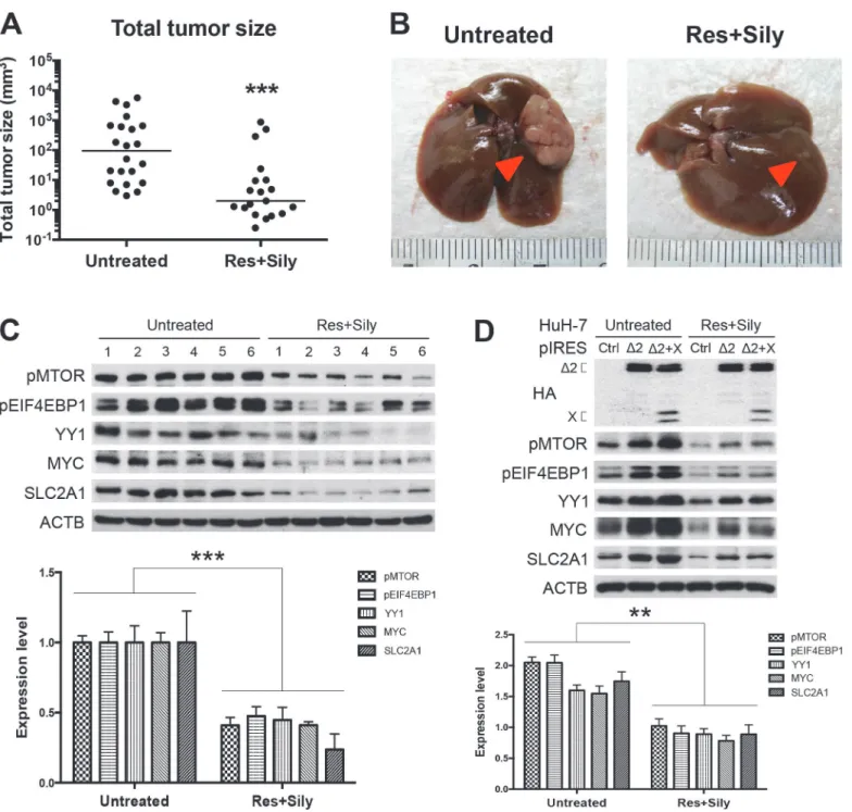
Resveratrol and silymarin are naturally occurring compounds that have anticarcinogenic ef-fects on various types of cancers [26,27]. In the model of chemopreventive study in transgenic mice harboring both HBV pre-S2 mutant and X proteins, the combined resveratrol and sily-marin product has been shown to inhibit HCC growth [28]. As shown inFig. 5A and 5B, the combined product of resveratrol and silymarin could significantly decrease the transgenic tumor size as compared with the untreated group. Western blot analysis performed on the ex-pression profiles of the MTOR signal cascade in the tumor adjacent tissues revealed that the treated group expressed a lower level of MTOR/EIF4EBP1/YY1/MYC/SLC2A1 signaling than the untreated group (Fig. 5C). We further examined thein vitroeffect of resveratrol and sily-marin on MTOR/EIF4EBP1/YY1/MYC/SLC2A1 signaling cascade in HBV tumorigenesis.
Fig 2. MTOR, YY1, MYC, and SLC2A1 signals were chronologically activated in pre-S2 mutant transgenic livers and HCCs: (A) Western blot analysis of the indicated biomarkers in different ages of pre-S2 mutant and non-transgenic livers, as well as paired nontumorous livers and tumors.Six livers were used in each group except the tumor stage due to small tumor size and low tumor formation rate. (B) Quantitative results were normalized by age-matched control livers.
Pre-S2 Mutant Induces Aerobic Glycolysis
Consistent with pre-S2 mutant alone, we observed that HuH-7 cells (Fig. 5D) and HepG2 cells (S6 Fig.) expressing both pre-S2 mutant and X proteins showed activation of the MTOR signal cascade, which could be inhibited by the combined resveratrol and silymarin treatment.
Discussion
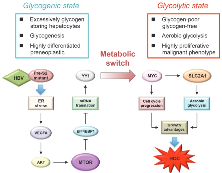
This study demonstrated the contributing role of HBV pre-S2 mutant in metabolic distur-bances of HBV-related HCC development mediated via ER stress-induced, MTOR-dependent glycolytic signal cascade. Our findings, together with other previous studies [19,29], support the important role of MTOR signaling as a molecular regulator linking metabolic disorder and cancer in chronic HBV infection. In this study, we demonstrated that pre-S2 mutant could stimulate aerobic glycolysis through activation of the MTOR/YY1/MYC signaling to upregu-late SLC2A1. Upon the activation of SLC2A1 at the advanced stage of tumorigenesis, hepato-cytes underwent a metabolic switch from the glycogen-storage state toward increased aerobic glycolysis. SLC2A1 belongs to the class I of SLC2A family of proteins (SLC2A1-4) whose ex-pression level usually correlates with the rate of cellular glucose metabolism [25]. The increased SLC2A1 expression in HCC does not only indicate an increased utilization of energy but can also directly cause tumorigenesis [30]. Therefore, our findings suggest that pre-S2 mutant may promote tumorigenesis by sustaining high activation rates of aerobic glycolysis through the MTOR signal cascade, as summarized inFig. 6.
One novel finding in this study is the upregulation of MYC by pre-S2 mutant-activated MTOR signal through YY1 to activate SLC2A1. In this pre-S2 mutant transgenic mouse model, we observed that MTOR/YY1 signaling was activated throughout the entire period in transgenic liver tissues, but MYC was overexpressed only until HCC developed. YY1 has been shown to regulate MYC expression in certain scenarios. In normal cells, YY1 forms a ternary complex with the transcriptional corepressors E1A binding protein p300 and histone deacety-lase 3 to repressMYCtranscription [31]. In transformed cells, YY1 may instead activateMYC
promoter by recruiting the transcriptional coactivator and histone acetyltransferase CREB binding protein [32]. This proposed model for YY1-mediated transcriptional regulation of MYC may possibly explain the delayed overexpression of MYC in pre-S2 mutant transgenic HCCs. As a critical regulator of cell proliferation, MYC regulates a number of genes involved in a variety of biological processes, including glycolysis, cell cycle, apoptosis, and cell adhesion [24]. However, which MYC target genes contribute to HBV-related tumorigenesis remains poorly defined. In this report, we demonstrated that the glycolytic geneSLC2A1and cell cycle progression genesCCNA2,CCNE1, andE2F2were upregulated by MYC through pre-S2 mu-tant-induced MTOR activation, providing a candidate signaling pathway driven by MYC in HBV-related HCCs.
In this study, we further clarified that MTOR-mediated YY1 upregulation by pre-S2 mutant was mediated through phosphorylating its downstream effector EIF4EBP1, a repressor of mRNA translation. As a master regulator of protein synthesis, recent studies have uncovered that MTOR regulates the selective translation of mRNAs with 5’terminal oligopyrimidine
Fig 3. Pre-S2 mutant activated MTOR/YY1/MYC/SLC2A1 signaling cascade to promote SLC2A1 translocation, aerobic glycolysis, and growth advantages in HuH-7 cells: (A and B) HuH-7 cells were transfected with pre-S2 mutant or control plasmid (Ctrl).After 24 hours (h), cells were left untreated or treated with rapamycin (Rapa),YY1siRNA (siYY1) andMYCsiRNA (siMYC) for another 24 hours, and analyzed by Western blot for the indicated biomarkers. (C) Western blots of the whole cell lysate fraction (CL) and the plasma membrane fraction (PM). SLC2A1 translocation represented the level of SLC2A1 in the PM fraction. (D) Confocal microscopy and IF staining of HA (green), SLC2A1 (red), and DAPI (blue). Arrows indicate the peripheral expression of SLC2A1. Original magnification, ×80. Scale bar, 20μm. (E) For functionalin vitroassays, HuH-7 cells transfected with pre-S2 mutant or control plasmid with or without further treatment were subjected to glucose uptake (a), lactate production (b), and cell proliferation (c) assays 48 hours after
transfection. Data in each experiment were presented as relative values to the untreated control cells. All the transfection experiments were performed in triplicate and repeated at least three times independently.
Pre-S2 Mutant Induces Aerobic Glycolysis
Fig 4. Activation of MTOR/YY1/MYC/SLC2A1 signaling by pre-S2 mutant was mediated by EIF4EBP1 and validated in human HBV-related HCCs: (A) Quantitative results of Western blots of pEIF4EBP1 and pRPS6KB1 were normalized by age-matched control livers.(B) The effect ofEIF4EBP1 andRPS6KB1siRNAs on MTOR signaling activation was determined by Western blot assay in HuH-7 cells. (C) Western blot analysis revealed enhanced expression of the indicated biomarkers in HBV-related HCCs at a level comparable to or even higher than that in the paired nontumorous livers. (D) The data were quantified and statistically analyzed.
doi:10.1371/journal.pone.0122373.g004
Fig 5. The MTOR/EIF4EBP1/YY1/MYC/SLC2A1 signaling mediated the chemopreventive effect of combined resveratrol and silymarin product on tumor growth: Total tumor size (A) and gross view of representative HCCs (B) with or without treatment of resveratrol (Res) and silymarin (Sily).
Arrows indicate tumors. (C and D) Western blot analysis revealed a lower expression level of MTOR signal cascade in the treated cells and mice group (six smallest tumor adjacent tissues) than the untreated cells and mice group (six biggest tumor adjacent tissues). Quantitative results by coexpression of pre-S2 mutant and X proteins were relative to the untreated control cells and mice.
(TOP) and TOP-like motifs through the EIF4EBP1-dependent translational control [33]. These reports support our findings, sinceYY1mRNA can be classified into the subset of TOP-like mRNAs according to the defined criteria [34], and provide a molecular explanation for the translational upregulation of YY1 by MTOR. Although YY1 has been reported to be highly ex-pressed in HCC [22], its exact role in HBV-related HCC development remains largely unclear. In this study, we demonstrated that YY1 overexpression by pre-S2 mutant-induced MTOR ac-tivation could stimulate aerobic glycolysis through MYC to upregulate SLC2A1, proposing a novel oncogenic role of YY1 in HCC development.
In this study, we demonstrated that SLC2A1 was overexpressed in pre-S2 mutant-expressed HCC cell lines and human HBV-related HCC tissues. In addition to SLC2A1, the effect of pre-S2 mutant on the expression of other class I SLC2A proteins was examined. As shown inS7 Fig., we observed no significant difference in SLC2A2, SLC2A3, and SLC2A4 expression
Fig 6. Schematic model for the upregulation of aerobic glycolysis by pre-S2 mutant in HBV tumorigenesis: Through the induction of ER stress-dependent VEGFA/AKT signaling, pre-S2 mutant activates MTOR signal, which then increases YY1 expression through the EIF4EBP1-mediated translational control.Upon the activation of MYC and SLC2A1 signals by YY1 at the advanced stage of HCC tumorigenesis, hepatocytes may undergo a metabolic switch toward an increase in aerobic glycolysis. The combined effects of aerobic glycolysis and cell cycle progression induced by MYC and SLC2A1 may contribute to growth advantages of hepatocytes and HCC development.
doi:10.1371/journal.pone.0122373.g006
Pre-S2 Mutant Induces Aerobic Glycolysis
between the pre-S2 mutant transgenic HCC and nontumorous liver tissues. Also, pre-S2 mu-tant showed no effect on the expression of these three SLC2A proteins in HCC cells.
The chemopreventive effects of resveratrol and silymarin compounds on liver pathology and HCC have been proposed [35,36]. In this study, we demonstrated that MTOR/EIF4EBP1/ YY1/MYC/SLC2A1 signaling cascade in pre-S2 mutant-mediated hepatocarcinogenesis could be inhibited by the combined treatment of resveratrol and silymarin. The results suggest that the downregulation of the MTOR signal cascade may be an important mechanism for the che-mopreventive effect of resveratrol and silymarin on HCC development. The MTOR inhibitor rapamycin has been shown to synergistically enhance the resveratrol-induced apoptotic cell death [37].
In conclusion, this study demonstrates that the pre-S2 mutant-induced MTOR signal cas-cade plays a crucial role in driving the metabolic alterations toward increased aerobic glycolysis through the activation of MYC and SLC2A1 signals in HBV-associated tumorigenesis. Target-ing the MTOR/EIF4EBP1/YY1/MYC/SLC2A1 signalTarget-ing pathway may provide avenues for HCC therapy.
Supporting Information
S1 Fig.MycandSlc2a1were identified as candidate genes regulating glycolysis in pre-S2 mutant transgenic HCCs: Microarray expression profiles of selected YY1-activated onco-genes (A) and MYC-activated glycolytic onco-genes (B) in different months (M) of pre-S2 mu-tant transgenic livers, nontumorous livers (NT), and tumors (T).All values were shown as fold changes relative to the data of age-matched non-transgenic livers. Numbers on a gray background represent1.5-fold increase. (C) Transcript levels ofMycandSlc2a1in each stage of transgenic livers relative to the control livers were measured by real-time PCR. Abbrevia-tions are:Myc, myelocytomatosis oncogene;Ptgs2, prostaglandin-endoperoxide synthase 2;
Tgfb1, transforming growth factor, beta 1;Hspa5, heat shock 70kDa protein 5;Erbb2, v-erb-b2 avian erythroblastic leukemia viral oncogene homolog 2;Snai1, snail family zinc finger 1;
Npm1, nucleophosmin;Otx2, orthodenticle homeobox 2;Msx2, msh homeobox 2; Slc2a1, sol-ute carrier family 2 (facilitated glucose transporter), member 1;Ldha, lactate dehydrogenase A;
Hk2, hexokinase 2;Pfkm, phosphofructokinase, muscle;Eno1, enolase 1. (TIF)
S2 Fig. Pre-S2 mutant activated MTOR/YY1/MYC/SLC2A1 signaling cascade in HepG2 cells: (A and B) HepG2 cells were transfected with pre-S2 mutant or control plasmid (Ctrl).
After 24 hours (h), cells were left untreated or treated with rapamycin (Rapa),YY1siRNA (siYY1) andMYCsiRNA (siMYC) for another 24 hours, and analyzed by Western blot for the indicated biomarkers. Data in each experiment were presented as relative values to the untreat-ed control cells.
(TIF)
S3 Fig. Pre-S2 mutant upregulated CCNA2, CCNE1, and E2F2 through MYC activation in transgenic livers, HuH-7 cells, and HepG2 cells: Expression profiles of the selected cell cycle genes in pre-S2 mutant transgenic livers (A and B), HuH-7 cells (C), and HepG2 cells (D) were determined by Western blot assay.Quantitative results were normalized by the age-matched control livers or the untreated cells.
(TIF)
(PM) of pre-S2 mutant- or control plasmid-transfected HepG2 cells.SLC2A1 translocation represented the level of SLC2A1 in the PM fraction. (C) For functionalin vitroassays, HepG2 cells transfected with pre-S2 mutant or control plasmid with or without further treatment were subjected to glucose uptake (a), lactate production (b), and cell proliferation (c) assays. Data in each experiment were presented as relative values to the untreated control cells.
(TIF)
S5 Fig. Pre-S2 mutant activated the MTOR signal cascade through EIF4EBP1: (A) Expres-sion profiles of pEIF4EBP1 and pRPS6KB1 in pre-S2 mutant and non-transgenic livers were established by Western blots.(B) The effect ofEIF4EBP1andRPS6KB1siRNAs on MTOR signaling activation was determined by Western blot assay in HepG2 cells. (TIF)
S6 Fig. Combined resveratrol and silymarin treatment inhibited the MTOR signal cascade in HepG2 cells overexpressing pre-S2 mutant and X proteins: Western blot assay was per-formed to examine the expression of the indicated biomarkers.Quantitative results by coex-pression of pre-S2 mutant and X proteins were relative to the untreated control cells.
(TIF)
S7 Fig. Pre-S2 mutant showed no effect on SLC2A2, SLC2A3, and SLC2A4 expression: The expression profiles of SLC2A2, SLC2A3, and SLC2A4 in pre-S2 mutant transgenic livers (A), HuH-7 cells (B), and HepG2 cells (C) were examined by Western blot assay.
(TIF)
S1 Table. Primers for real-time PCR.
(DOC)
Acknowledgments
We are grateful for the support from the Human Biobank, Research Center of Clinical Medi-cine, National Cheng Kung University Hospital.
Author Contributions
Conceived and designed the experiments: CFT IJS. Performed the experiments: CFT. Analyzed the data: CFT. Contributed reagents/materials/analysis tools: WCH HCW YJL HWT WH. Wrote the paper: CFT IJS.
References
1. Beasley RP, Hwang LY, Lin CC, Chien CS. Hepatocellular carcinoma and hepatitis B virus. A prospec-tive study of 22 707 men in Taiwan. Lancet. 1981; 2: 1129–1133. PMID:6118576
2. Sitia G, Aiolfi R, Di Lucia P, Mainetti M, Fiocchi A, Mingozzi F, et al. Antiplatelet therapy prevents hepa-tocellular carcinoma and improves survival in a mouse model of chronic hepatitis B. Proc Natl Acad Sci U S A. 2012; 109: E2165–2172. doi:10.1073/pnas.1209182109PMID:22753481
3. Su IJ, Hsieh WC, Tsai HW, Wu HC. Chemoprevention and novel therapy for hepatocellular carcinoma associated with chronic hepatitis B virus infection. Hepatobiliary Surg Nutr. 2013; 2: 37–39. doi:10.
3978/j.issn.2304-3881.2012.10.08PMID:24570914
4. Wang HC, Huang W, Lai MD, Su IJ. Hepatitis B virus pre-S mutants, endoplasmic reticulum stress and hepatocarcinogenesis. Cancer Sci. 2006; 97: 683–688. PMID:16863502
5. Wang HC, Chang WT, Chang WW, Wu HC, Huang W, Lei HY, et al. Hepatitis B virus pre-S2 mutant upregulates cyclin A expression and induces nodular proliferation of hepatocytes. Hepatology. 2005; 41: 761–770. PMID:15726643
Pre-S2 Mutant Induces Aerobic Glycolysis
6. Chen CH, Hung CH, Lee CM, Hu TH, Wang JH, Wang JC, et al. Pre-S deletion and complex mutations of hepatitis B virus related to advanced liver disease in HBeAg-negative patients. Gastroenterology. 2007; 133: 1466–1474. PMID:17915220
7. Tsai HW, Lin YJ, Lin PW, Wu HC, Hsu KH, Yen CJ, et al. A clustered ground-glass hepatocyte pattern represents a new prognostic marker for the recurrence of hepatocellular carcinoma after surgery. Can-cer. 2011; 117: 2951–2960. doi:10.1002/cncr.25837PMID:21692054
8. Wu HC, Tsai HW, Teng CF, Hsieh WC, Lin YJ, Wang LH, et al. Ground-glass hepatocytes co-express-ing hepatitis B virus X protein and surface antigens exhibit enhanced oncogenic effects and tumorigen-esis. Hum Pathol. 2014; 45:1294–1301. doi:10.1016/j.humpath.2013.10.039PMID:24767856
9. Su IJ, Wang HC, Wu HC, Huang WY. Ground glass hepatocytes contain pre-S mutants and represent preneoplastic lesions in chronic hepatitis B virus infection. J Gastroenterol Hepatol. 2008; 23: 1169– 1174. doi:10.1111/j.1440-1746.2008.05348.xPMID:18505413
10. Pollicino T, Cacciola I, Saffioti F, Raimondo G. Hepatitis B virus PreS/S gene variants: Pathobiology and clinical implications. J Hepatol. 2014; 61: 408–417. doi:10.1016/j.jhep.2014.04.041PMID:
24801416
11. Cantor JR, Sabatini DM. Cancer cell metabolism: one hallmark, many faces. Cancer Discov. 2012; 2: 881–898. doi:10.1158/2159-8290.CD-12-0345PMID:23009760
12. Hsu PP, Sabatini DM. Cancer cell metabolism: Warburg and beyond. Cell. 2008; 134: 703–707. doi:
10.1016/j.cell.2008.08.021PMID:18775299
13. Bannasch P. Glycogenotic hepatocellular carcinoma with glycogen-ground-glass hepatocytes: a heu-ristically highly relevant phenotype. World J Gastroenterol. 2012; 18: 6701–6708. doi:10.3748/wjg.v18.
i46.6701PMID:23239906
14. Beyoglu D, Imbeaud S, Maurhofer O, Bioulac-Sage P, Zucman-Rossi J, Dufour JF, et al. Tissue meta-bolomics of hepatocellular carcinoma: Tumor energy metabolism and the role of transcriptomic classifi-cation. Hepatology. 2013; 58: 229–238. doi:10.1002/hep.26350PMID:23463346
15. Huang Q, Tan Y, Yin P, Ye G, Gao P, Lu X, et al. Metabolic characterization of hepatocellular carcino-ma using nontargeted tissue metabolomics. Cancer Res. 2013; 73: 4992–5002. doi:
10.1158/0008-5472.CAN-13-0308PMID:23824744
16. Toshkov I, Chisari FV, Bannasch P. Hepatic preneoplasia in hepatitis B virus transgenic mice. Hepatol-ogy. 1994; 20: 1162–1172. PMID:7927248
17. Laplante M, Sabatini DM. mTOR signaling at a glance. J Cell Sci. 2009; 122: 3589–3594. doi:10.1242/ jcs.051011PMID:19812304
18. Cornu M, Albert V, Hall MN. mTOR in aging, metabolism, and cancer. Curr Opin Genet Dev. 2013; 23: 53–62. doi:10.1016/j.gde.2012.12.005PMID:23317514
19. Duvel K, Yecies JL, Menon S, Raman P, Lipovsky AI, Souza AL, et al. Activation of a metabolic gene regulatory network downstream of mTOR complex 1. Mol Cell. 2010; 39: 171–183. doi:10.1016/j. molcel.2010.06.022PMID:20670887
20. Yang JC, Teng CF, Wu HC, Tsai HW, Chuang HC, Tsai TF, et al. Enhanced expression of vascular en-dothelial growth factor-A in ground glass hepatocytes and its implication in hepatitis B virus hepatocar-cinogenesis. Hepatology. 2009; 49: 1962–1971. doi:10.1002/hep.22889PMID:19475690
21. Teng CF, Wu HC, Tsai HW, Shiah HS, Huang W, Su IJ. Novel feedback inhibition of surface antigen synthesis by mammalian target of rapamycin (mTOR) signal and its implication for hepatitis B virus tu-morigenesis and therapy. Hepatology. 2011; 54: 1199–1207. doi:10.1002/hep.24529PMID:21735472 22. Zhang Q, Stovall DB, Inoue K, Sui G. The oncogenic role of Yin Yang 1. Crit Rev Oncog. 2011; 16:
163–197. PMID:22248053
23. Wu BK, Li CC, Chen HJ, Chang JL, Jeng KS, Chou CK, et al. Blocking of G1/S transition and cell death in the regenerating liver of Hepatitis B virus X protein transgenic mice. Biochem Biophys Res Commun. 2006; 340: 916–928. PMID:16403455
24. Miller DM, Thomas SD, Islam A, Muench D, Sedoris K. c-Myc and Cancer Metabolism. Clin Cancer Res. 2012; 18: 5546–5553. doi:10.1158/1078-0432.CCR-12-0977PMID:23071356
25. Amann T, Hellerbrand C. GLUT1 as a therapeutic target in hepatocellular carcinoma. Expert Opin Ther Targets. 2009; 13: 1411–1427. doi:10.1517/14728220903307509PMID:19874261
26. Joe AK, Liu H, Suzui M, Vural ME, Xiao D, Weinstein IB. Resveratrol induces growth inhibition, S-phase arrest, apoptosis, and changes in biomarker expression in several human cancer cell lines. Clin Cancer Res. 2002; 8: 893–903. PMID:11895924
28. Hsieh WC, Yang CW, Huang YS, Chao TW, Tsai TF, Su IJ. Chemoprevention of HBV-related hepato-cellular carcinoma by the combined product of resveratrol and silymarin in transgenic mice. Functional Foods in Health and Disease. 2013; 3: 341–352.
29. Zha X, Sun Q, Zhang H. mTOR upregulation of glycolytic enzymes promotes tumor development. Cell Cycle. 2011; 10: 1015–1016. PMID:21393998
30. Amann T, Maegdefrau U, Hartmann A, Agaimy A, Marienhagen J, Weiss TS, et al. GLUT1 expression is increased in hepatocellular carcinoma and promotes tumorigenesis. Am J Pathol. 2009; 174: 1544– 1552. doi:10.2353/ajpath.2009.080596PMID:19286567
31. Sankar N, Baluchamy S, Kadeppagari RK, Singhal G, Weitzman S, Thimmapaya B. p300 provides a corepressor function by cooperating with YY1 And HDAC3 to repress c-Myc. Oncogene. 2008; 27: 5717–5728. doi:10.1038/onc.2008.181PMID:18542060
32. Hu HM, Kanda K, Zhang L, Boxer LM. Activation of the c-myc p1 promoter in Burkitt's lymphoma by the hs3 immunoglobulin heavy-chain gene enhancer. Leukemia. 2007; 21: 747–753. PMID:17287852 33. Hsieh AC, Liu Y, Edlind MP, Ingolia NT, Janes MR, Sher A, et al. The translational landscape of mTOR
signalling steers cancer initiation and metastasis. Nature. 2012; 485: 55–61. doi:10.1038/nature10912 PMID:22367541
34. Thoreen CC, Chantranupong L, Keys HR, Wang T, Gray NS, Sabatini DM. A unifying model for mTORC1-mediated regulation of mRNA translation. Nature. 2012; 485: 109–113. doi:10.1038/
nature11083PMID:22552098
35. Bishayee A, Politis T, Darvesh AS. Resveratrol in the chemoprevention and treatment of hepatocellular carcinoma. Cancer Treat Rev. 2010; 36: 43–53. doi:10.1016/j.ctrv.2009.10.002PMID:19910122
36. Feher J, Lengyel G. Silymarin in the prevention and treatment of liver diseases and primary liver cancer. Curr Pharm Biotechnol. 2012; 13: 210–217. PMID:21466434
37. Jiang H, Shang X, Wu H, Gautam SC, Al-Holou S, Li C, et al. Resveratrol downregulates PI3K/Akt/ mTOR signaling pathways in human U251 glioma cells. J Exp Ther Oncol. 2009; 8: 25–33. PMID:
19827268
Pre-S2 Mutant Induces Aerobic Glycolysis