Recognizing the LU4 Blood Type Variant Inhibits Cell
Adhesion to Laminin
a
5
Yamato Kikkawa*, Takahiro Miwa, Yukiko Tohara, Takayuki Hamakubo, Motoyoshi Nomizu
Laboratory of Clinical Biochemistry, Tokyo University of Pharmacy and Life Sciences, Tokyo, Japan
Abstract
Background:The Lutheran blood group glycoprotein (Lu), an Ig superfamily (IgSF) transmembrane receptor, is also known as basal cell adhesion molecule (B-CAM). Lu/B-CAM is a specific receptor for laminina5, a major component of basement membranes in various tissues. Previous reports have shown that Lu/B-CAM binding to laminina5 contributes to sickle cell vaso-occlusion. However, as there are no useful tools such as function-blocking antibodies or drugs, it is unclear how epithelial and sickled red blood cells adhere to laminina5 via Lu/B-CAM.
Methodology/Principal Findings:In this study, we discovered a function-blocking antibody that inhibits Lu binding to laminina5 using a unique binding assay on tissue sections. To characterize the function-blocking antibody, we identified the site on Lu/B-CAM recognized by this antibody. The extracellular domain of Lu/B-CAM contains five IgSF domains, D1-D2-D3-D4-D5. The antibody epitope was localized to D2, but not to the D3 domain containing the major part of the laminina5 binding site. Furthermore, mutagenesis studies showed that Arg175, the LU4 blood group antigenic site, was crucial for forming the epitope and the antibody bound sufficiently close to sterically hinder the interaction witha5. Cell adhesion assay using the antibody also showed that Lu/B-CAM serves as a secondary receptor for the adhesion of carcinoma cells to laminina5.
Conclusion/Significance:This function-blocking antibody against Lu/B-CAM should be useful for not only investigating cell adhesion to laminina5 but also for developing drugs to inhibit sickle cell vaso-occlusion.
Citation:Kikkawa Y, Miwa T, Tohara Y, Hamakubo T, Nomizu M (2011) An Antibody to the Lutheran Glycoprotein (Lu) Recognizing the LU4 Blood Type Variant Inhibits Cell Adhesion to Laminina5. PLoS ONE 6(8): e23329. doi:10.1371/journal.pone.0023329
Editor:Ivan Cruz Moura, Institut National de la Sante´ et de la Recherche Me´dicale (INSERM), France ReceivedApril 20, 2011;AcceptedJuly 13, 2011;PublishedAugust 12, 2011
Copyright:ß2011 Kikkawa et al. This is an open-access article distributed under the terms of the Creative Commons Attribution License, which permits unrestricted use, distribution, and reproduction in any medium, provided the original author and source are credited.
Funding:This work was funded by the Ministry of Education, Sciences, Sports and Culture, Japan. The funders had no role in study design, data collection and analysis, decision to publish, or preparation of the manuscript.
Competing Interests:The authors have declared that no competing interests exist. * E-mail: [email protected]
Introduction
The Lutheran glycoprotein (Lu) carries the antigen of the Lutheran blood group system. Lu is an Ig superfamily (IgSF) transmembrane protein in which the extracellular domain contains one variable, one constant-1 and three intermediate Ig-like domains, V-C1-I-I-I [1,2,3]. A splice variant of Lu known as basal cell adhesion molecule (B-CAM) [4] has the same extracellular and transmembrane domains as Lu, but it lacks the COOH-terminal 40 amino acids of the cytoplasmic tail. The cytoplasmic domain of Lu carries an SH3 binding motif, a dileucine motif and potential phosphorylation sites that could be involved in intracellular signaling pathways [1]. A dileucine motif at 608–609 mediates basolateral sorting of Lu in epithelial cells [5]. Protein kinase A phosphorylates Ser621in the cytoplasmic tail and stimulates adhesion of sickled red blood cells to laminin under flow conditions [6]. Erythroid spectrin attaches to the Arg573Lys574 motif in the cytoplasmic tail of Lu and regulates its adhesive activity [7,8]. Recent studies have shown that adrenergic stimuli increase Lu/B-CAM binding to laminin via both a PKA-dependent pathway and exchange proteins activated by the
cAMP-dependent Rap1 pathway [9,10]. However it is unclear if there is synergistic cross-talk between these pathways.
Lu/B-CAM binds with high affinity to laminina5 but not to the other four lamininachains [11,12,13,14,15]. Laminina5 is widely expressed during development and in adult tissues and is the major
a chain in many basement membranes [16]. The a5 chain associates with thec1 chain and eitherb1 orb2 chains to form laminin-511 and -521, respectively [16]. Laminin-511/521 trimers are bound by several different receptors, including not only Lu/B-CAM but also integrins a3b1, a6b1, and a6b4 [17,18] and a -dystroglycan [19]. These receptors bind to a laminin type globular (LG) domain found at the COOH-terminus of laminin a5 and consisting of five homologous subdomains in tandem (LG1–LG5) [19,20]. a-dystroglycan binds primarily to the LG4–5 tandem [21], whereas the binding sites for Lu/B-CAM anda3b1/a6b1 integrins are localized on LG1–3 [20,22]. Recently we showed that Lu/B-CAM anda3b1/a6b1 integrins bind competitively to thea5LG1–3 tandem [20].
two groups localized the laminina5 binding site to the first three IgSF domains (D1-D2-D3) [13,24] and suggested that a specific spatial arrangement of D1-D2-D3 is required for the interaction. Recently, Mankelow et al reported the crystallographic structure of Lu/B-CAM using a combined approach of modeling and small angle X-ray scattering (SAXS) [2]. They found by site-directed mutagenesis that the laminin a5 binding site is formed from a patch of negatively-charged residues at the base of the D2 domain and the top of the D3 domain. Thus, it is likely that the binding site for laminin a5 is formed by the D2 and D3 domains. However, it remains a possibility that Lu/B-CAM could have two distinct binding sites for laminina5.
The Lutheran glycoprotein (Lu) has been studied not only as the antigen of the Lutheran blood group system but also in the context of sickle cell disease. So far, several groups have shown that Lu/B-CAM binding to laminin a5 contributes to sickle cell vaso-occlusion [25,26]. Therefore, development of a drug or antibody to interrupt this interaction has potential therapeutic implications [27]. A recent study showed that hydroxyurea reduced the adhesion of sickled red blood cells to laminina5 via Lu [28]. On the other hands, there have been no antibodies to modulate the adhesion of sickled red blood cells to laminina5.
Here we developed a soluble Lu binding assay using tissue sections. This allowed us to examine whether monoclonal antibodies to Lu/B-CAM could block its binding to laminin a5 in bona fide basement membranes. Of the tested commercial antibodies, we found that one monoclonal antibody against Lu/B-CAM could inhibit its binding to laminin a5. We hypothesized that the epitope of the function-blocking antibody might be near the laminin a5 binding region. To narrow the region on Lu/B-CAM recognized by this antibody, we produced a series of mutated recombinant Lu/B-CAM fragments. Furthermore, we found that the epitope contains an amino acid that comprises an antigenic site-related to Lu blood type. We also clarified the inhibitory mechanism whereby the antibody inhibited the binding of laminina5 and examined the inhibitory effects of the antibody on the adhesion of hepatocellular carcinoma cells to the laminin
a5 chain.
Materials and Methods
Proteins and antibodies
Recombinant proteins containing the Lu/B-CAM extracellular domain fused with a 66His-Tag (Sol-Lu) or a Fc-Tag (Lu-Fc) were produced and characterized as described previously [15,20]. Monoclonal antibodies against Lu/B-CAM are listed in Table 1. Rabbit antibody against domain LEb/L4a of mouse laminin a5 was a gift from Dr. Jeffrey Miner (Washington University School of Medicine, St Louis, MO). Polyclonal antibody against the globular (G) domain of laminina2 was a gift from Dr. Peter D. Yurchenco (Robert Wood Johnson Medical School, Piscataway,
NJ). Monoclonal antibodies against human integrina1 (FB12),a3 (P1B5),a6 (GoH3) andb1 (6S6) were purchased from Millipore (Temecula, CA).
Lu-Fc binding assay on tissue sections
Male C57BL/6J mouse (10–12 weeks old) were purchased from Nihon Charles River (Yokohama, Japan). Animal studies were permitted by Animal Research Committee of Tokyo University of Pharmacy and Life Sciences (Yaku 10–21). Mouse kidney and skeletal muscle were frozen whole by immersing in OCT compound and quick-freezing in 2-methylbutane cooled in a dry ice-ethanol bath. Sections were cut at 7mm in a cryostat and
air-dried. The sections were blocked in 10% normal goat serum. For Lu-Fc binding assays, Lu-Fc was adjusted to 1mg/ml with Ca2+
and Mg2+
-free phosphate-buffered saline (PBS(2)) and placed on the sections. All incubations and washes were in PBS(2). Bound Lu-Fc was detected with anti-human IgG antibody conjugated to Alexa488 (Invitrogen, Carlsbad, CA, USA). Anti-laminin a5 polyclonal antibody was used for counter staining. Anti-rabbit IgG antibody conjugated to Alexa594 (Invitrogen, Carlsbad, CA) was used as secondary antibody. After several washes, sections were mounted in 90% glycerol containing 0.16PBS(2) and 1 mg/ml
p-phenylenediamine. Images were captured using Biozero (Key-ence, Osaka, Japan). Fluorescence intensities of the bound recombinant proteins and laminina5 were measured in the same areas and quantitated by using a BZ-analyzer (Keyence, Osaka, Japan). Fluorescence intensity of laminina5 was used to normalize for differences in the amount ofa5 chain in each section. For Lu-Fc inhibition assays, Lu-Lu-Fc was diluted with PBS(2) containing 10mg/ml or 1:10 dilution of anti-Lu/B-CAM monoclonal antibody. Immunocomplexes containing Lu-Fc and anti-Lu/B-CAM mAb were incubated on kidney sections, and Lu-Fc binding to laminina5 was detected as described above.
Construction of expression vectors
An expression vector containing deleted Lu/B-CAM fused with a Fc-tag was prepared as follows. The CD4-Ig vector was used as template for polymerase chain reaction (PCR) [29]. A DNA fragment encoding human IgG1Fc was amplified using a specific
primer set: 59
-GGAATTCCTTCTAGACGATCCCGAGGGT-GAGTAGTACTAA-39; 59
-TCCCTGTCTCCGGGTAAATG-ACCTAGG-39. The PCR products were digested with EcoRI and AvrII and inserted into the EcoRI-XbaI sites of pcDNA3.1-Neo (+). The full-length cDNAs for human Lu and melanoma cell adhesion molecule (Mel-CAM) were purchased from Invitrogen (Carlsbad, CA, USA) and used as template for PCR. cDNAs encoding the mutant proteins were generated by PCR using the primer sets listed Table S1 and inserted into the EcoRI and XbaI sites of the above human IgG1 Fc expression vector. For all PCR, KOD plus DNA polymerase (TOYOBO, Osaka, Japan) was used according to the manufacturer’s instructions.
Table 1.Monoclonal antibodies to Lutheran/B-CAM.
Clone Ig class Function blocking Antigen domain Source Reference
BRIC108 IgG1 2 D1 Biogenesis Parsons et al., PNAS 1995, and Blood 1997
BRIC221 IgG2b 2 D4 SEROTEC Parsons et al., PNAS 1995, and Blood 1997
87202 IgG1 2 D1 R&D system
-87207 IgG2a + D2 R&D system
Expression and purification of recombinant proteins
HEK293 cells were purchased from ATCC (Manassas, VA, USA) and maintained in DMEM containing 10% FBS. HEK293 cells were transfected with the deletion mutant Lu-Fc expression vectors using Lipofectamine 2000 (Invitrogen), and stable clones were selected using 400mg/ml Zeocin (Invitrogen). All further cell culture was carried out in the presence of the antibiotic. The selected cells were grown to confluency in culture dishes with DMEM containing 10% FBS. The confluent cells were incubated in serum free DMEM for 4 days. The conditioned media were harvested and clarified by sequential centrifugation at 500 rpm for 10 min and 10,000 rpm for 20 min. The collected media were precipitated with ammonium sulfate at 80% saturation. The resulting precipitates were collected by centrifugation at 10,000 rpm for 30 min, and then dissolved in and dialyzed against PBS(2). The 30-fold concentrated media were used for purification. The recombinant proteins were purified from the conditioned culture media by Protein A sepharose (GE Health Care Bio-Science, Piscataway, NJ, USA). The eluted fractions were pooled and dialyzed against PBS(2). The purified proteins were separated by SDS-PAGE using 7.5% gels under reducing conditions. The separated proteins were stained with Coomassie Brilliant Blue.
Antigen capture ELISA
ELISA plates were coated with 3mg/ml of Sol-Lu and blocked with 1% bovine serum albumin (BSA) in PBS(2). Diluted monoclonal antibodies were incubated with recombinant proteins for 1 hour at room temperature and then transferred into the Sol-Lu-coated wells. After a further 1 hour incubation and washing with PBS(2) containing 0.05% Tween 20, bound antibodies were detected by addition of horseradish-peroxidase conjugated anti-mouse IgG (GE Healthcare, Piscataway, NJ, USA), followed by addition of 0.4 mg/ml o-phenylendiamine and 0.01% H2O2. The optical density at 450 nm was measured
in a model 550 microplate reader (Bio-Rad Laboratories, Hercules, CA, USA).
Cell adhesion inhibition assays
Human hepatocellular carcinoma cell line HuH-7 was purchased from Health Science Research Resources Bank (Osaka, Japan) and maintained in DMEM containing 10% FBS. Cell adhesion assays were performed as described previously [30]. Briefly, 96-well microtiter plates (Nunc, Roskilde, Denmark) were incubated with 2.5mg/ml of recombinant laminin-511 at
37uC for 1 h, and then blocked with PBS(2) containing 1% BSA for another hour. HuH-7 cells were suspended in serum-free DMEM at a density of 46105 cells/ml and preincubated with 1mg/ml of antibody at room temperature for 10 min. 50ml of
cell suspension were added to wells coated with recombinant laminin-511. After incubation at 37uC for 1 h, the attached cells were fixed with 4% formaldehyde, stained with Diff-Quik (International Regents Corp., Kobe, Japan), and counted under the microscope.
Results
Inhibitory effects of monoclonal antibodies on the binding of Lu-Fc to laminina5 in mouse tissue sections
In a previous study we prepared a dimerized soluble recombinant protein (Lu-Fc) composed of the Lu extracellular domain and human IgG1Fc region [20]. Lu-Fc dimerizes via the
Fc region and should bind to laminina5 more effectively than the monomeric Lu (Sol-Lu) used in our earlier study [15]. In
addition, although Sol-Lu worked effectively as a histochemical probe on fetal tissue sections [15], the need to use a mouse monoclonal antibody to detect it restricts its usefulness on adult mouse tissues containing endogenous immunoglobulins. There-fore, here we used Lu-Fc as a probe on adult mouse tissue sections because it could be detected by anti-human IgG antibody conjugated to Alexa488. Lu-Fc, but not Fc alone, bound essentially all basement membranes containing laminin a5 in the adult mouse kidney tissues (Fig. 1A). In adult mouse skeletal muscle basement membranes, laminin a5 is found primarily at neuromuscular junctions [31]. Lu-Fc bound to neuromuscular junctions but not to the extrasynaptic basement membrane, suggesting that Lu/B-CAM is a specific receptor fora5 (Fig. 1B). The binding of Lu-Fc to laminina5 in tissue sections also allowed us to examine whether anti-Lu/B-CAM monoclonal antibodies could block binding (Fig. 1C). Before incubation on kidney sections, Lu-Fc was incubated with anti-Lu/B-CAM monoclonal antibodies. Antibody 87207 inhibited binding to laminina5, but BRIC221, 87202, and BRIC108 antibodies did not (Fig. 1C and Table 1).
Epitope mapping of antibodies on Lu/B-CAM
It is possible that the function-blocking antibody 87207 recognized an epitope of Lu/B-CAM involved in binding laminin
Analysis of Lu-Fc mutants with reduced laminina5 interaction
Recently, Mankelow et al. reported that a cluster of negatively charged residues in the D2 and D3 domains forms the laminin
a5-binding site [2]. Therefore, these negatively charged residues may be part of the epitope of antibody 87207, which inhibited binding to laminina5. Lu-Fc mutants with one or two of these charged amino acids substituted with alanine were produced in HEK293 cells (Fig. 4A). The purified recombinant proteins were subjected to SDS-PAGE under reducing conditions (Fig. 4B). When 87207 was adsorbed to these Lu-Fc mutants, it could not bind to Sol-Lu-coated wells (Fig. 4C). 87207 did not recognize these acidic residues in the D2 and D3 domains. These results suggest that 87207 binding to its epitope covers and masks the acidic amino acid residues contributing to the interaction of Lu/B-CAM with laminina5.
Fine mapping of the 87207’s epitope on the D2 domain of Lu/B-CAM
We sought to more finely identify the epitope of 87207 on the D2 domain of Lu/B-CAM. The antibody 87207 was produced in a mouse immunized with human Lutheran. Therefore, the differences between the human and mouse Lu/B-CAM amino acid sequences (Fig. 5A) should be responsible for its immunoge-nicity. Lutheran, as a determinant of the blood group system, consists of 19 documented antigens [27] deriving from changes in amino acid residues that are likely exposed on the surface of Lu/B-CAM. Three of these antigenic sites, Arg175, Met204, and Arg227, are localized in the D2 domain. These amino acid residues also differ from the corresponding mouse Lu/B-CAM residues. We considered the possibility that these amino acid residues could be part of the 87207 epitope. To test this, Arg175, Met204, and Arg227 were individually substituted with the corresponding amino acids found in mouse Lu/B-CAM: Asn175, Ile204, and His227, respec-tively. The mutated recombinant proteins fused with the Fc tag were generated by site-directed mutagenesis. The recombinant proteins were purified from the culture media using Protein A Sepharose and subjected to SDS-PAGE under reducing condi-tions, which showed the appropriate size (Fig. 5B). Preincubation with 87207 showed that although the antibody reacted with M204I-Fc and R227H-Fc, R175N-Fc was not immunoreactive (Fig. 5C). Moreover, to confirm if Arg175was essential for forming the epitope, we produced mutant protein substituted with Ala. R175A-Fc also lacked the immunoreactivity of 87207. This indicates that Arg175is critical for forming the 87207 epitope.
The mechanism of 87207-mediated inhibition of Lu/ B-CAM binding to laminina5
To examine if Arg175was involved in the binding of laminina5, the binding assay on mouse kidney tissue sections was performed using R175A-Fc (Fig. 6A). Fluorescence intensity was quantitated to measure the binding of the mutant proteins to laminina5 (Fig. 6B). Although Arg175 was a part of the epitope recognized by the function-blocking antibody, it was not involved in the binding of laminina5. Furthermore, to clarify a mechanism whereby 87207 inhibits the binding of Lu to laminina5, the binding of mutant proteins was examined on mouse kidney tissue sections. Lu-Fc containing the mutations in the D2 domain bound to endogenous laminina5 on mouse kidney sections (Fig. 6A), but they reduced the interaction with laminin a5 (Fig. 6B). Furthermore, E340A/ D341A-Fc and D343A-Fc (mutations in the D3 domain) exhibited little or no binding activity (Fig. 6A and B). As shown in Fig. 3, M2Lu-Fc, for which the D1–D2 tandem of Lu was replaced with the analogous tandem of Mel-CAM, lost the epitope of the function-blocking antibody and did not bind to laminin a5. The straightforward interpretation was that the D2 domain was involved in the binding of laminina5. However, our mutagenesis study also Figure 1. Lu/B-CAM binding assay on tissue sections.(A) Binding
of Lu-Fc to laminina5 in adult mouse kidney sections. Sections were incubated with Lu-Fc (upper panel) or Fc (lower panel) at room temperature for 1 hour. Endogenous laminina5 and bound Lu-Fc were detected with anti-laminina5 domain LEb/L4b and anti-human IgG, as indicated. Merged pictures showed that laminin a5 and Lu-Fc co-localized at basement membranes. Arrows indicate glomeruli, laminin
showed the D3 domain to be a major region for laminina5 binding, as described in Mankelow et al [2]. These results suggest that the D2 domain is essential for formation of the binding site but not the interaction with the laminina5 chain. Therefore, we conclude that, because the majora5 binding sites are localized in the D3 but not the D2 domain, antibody 87207 seems to sterically hinder Lu/B-CAM binding to laminina5 (Fig. 6C).
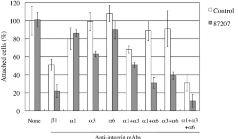
Inhibitory effects of 87207 on adhesion of HuH-7 cells to laminina5
To examine if antibody 87207 can inhibit Lu/B-CAM-mediated cell adhesion to laminin a5 in vitro, cell adhesion assays were performed using HuH-7 cells derived from a human hepatocellular carcinoma. HuH-7 cells express a1b1/a3b1/a6b1 integrins and Lu/B-CAM that can bind to laminin-511 [30]. After incubation Figure 2. Identifying the IgSF domains recognized by anti-Lu/B-CAM antibodies.(A) Diagram of the deletion and chimeric mutant proteins designed to narrow the epitopes of anti-Lu/B-CAM antibodies. For chimeric mutant proteins, the D1 or D1–D2 domains of Lu-Fc were replaced with the analogous domains of melanoma cell adhesion molecule (Mel-CAM). The deletion and chimeric mutant proteins were fused with an IgG Fc domain tag. (B) The mutant proteins purified from conditioned media of HEK293 transfectants were subjected to SDS-PAGE on a 7.5% gel under reducing conditions. Protein was stained with Coomassie Brilliant Blue. Molecular mass standards are indicated. (C) ELISA using the indicated anti-Lu/ B-CAM antibodies absorbed with the various recombinant proteins. Wells were coated with 3mg/ml of Sol-Lu. The mutant proteins were mixed with the diluted antibodies at 1mg/ml. Each bar represents the mean of triplicate assays. Error bars indicate standard deviation.
with antibodies, the cells were plated on dishes coated with recombinant laminin-511 (a5b1c1). Although antibody 87207 alone had no effect on adhesion of HuH-7 cells to laminin-511 (Fig. 7), the remaining adhesion of HuH-7 cells to laminina5 in the presence of an inhibitory anti-integrinb1 antibody was significantly reduced by addition of antibody 87207. Moreover, combining antibody 87207 with inhibitory antibodies toa1/a3/a6 integrins dramatically reduced adhesion of HuH-7 cells to laminin-511.
Discussion
Soluble receptor binding assays on tissues is a proven method to reveal the presence of ligands [32]. To visualize and localize soluble
receptors bound to ligands, they have been conjugated with alkaline phosphatase or detected with a specific monoclonal antibody [15,32]. In the present study, we developed a binding assay on tissue sections using Lu-Fc, consisting of the ectodomain of Lu/B-CAM fused with a human Fc-tag. The staining of laminina5 and Lu-Fc overlapped, indicating that Lu-Fc bound to endogenous laminina5 in tissue sections. Therefore, it is unlikely that an unknown ligand for Lu/B-CAM exists in adult mouse kidney and skeletal muscle. The use of a Fc-tagged soluble receptor in binding assays on tissue sections was advantageous, as Lu-Fc was easily purified by Protein A Sepharose and detected with anti-human IgG antibody conjugated to Alexa488. Fc-tagged soluble receptors should be useful tools to identify unknown ligands in adult tissue sections.
Figure 3. The binding of Lu-Fc and chimeric mutant proteins to laminina5 in tissue sections.(A) Sections of adult mouse kidney were incubated with the Lu-Fc, and chimeric mutants, as indicated. The recombinant proteins bound to endogenous laminina5 were detected with an antibody against human IgG. Bar, 100mm. M1Lu-Fc bound to laminina5 but not M2Lu-Fc did. (B) Quantitation of the recombinant proteins bound to laminina5. Fluorescence intensities were measured as described in Materials and Methods. The binding of Lu-Fc was set to 100. Each bar represents the mean of triplicate assays. Error bars indicate standard deviation.
The binding assays also allowed us to characterize monoclonal antibodies to Lu/B-CAM. The results showed that antibody 87207 inhibited the binding of Lu-Fc to laminina5. The adhesion assays also showed that combining antibody 87207 with anti-integrin antibodies inhibited adhesion of hepatocellular carcinoma cells to laminin-511. In our previous studies, recombinant proteins containing Lu extracellular domains were used to prevent the binding of Lu/B-CAM to laminina5 [20]. However, since Lu/-CAM anda3b1/a6b1 integrins bind competitively to the LG1–3 tandem of laminin a5, the possibility that they inhibited the
binding of integrins to laminina5 was excluded. Because antibody 87207 directly inhibited the binding of Lu/B-CAM to laminina5, it could be a useful tool to clarify the role of Lu/B-CAM in cell-adhesion to laminina5. In future experiments it will be crucial to examine whether 87207 inhibits the adhesion of human sickle cells to laminin a5. Our results also showed that cell adhesion to laminina5 was not inhibited in the presence of Lu antibody alone. In a previous study, we showed that Lu and integrins competitively bind to the LG1–3 tandem of laminina5 [20]. When Lu binding was inhibited by the 87207 antibody, free integrins on the surface Figure 4. Lu-Fc mutants with reduced interaction with laminina5 still bind antibody 87207.(A) Diagrams of Lu-Fc variants mutated at acidic residues involved in laminina5 binding. Glu163Asp164, Glu211, and Asp229Asp230were localized in the D2 domain, and Glu340, Asp341, and Asp343 in the D3 domain. Each of these amino acids was substituted with Ala. (B) The purified mutant proteins were subjected to SDS-PAGE on a 7.5% gel under reducing conditions and stained with Coomassie Brilliant Blue. Molecular mass standards are indicated. (C) ELISA using 87207 absorbed with the mutant recombinant proteins before addition to the wells of microtiter plates coated with Sol-Lu. Each bar represents the mean of triplicate assays. Error bars indicate standard deviation.
of HuH-7 cells were apparently able to interact with the LG1–3 tandem and mediate binding. On the other hand, Lu binding could not compensate for inhibition by anti-integrin antibodies, which could block cell adhesion to laminin a5. Therefore we conclude that Lu serves as a secondary receptor for laminina5 in adhesion of HuH-7 cells.
Recently, the laminin a5 binding site on Lu/B-CAM was identified using x-ray crystallography, small-angle x-ray scattering,
D2 domain of Lu lost binding activity, and 87207 completely abolished Lu binding to laminin a5. These results led us to consider that the D2 domain was the major part of thea5 binding site. Therefore we examined whether Glu163, Asp164, Glu211, Asp229, and Asp230 in the D2 domain, which contribute to the interaction with laminin a5, were immunoreactive with 87207. However these acidic amino acid residues did not form the epitope of 87207. Instead, antibody 87207 may cover the laminin a5 binding site on the D2 domain of Lu/B-CAM. On the other hand,
our binding assay on tissue sections showed that Glu340, Asp341, and Asp343in the D3 domain was the major part of thea5 binding site, as described in Mankelow et al [2]. As shown in Fig. 5C, the inhibitory mechanism of 87207 seems to involve steric hindrance of Lu/B-CAM binding to laminina5. The D2 domain may also be essential for exposing the binding site of laminina5 on the D3 domain rather than directly interacting witha5. The Lu/B-CAM model predicts a rod-like structure with a flexible hinge region of 8 residues between the D2 and D3 domains. Mutagenesis studies Figure 6. Analysis of Lu-Fc mutant binding to laminina5 in tissue sections.(A) Sections of adult mouse kidney were incubated with the Lu-Fc, and Lu-Fc mutants, as indicated. The recombinant proteins bound to endogenous laminina5 were detected with an antibody against human IgG. Bar, 100mm. (B) Quantitation of the recombinant proteins bound to laminina5. Fluorescence intensities were measured as described in Materials and Methods. The binding of Lu-Fc was set to 100. Each bar represents the mean of triplicate assays. Error bars indicate standard deviation. In comparison with wild type, the mutations in the D2 domain reduced the interaction with laminina5. The binding of E340A/D341A-Fc and D343A-Fc to endogenous laminina5 was mostly abolished. (C) Schema of the inhibitory effects of antibody 87207 on the binding of Lutheran to laminina5. The D3 domain, rather than the D2 domain, is most important fora5 binding. Antibody 87207 recognizes the D2 domain but spatially inhibits the binding of laminina5 to the D3 domain that is adjacent.
show that a small deletion in the hinge region also abolished the binding to laminina5 [2], suggesting that the flexible junction is essential for ligand binding. Although the epitope was not localized adjacent to this hinge region, the bound antibody may impact its flexibility.
Lu has been studied as the antigen of the Lutheran blood group system, which consists of 19 antigens numbered from LU1 to LU21, with two of these declared obsolete [27]. 87207 recognized the D2 domain, which contains the antigens for human alloantibodies LU4, LU8, LU14, and LU16. We suspected that the amino acids relevant to these determinants would be exposed on the surface of human Lu/B-CAM and would be particularly immunogenic in mice. In fact, none of these amino acids is conserved in mouse Lu/B-CAM. Our mutagenesis studies showed that Arg175, the site of LU4/LU-4, was a pivotal amino acid in the epitope recognized by the function-blocking antibody 87207. We also examined whether Arg175contributed to laminina5 binding. As shown in Fig. 5B, substitution with Ala did not affect the binding of laminina5 to Lu/B-CAM. Arg175seemed to be only a part of the epitope. The epitope recognized by a typical monoclonal antibody is formed by several residues. In future experiments we will identify the other residues required for formation of the epitope recognized by 87207.
Lu has also been studied in the context of sickle cell disease [27,33]. The increased adhesion of sickled red cells to laminina5
via Lutheran binding is suspected to contribute to vaso-occlusion in patients [25,26]. To prove this hypothesis, the generation of inhibitory antibodies or small molecules is required. Here we showed that a monoclonal antibody can inhibit the binding of Lu to laminina5. Although the epitope of 87207 was not part of the laminin a5 binding site, this epitope may still be useful for developing drugs and humanized antibodies to inhibit the vaso-occlusion common in sickle cell disease.
Supporting Information
Table S1 Specific primer sets for mutant proteins are listed in Table S1.
(DOC)
Acknowledgments
We thank Dr. Jeffrey H. Miner for comments on the manuscript.
Author Contributions
Conceived and designed the experiments: YK MN. Performed the experiments: YK TM YT TH. Analyzed the data: YK TM YT TH. Contributed reagents/materials/analysis tools: YK TM YT TH. Wrote the paper: YK MN.
References
1. Parsons SF, Mallinson G, Holmes CH, Houlihan JM, Simpson KL, et al. (1995) The Lutheran blood group glycoprotein, another member of the immunoglob-ulin superfamily, is widely expressed in human tissues and is developmentally regulated in human liver. Proc Natl Acad Sci U S A 92: 5496–5500. 2. Mankelow TJ, Burton N, Stefansdottir FO, Spring FA, Parsons SF, et al. (2007)
The Laminin 511/521-binding site on the Lutheran blood group glycoprotein is located at the flexible junction of Ig domains 2 and 3. Blood 110: 3398–3406. 3. Burton NM, Brady RL (2008) Molecular structure of the extracellular region of Lutheran blood group glycoprotein and location of the laminin binding site. Blood Cells Mol Dis 40: 446–448.
4. Campbell IG, Foulkes WD, Senger G, Trowsdale J, Garin-Chesa P, et al. (1994) Molecular cloning of the B-CAM cell surface glycoprotein of epithelial cancers: a novel member of the immunoglobulin superfamily. Cancer Res 54: 5761–5765.
5. El Nemer W, Colin Y, Bauvy C, Codogno P, Fraser RH, et al. (1999) Isoforms of the Lutheran/basal cell adhesion molecule glycoprotein are differentially delivered in polarized epithelial cells. Mapping of the basolateral sorting signal to a cytoplasmic di-leucine motif. J Biol Chem 274: 31903–31908.
6. Gauthier E, Rahuel C, Wautier MP, El Nemer W, Gane P, et al. (2005) Protein kinase A-dependent phosphorylation of Lutheran/basal cell adhesion molecule glycoprotein regulates cell adhesion to laminin alpha5. J Biol Chem 280: 30055–30062. 7. Kroviarski Y, El Nemer W, Gane P, Rahuel C, Gauthier E, et al. (2004) Direct
interaction between the Lu/B-CAM adhesion glycoproteins and erythroid spectrin. Br J Haematol 126: 255–264.
8. An X, Gauthier E, Zhang X, Guo X, Anstee DJ, et al. (2008) Adhesive activity of Lu glycoproteins is regulated by interaction with spectrin. Blood 112: 5212–5218.
Figure 7. Inhibitory effects of antibody 87207 on adhesion of HuH-7 cells to laminina5.HuH-7 cells pre-incubated with antibody 87207 and function-blocking antibodies against the indicated integrin subunits were added to laminin-511-coated wells. After incubation for 1 hr, the attached cells were stained and counted. Values are expressed as percentages of the number of cells adhering in the absence of antibody. Each column represents the mean of triplicate assays. Bars, standard deviation. Antibody 87207 alone had no effect, but cooperated with anti-integrinb1 or a combination of the three integrinachain antibodies to inhibit adhesion by,90%.
9. Hines PC, Zen Q, Burney SN, Shea DA, Ataga KI, et al. (2003) Novel epinephrine and cyclic AMP-mediated activation of BCAM/Lu-dependent sickle (SS) RBC adhesion. Blood 101: 3281–3287.
10. Murphy MM, Zayed MA, Evans A, Parker CE, Ataga KI, et al. (2005) Role of Rap1 in promoting sickle red blood cell adhesion to laminin via BCAM/LU. Blood 105: 3322–3329.
11. El Nemer W, Gane P, Colin Y, Bony V, Rahuel C, et al. (1998) The Lutheran blood group glycoproteins, the erythroid receptors for laminin, are adhesion molecules. J Biol Chem 273: 16686–16693.
12. Udani M, Zen Q, Cottman M, Leonard N, Jefferson S, et al. (1998) Basal cell adhesion molecule/lutheran protein. The receptor critical for sickle cell adhesion to laminin. J Clin Invest 101: 2550–2558.
13. Parsons SF, Lee G, Spring FA, Willig TN, Peters LL, et al. (2001) Lutheran blood group glycoprotein and its newly characterized mouse homologue specifically bind alpha 5 chain-containing human laminin with high affinity. Blood 97: 312–320.
14. Moulson CL, Li C, Miner JH (2001) Localization of Lutheran, a novel laminin receptor, in normal, knockout, and transgenic mice suggests an interaction with laminin alpha5 in vivo. Dev Dyn 222: 101–114.
15. Kikkawa Y, Moulson CL, Virtanen I, Miner JH (2002) Identification of the binding site for the Lutheran blood group glycoprotein on laminin alpha 5 through expression of chimeric laminin chains in vivo. J Biol Chem 277: 44864–44869.
16. Miner JH, Patton BL, Lentz SI, Gilbert DJ, Snider WD, et al. (1997) The laminin a chains: expression, developmental transitions, and chromosomal locations of alpha 1–5, identification of heterotrimeric laminins 8–11, and cloning of a novel alpha 3 isoform. J Cell Biol 137: 685–701.
17. Kikkawa Y, Sanzen N, Fujiwara H, Sonnenberg A, Sekiguchi K (2000) Integrin binding specificity of laminin-10/11: laminin-10/11 are recognized by alpha 3 beta 1, alpha 6 beta 1 and alpha 6 beta 4 integrins. J Cell Sci 113(Pt 5): 869–876. 18. Kikkawa Y, Sanzen N, Sekiguchi K (1998) Isolation and characterization of laminin-10/11 secreted by human lung carcinoma cells. laminin-10/11 mediates cell adhesion through integrin alpha3 beta1. J Biol Chem 273: 15854–15859. 19. Shimizu H, Hosokawa H, Ninomiya H, Miner JH, Masaki T (1999) Adhesion of
cultured bovine aortic endothelial cells to laminin-1 mediated by dystroglycan. J Biol Chem 274: 11995–12000.
20. Kikkawa Y, Sasaki T, Nguyen MT, Nomizu M, Mitaka T, et al. (2007) The LG1–3 tandem of laminin alpha5 harbors the binding sites of Lutheran/basal
cell adhesion molecule and alpha3beta1/alpha6beta1 integrins. J Biol Chem 282: 14853–14860.
21. Yu H, Talts JF (2003) Beta1 integrin and alpha-dystroglycan binding sites are localized to different laminin-G-domain-like (LG) modules within the laminin alpha5 chain G domain. Biochem J 371: 289–299.
22. Ido H, Harada K, Futaki S, Hayashi Y, Nishiuchi R, et al. (2004) Molecular dissection of the alpha-dystroglycan- and integrin-binding sites within the globular domain of human laminin-10. J Biol Chem 279: 10946–10954. 23. Zen Q, Cottman M, Truskey G, Fraser R, Telen MJ (1999) Critical factors in
basal cell adhesion molecule/lutheran-mediated adhesion to laminin. J Biol Chem 274: 728–734.
24. El Nemer W, Gane P, Colin Y, D’Ambrosio AM, Callebaut I, et al. (2001) Characterization of the laminin binding domains of the Lutheran blood group glycoprotein. J Biol Chem 276: 23757–23762.
25. Wick TM, Eckman JR (1996) Molecular basis of sickle cell-endothelial cell interactions. Curr Opin Hematol 3: 118–124.
26. Hebbel RP (1997) Perspectives series: cell adhesion in vascular biology. Adhesive interactions of sickle erythrocytes with endothelium. J Clin Invest 99: 2561–2564.
27. Eyler CE, Telen MJ (2006) The Lutheran glycoprotein: a multifunctional adhesion receptor. Transfusion 46: 668–677.
28. Bartolucci P, Chaar V, Picot J, Bachir D, Habibi A, et al. (2010) Decreased sickle red blood cell adhesion to laminin by hydroxyurea is associated with inhibition of Lu/BCAM protein phosphorylation. Blood 116: 2152–2159.
29. Zettlmeissl G, Gregersen JP, Duport JM, Mehdi S, Reiner G, et al. (1990) Expression and characterization of human CD4:immunoglobulin fusion proteins. DNA Cell Biol 9: 347–353.
30. Kikkawa Y, Sudo R, Kon J, Mizuguchi T, Nomizu M, et al. (2008) Laminin alpha 5 mediates ectopic adhesion of hepatocellular carcinoma through integrins and/or Lutheran/basal cell adhesion molecule. Exp Cell Res 314: 2579–2590. 31. Patton BL, Miner JH, Chiu AY, Sanes JR (1997) Distribution and function of laminins in the neuromuscular system of developing, adult, and mutant mice. J Cell Biol 139: 1507–1521.
32. Muller U, Wang D, Denda S, Meneses JJ, Pedersen RA, et al. (1997) Integrin alpha8beta1 is critically important for epithelial-mesenchymal interactions during kidney morphogenesis. Cell 88: 603–613.