Antiplasmin Gene
Eri Kawashita1*, Yosuke Kanno1, Kanako Ikeda1, Hiromi Kuretake1, Osamu Matsuo2, Hiroyuki Matsuno1
1Department of Clinical Pathological Biochemistry, Faculty of Pharmaceutical Science, Doshisha Women’s College of Liberal Arts, Kyo-tanabe, Kyoto, Japan,2Department of Physiology II. Kinki University School of Medicine, Osakasayama, Osaka, Japan
Abstract
Background: The a2-antiplasmin (a2AP) protein is known to be a principal physiological inhibitor of plasmin, and is
expressed in various part of the brain, including the hippocampus, cortex, hypothalamus and cerebellum, thus suggesting a potential role fora2AP in brain functions. However, the involvement ofa2AP in brain functions is currently unclear.
Objectives:The goal of this study was to investigate the effects of the deletion of thea2AP gene on the behavior of mice.
Methods:The motor function was examined by the wire hang test and rotarod test. To evaluate the cognitive function, a repeated rotarod test, Y-maze test, Morris water maze test, passive or shuttle avoidance test and fear conditioning test were performed. An open field test, dark/light transition test or tail suspension test was performed to determine the involvement ofa2AP in anxiety or depression-like behavior.
Results and Conclusions:Thea2AP knockout (a2AP2/2) mice exhibited impaired motor function compared witha2AP+/+
mice. The a2AP2/2 mice also exhibited impairments in motor learning, working memory, spatial memory and fear
conditioning memory. Furthermore, the deletion ofa2AP induced anxiety-like behavior, and caused an anti-depression-like
effect in tail suspension. Therefore, our findings suggest that a2AP is a crucial mediator of motor function, cognitive
function, anxiety-like behavior and depression-like behavior, providing new insights into the role of a2AP in the brain
functions.
Citation:Kawashita E, Kanno Y, Ikeda K, Kuretake H, Matsuo O, et al. (2014) Altered Behavior in Mice with Deletion of the Alpha2-Antiplasmin Gene. PLoS ONE 9(5): e97947. doi:10.1371/journal.pone.0097947
Editor:Kenji Hashimoto, Chiba University Center for Forensic Mental Health, Japan
ReceivedJanuary 22, 2014;AcceptedApril 27, 2014;PublishedMay 29, 2014
Copyright:ß2014 Kawashita et al. This is an open-access article distributed under the terms of the Creative Commons Attribution License, which permits unrestricted use, distribution, and reproduction in any medium, provided the original author and source are credited.
Funding:This study was supported by Grants-in-Aid for Young Scientists (B, Grant Number 25830057), provided by the Ministry of Education, Culture, Sports, Science and Technology, Japan Society for the Promotion of Science. The funders had no role in study design, data collection and analysis, decision to publish, or preparation of the manuscript.
Competing Interests:The authors have declared that no competing interests exist. * E-mail: [email protected]
Introduction
a2-Antiplasmin (a2AP), a member of the serine protease
inhibitor (serpin) family, is a glycoprotein with a molecular weight of approximately 70 kDa, and is a principal physiological plasmin inhibitor [1–5]. Lysines at the C-terminus ofa2AP bind to
lysine-binding sites in the kringle domains of plasmin and its precursor, plasminogen (Plg), thus regulating in fibrinolysis and proteolysis.
a2AP also regulates myofibroblast differentiation and neuronal
morphology, independent of plasmin [6–9].
Many studies have reported that the extracellular proteolysis by plasmin or tissue plasminogen activator (tPA) regulates the synaptic plasticity, cognitive function and anxiety [4,10–16]. The deletion of the tPA gene of mice or the treatment with tPA inhibitor in the hippocampus of mice shows an interference with late-phase long-term potentiation (L-LTP) [4,10]. The neuronal expression of plasminogen or tPA is involved in hippocampus-dependent learning or stress-induced response, including cognitive decline, depression- and anxiety-like behaviors [11–16]. On the other hand, our previous study demonstrated thata2AP induces
filopodia formation, dendritic elongation and branching in hippocampal neurons, independent of its effects on plasmin [9].
a2AP is mainly produced by the liver and kidneys; however, it is
also expressed in various regions in the brain, including the hippocampus, cortex and cerebellum [17–18]. These findings suggest thata2AP might play important roles in brain functions in
both a plasmin-dependent and plasmin-independent manner. However, the role ofa2AP in the brain has not been sufficiently
addressed. In this study, we demonstrate that a2AP may be a
crucial regulator of motor function, cognitive function, anxiety-like and depression-like behavior.
Results and Discussion
Impairments in Motor Function and Motor Learning in a2ap2/2Mice Compared with WT Mice
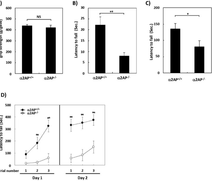
The a2AP2/2 mice exhibited no evident abnormalities in a
top or a wire indicated that the latency to fall was significantly shorter in a2AP2/2 mice than in WT mice (Figs. 1B and C).
Moreover, in the first trial of the rotarod test for motor coordination and balance, the latency to fall tended to be shorter in a2AP2/2 mice compared with WT mice (Fig. 1D). These
results suggest that the deletion of thea2AP gene causes impaired
motor function, but no major differences in muscle strength. The rotarod test is also used to evaluate motor learning by repeating trials. Both types of mice exhibited increased latencies to fall, but the latency of thea2AP2/2mice was obviously shorter
than that of the WT mice, suggesting that the deletion of thea2AP
gene also causes impaired motor learning.
Impaired Cognitive Function ina2ap2/2Mice Compared with WT Mice
To determine the effects of a2AP deficiency on the cognitive
function, the Y-maze test and Morris water maze (MWM) test
were performed. In the Y-maze test, there were minimal differences in the total numbers of arm entries, which indicates the amount of spontaneous behavior, between thea2AP2/2and
WT mice (Fig. 2A). However, the alteration behavior, indicating the working memory, was significantly lower in thea2AP2/2mice
compared with the WT mice (Fig. 2B). In the training sessions of the MWM test, the latency to reach the platform gradually decreased in both types of mice by the repeated training sessions, but the latency of thea2AP2/2mice was a couple of fold longer
than that of the WT mice (Fig. 2C). The swimming speeds of the WT and a2AP2/2 mice were 17.760.4 and 15.460.7 cm/sec,
respectively. However, the subtle difference in the swimming speeds could not account for the more than 2 fold difference in the escape latency between the WT anda2AP2/2mice. Furthermore,
in the probe test, the number of crossings in the quadrant where the platform had been and the number of crossings over the platform area were significantly lower in thea2AP2/2mice than
Figure 1. The impaired motor function ina2AP2/2mice compared with WT mice.The grip strength was measured by the traction test (A).
There were no differences between thea2AP2/2anda2AP+/+mice (WT anda2AP2/2mice, n = 26 and 15, respectively). The wire hang test using a
cage top (B) and wire hang test using a wire (C) showed impaired motor function ina2AP2/2mice compared with WT mice (WT anda2AP2/2mice,
n = 14 and 10, respectively). The rotarod test showed that thea2AP2/2mice exhibited impaired motor function and learning (WT anda2AP2/2mice,
n = 16 and 14, respectively) (D). The values represent the means6S.E. Significance was evaluated using Student’st-test or an ANOVA with a LSD
post-hoc test. *P,0.05, **P,0.01.
doi:10.1371/journal.pone.0097947.g001
in the WT mice (Figs. 2D and E, respectively). These results indicate that the deletion of thea2AP gene also caused impaired
cognitive function.
Next, we assessed the effects of a2AP deficiency on affect
memory. We confirmed that there was no significant difference in the pain sensitivity betweena2AP2/2and WT mice by the hot
Figure 2. The impaired working memory and spatial memory ina2AP2/2mice compared with WT mice.The Y-maze test showed that
there was little effect of thea2AP deficiency on the spontaneous behavior of the mice (A), but the working memory was impaired in thea2AP2/2
mice compared with WT mice (B) (WT anda2AP2/2mice, n = 12 and 8, respectively). The results of the training sessions are shown in C. The number
of crossings in each quadrant and the number of crossings over the platform in the probe test are shown in D and E, respectively. The Morris water
maze test showed thata2AP2/2mice exhibited impairments in their spatial memory (C–E) (n = 8). The values represent the means6S.E. Significance
was evaluated using Student’st-test or an ANOVA with a LSD post-hoc test. *P,0.05, **P,0.01.
plate test (Figure S1). In the passive avoidance test, the latency to enter the dark compartment was significantly shorter ina2AP2/2
mice than that of WT mice after the electric shock (Fig. 3A). A similar result was found in the shuttle avoidance test. The total escape scores of thea2AP2/2mice were lower than those of the
WT mice on both days (Fig. 3B). Interestingly, the escape scores in the first half of the test were significantly lower in thea2AP2/2
mice compared with the WT mice, but in the second half of the test, there was no significant difference between the types of mice, suggesting that the deletion of thea2AP causes a cognitive delay.
Moreover, in the contextual fear conditioning task, the freezing time and the number of occurrences of tail-rattling were remarkably lower in thea2AP2/2mice compared with the WT
mice (Figs. 3C and D). In addition, in the cued fear conditioning task, the freezing time of the a2AP2/2 mice was significantly
lower than that of the WT mice (Fig. 3E). We also confirmed that
a2AP2/2 mice have an intact electric shock-induced acute
freezing response (Figure S2). These results suggest that a2AP
plays an important role in affect memory, and possibly in the etiology of posttraumatic stress disorder (PTSD) related to the fear-based memory formation [20].
The Effects of the Deletion of thea2ap Gene on Anxiety-like or Depression-Anxiety-like Behavior
To determine the effects of a2AP deficiency on anxiety-like
behavior, the open field test and dark/light transition test were performed ina2AP2/2and WT mice. In the open filed test, the
time spent in the center of the field was significantly shorter in the
a2AP2/2mice than in the WT mice (Fig. 4A), while there was no
significant differences in the distance in the center nor the total distance moved in the field (Figs. 4B and C). In the dark/light transition test, there was little difference in the time spent in the dark compartment (Fig. 4D), but the first latency for thea2AP2/2
mice to move to the light compartment was remarkably longer than that of the WT mice (Fig. 4E). In addition, the number of transitions was significantly lower in thea2AP2/2than in the WT
mice (Fig. 4F). These results suggest that the deletion of a2AP
induces anxiety-like behavior.
Next, to examine the involvement ofa2AP in depression-like
behavior, the tail suspension test was performed. The a2AP2/2
mice showed a longer first latency to immobility and a shorter immobility time compared with WT mice (Figs. 5A and B), indicating that a2AP2/2 mice exhibit anti-depression-like
reac-tion.
The tPA/plasmin proteolytic cascade is known for its throm-bolytic ability [21], while the extracellular proteolysis involved in the tPA/plasmin cascade has been reported to extend to synaptic plasticity in the CNS. The cleavage of brain-derived neurotrophic factor (BDNF) by tPA/plasmin cascade is critical for the production of L-LTP in the hippocampus [4]. However, the laminin degradation induced by plasmin results in the impairment of LTP in the hippocampus [22]. Plasmin also disrupts mossy fiber axon guidance [23], and excess tPA/plasmin suppresses dendri-togenesis and synapdendri-togenesis [24]. These reports imply that the tPA/plasmin cascade affects synaptic plasticity both positively and negatively. The proteolysis induced by plasmin potentiates N-methyl-D-aspartate receptor responses [25,26], which may be involved in the enhancement of synaptic plasticity and/or neuronal toxicity. The degradation of the extracellular matrix activated by the tPA/plasmin cascade upregulates the motility of dendritic spine [27], and induces neuronal detachment and apoptosis [28]. These molecular mechanisms may underlie the effects of tPA/plasmin on neuronal remodeling. a2AP is widely
known to be a principal physiological inhibitor of plasmin in the
thrombolytic system [1], and is expressed in various regions in the brain, including the hippocampus, cortex and cerebellum [18]. However, only a few studies have focused on the role ofa2AP in
the CNS. One study demonstrated that chronic injection ofa2AP
into the medial prefrontal cortex inhibits the NGF maturation induced by plasmin, causing cholinergic degeneration and cognitive impairment [29]. On the other hand, we previously demonstrated that a2AP induces dendritic elongation and
branching, which are essential for synaptic plasticity and memory formation, independent of plasmin [9]. Therefore, fibrinolytic factors, including tPA, plasmin and a2AP, regulate synaptic
plasticity, both positively and negatively, in accordance with physiological and pathological conditions.
In this study, we demonstrated that deletion of thea2AP gene
results in an impaired cognitive function. Such failure in the regulation of plasmin activity in the brain and/or the loss ofa
2AP-regulating neuronal outgrowth may lead to impaired synaptic plasticity in a2AP2/2 mice. In addition, recent studies have
demonstrated that tPA/plasmin plays a role in the disruption of the blood-brain barrier [30,31], and that plasminogen potentiates thrombin neurotoxicity in cases of intracerebral hemorrhage [32]. The increase in the permeability of the blood-brain barrier induced by plasmin is possibly involved in the impaired cognitive function observed in a2AP2/2 mice. A previous clinical study
showed that the plasma levels ofa2AP are lower in elderly people
[33], suggesting that a2AP may play a role in the age-related
cognitive decline. We also demonstrated thata2AP is involved in
the development of anxiety- and depression-like behaviors. Deletion of the tPA gene affects anxiety-like behavior [14,16], although it has not been sufficiently addressed whether plasmin plays a role in this effect. BDNF, which is converted to the mature form by extracellular proteases, including plasmin [34], has an antidepressant-like effect [35,36]. Hence, free plasmin may mediate the anti-depression-like reactions noted ina2AP2/2mice.
In summary, we herein demonstrated, for the first time, that
a2AP is a crucial mediator of the motor and cognitive functions as
well as anxiety- and depression-like behaviors. tPA, plasmin and
a2AP are each involved in the processes of neuronal migration,
dendritic growth and synaptic plasticity [4,9–16,21–29,37], suggesting that a2AP has an effect on both brain development
and neuronal plasticity during behavior. Although further research is needed to elucidate the timing at whicha2AP regulates neuronal
functions and the molecular mechanisms underlying the regula-tory processes controlled by a2AP, our findings provide new
insight into the physiological and pathological roles ofa2AP in the
brain.
Materials and Methods
Animals
The a2AP-deficient (a2AP2/2) mice were generated by
homologous recombination using 129/SvJ RW4 embryonic stem cells, as described previously [19]. To minimize the variability in the genetic background of the mice, we repeatedly backcrossed
a2AP2/2mice to C57BL/6J mice for more than 10 generations
($99.9% of C57BL/6J genomic background). Thea2AP2/2and
control a2AP+/+ (wild-type, WT) mice used for behavioral tests
were homozygously bred. All experiments were approved by the institutional animal care and use committee of Doshisha Women’s College (Permit number: Y13-022), and were performed in accordance with the institutional guidelines. All efforts were made to minimize suffering.
Experimentally-naive mice were used for the Morris water maze test, rotarod test, passive avoidance test and shuttle avoidance test.
The other behavioral tests were performed with the same group of mice in accordance with the behavioral test battery. The order of the behavioral tests was as follows: open field test, dark/light transition test, wire hang test, traction test, hot plate test, Y-maze test, fear conditioning test and tail suspension test.
Traction Test
The grip strength of the mice was measured with a traction meter (BrainScience idea.Co., Ltd., Osaka, Japan). Mice were made to grasp metal mesh with all four limbs, and were slowly pulled back using the tail. The maximum tension was recorded and normalized to the body weight.
Wire Hang Test
Mice were placed on a cage top or a wire. The cage top was slightly shaken to encourage gripping of the bars, and then was slowly inverted. The latency to fall was then measured up to 60 sec.
Rotarod Test
The rotarod test was performed using a Rota-Rod Treadmill (Muromachi Kikai, Tokyo, Japan). Mice were made to walk for a maximum of 500 sec. The time it took for a mouse to lose its balance on the rod was measured. Mice received three trials at 20 min intervals per day, and the trials were repeated on the next day.
Figure 3. The impaired affect memory ina2AP2/2mice compared with WT mice.The passive avoidance test (A) (WT anda2AP2/2mice,
n = 24 and 31, respectively), and shuttle avoidance test (B) (n = 4 for both groups) showed that the deletion ofa2AP results in impaired affect
memory. Furthermore,a2AP2/2mice exhibited impaired memory in the contextual fear conditioning test (C and D), and the cued fear conditioning
task (E) (WT anda2AP2/2mice, n = 14 and 10, respectively). The values represent the means6S.E. Significance was evaluated using Student’st-test or
Y-maze Test
The Y-maze apparatus consisted of three arms whose walls had different markings. Mice were placed in the center and allowed to explore the apparatus for 8 min, while being monitored by a video-tracking system (SMART, Panlab, Barcelona, Spain). The alteration behavior was calculated as the ratio of the number of alterations to the total number of arm entries minus 2.
Morris Water Maze Test
Mice received visible platform pre-training on the first day, followed by hidden platform training for two days. In the hidden platform training, five sessions consisting of four trials were performed on two days. Mice were placed into the pool from four different directions in each of the four trials. The escape latency was measured. In the probe test, mice were allowed to swim for 60 sec. The number of crossings in each quadrant and the number of crossings over the platform area was analyzed by a video-tracking system (SMART, Panlab).
Passive Avoidance Test
A chamber was divided into bright and dark compartments by a partition with a window. The floor of the dark compartment was composed of stainless steel rods connected to an electric shock generator. Mice were placed into the bright compartment, allowed to freely explore the chamber until they entered the dark compartment, and then were returned to the home cage. The third time the mice entered the dark compartment, they received an electric shock. On the second and third days, the time taken to enter the dark compartment was measured, up to a maximum of 5 min.
Shuttle Avoidance Test
A chamber composed of two compartments, Compartments A and B, was divided by a partition with an openable gate. The floor of Compartment A was made of a stainless steel electric shock generator. Mice were placed in Compartment A with the gate closed, and allowed to explore for 5 min. A conditional stimulus (CS), 5 sec of tone, was given to the mice, while the gate was
Figure 4. The effects ofa2AP deficiency on anxiety-like behavior.The open field test showed thata2AP2/2mice exhibited a shorter time in
the center, indicating anxiety-like behavior (A), while there was little difference in the distance moved (B and C) (WT anda2AP2/2mice, n = 11 and 10,
respectively). In the dark/light transition test, there was little difference in the time spent in the dark compartment (D). However, the first latency to
enter the light compartment was longer (E), and the number of transitions in thea2AP2/2mice was lower than in the WT mice (F), indicating that the
deletion ofa2AP caused anxiety-like behavior (WT anda2AP2/2mice, n = 11 and 10, respectively). The values represent the means6S.E. Significance
was evaluated using Student’st-test or an ANOVA with a LSD post-hoc test. *P,0.05, **P,0.01.
doi:10.1371/journal.pone.0097947.g004
opened. The mice received the unconditional stimulus (US), a 5-sec electric shock, 10 5-sec after the CS (CS-US trial). The number of entries into Compartment B was counted during the CS presentation and 10 sec before the US. Each mouse received 20 CS-US trials each day, for two days. The number of successful responses, where the mice moved to Compartment B, was defined escape score.
Fear Conditioning Test
Mice were habituated in a chamber for 2 min, followed by a 30-sec light stimulus (CS) paired with an electric shock during the last 3 sec of the CS (US). The mice were allowed to stay in the chamber for another 2 min. The CS-US pairing was repeated four times, and then the mice were returned to the home cage. The mice were placed back into the fear conditioning chamber 24 h later without the light cue, and the freezing time and the number of occurrences of tail-rattling were measured for 5 min (contextual conditioning). The mice were placed into a columnar chamber 1 hour later, and given a light stimulus (CS), and the freezing time was measured (cued conditioning). The assessment was performed by an observer blinded to the mouse genotype.
Open Field Test
Mice were placed into the center of a circular open field, and allowed to explore for 30 min. The total distance moved and time spent in the center area were analyzed by a video-tracking system (SMART, Panlab).
Dark/Light Transition Test
Mice were placed into a dark compartment of a two-compartment chamber, and were allowed to explore for 10 min. while being monitored by a video-tracking system (SMART, Panlab).
Tail Suspension Test
The tails of mice were fastened to a bar, and then the mice were observed for 6 min. The first latency to immobility and immobility
time were measured. The assessment was performed by an observer blinded to the mouse genotype.
Statistical Analysis
Student’s ttest or an ANOVA with a LSD post-hoc test was performed to evaluate the significance of the data.
Supporting Information
Figure S1 No difference in the reaction to the heat
betweena2AP2/2and WT mice.Mice were placed on a 55 or
58uC hot plate, and the first latency for them to lick their paws was measured. There was no significant difference in the reaction to the heat between a2AP2/2 and WT mice (WT and a2AP2/2
mice, n = 14 and 10, respectively). The values represent the means
6S.E. Significance was evaluated using Student’st-test.
(TIF)
Figure S2 The intact electric shock-induced acute
freezing response ina2AP2/2 mice.Mice were habituated
in a different shape of chamber from the one used for the fear conditioning test for 2 min, followed by an electric shock during the last 3 sec. The freezing time was measured while the mice were allowed to stay in the chamber for another 2 min. This trial was repeated four times. There was no difference in the freezing time after each electric shock. (WT anda2AP2/2mice, n = 14 and 10,
respectively). Significance was evaluated using an ANOVA with a LSD post-hoc test.
(TIF)
Author Contributions
Conceived and designed the experiments: EK YK. Performed the experiments: EK KI HK. Analyzed the data: EK KI HK. Contributed reagents/materials/analysis tools: EK. Wrote the paper: EK. Interpreta-tion of the data: EK YK OM HM.
Figure 5. The effects ofa2AP deficiency on depression-like behavior.The tail suspension test showed thata2AP2/2mice exhibited an
anti-depression-like reaction (A and B) (WT anda2AP2/2 mice, n = 14 and 9, respectively). The values represent the means6S.E. Significance was
evaluated using Student’st-test or an ANOVA with a LSD post-hoc test. *P,0.05, **P,0.01.
References
1. Collen D (1976) Identification and some properties of a new fast-reacting plasmin inhibitor in human plasma. Eur J Biochem 69: 209–216.
2. Hortin GL, Gibson BL and Fok KF (1988) Alpha 2-antiplasmin’s carboxy-terminal lysine residue is a major site of interaction with plasmin. Biochem Biophys Res Commun 155: 591–596.
3. Baricos WH, Cortez SL, el-Dahr SS, Schnaper HW (1995) ECM degradation by cultured human mesangial cells is mediated by a PA/plasmin/MMP-2 cascade. Kidney Int 47: 1039–1047.
4. Pang PT, Teng HK, Zaitsev E, Woo NT, Sakata K, et al. (2004) Cleavage of proBDNF by tPA/plasmin is essential for long-term hippocampal plasticity. Science 306: 487–491.
5. Lu BG, Sofian T, Law RH, Coughlin PB and Horvath AJ (2011) Contribution of conserved lysine residues in the alpha2-antiplasmin C terminus to plasmin binding and inhibition. J Biol Chem 286: 24544–24552.
6. Kanno Y, Kuroki A, Okada K, Tomogane K, Ueshima S, et al. (2007) Alpha2-antiplasmin is involved in the production of transforming growth factor beta1 and fibrosis. J Thromb Haemost 5: 2266–2273.
7. Kanno Y, Kawashita E, Minamida M, Kaneiwa A, Okada K, et al. (2010) alpha2-antiplasmin is associated with the progression of fibrosis. Am J Pathol 176: 238–245.
8. Kanno Y, Kawashita E, Kokado A, Okada K, Ueshima S, et al. (2013) Alpha2-antiplasmin regulates the development of dermal fibrosis by PGF(2)asynthesis
through ATGL/iPLA(2). Arthritis Rheum 65: 492–502.
9. Kawashita E, Kanno Y, Asayama H, Okada K, Ueshima S, et al. (2013) Involvement ofa2-antiplasmin in dendritic growth of hippocampal neurons.
J Neurochem 126: 58–69.
10. Huang YY, Bach ME, Lipp HP, Zhuo M, Wolfer DP, et al. (1996) Mice lacking the gene encoding tissue-type plasminogen activator show a selective interference with late-phase long-term potentiation in both Schaffer collateral and mossy fiber pathways. Proc Natl Acad Sci U S A 93: 8699–8704. 11. Baranes D, Lederfein D, Huang YY, Chen M, Bailey CH, et al. (1998) Tissue
plasminogen activator contributes to the late phase of LTP and to synaptic growth in the hippocampal mossy fiber pathway. Neuron 21: 813–825. 12. Madani R, Hulo S, Toni N, Madani H, Steimer T, et al. (1999) Enhanced
hippocampal long-term potentiation and learning by increased neuronal expression of tissue-type plasminogen activator in transgenic mice. EMBO J 18: 3007–3012.
13. Pawlak R, Nagai N, Urano T, Napiorkowska-Pawlak D, Ihara H, et al. (2002) Rapid, specific and active site-catalyzed effect of tissue-plasminogen activator on hippocampus-dependent learning in mice. Neuroscience 113: 995–1001. 14. Pawlak R, Magarinos AM, Melchor J, McEwen B, Strickland S (2003) Tissue
plasminogen activator in the amygdala is critical for stress-induced anxiety-like behavior. Nat Neurosci 6: 168–174.
15. Pawlak R, Rao BS, Melchor JP, Chattarji S, McEwen B, et al. (2005) Tissue plasminogen activator and plasminogen mediate stress-induced decline of neuronal and cognitive functions in the mouse hippocampus. Proc Natl Acad Sci U S A 102: 18201–18206.
16. Bahi A, Dreyer JL (2012) Hippocampus-specific deletion of tissue plasminogen activator ‘‘tPA’’ in adult mice impairs depression- and anxiety-like behaviors. Eur Neuropsychopharmacol 22: 672–682.
17. Saito H, Goodnough LT, Knowles BB and Aden DP (1982) Synthesis and secretion of alpha 2-plasmin inhibitor by established human liver cell lines. Proc Natl Acad Sci U S A 79: 5684–5687.
18. Menoud PA, Sappino N, Boudal-Khoshbeen M, Vassalli JD and Sappino AP (1996) The kidney is a major site of alpha(2)-antiplasmin production. J Clin Invest 97: 2478–2484.
19. Okada K, Lijnen HR, Dewerchin M, Belayew A, Matsuo O, et al. (1997) Characterization and targeting of the murine alpha2-antiplasmin gene. Thromb Haemost 78: 1104–1110.
20. Parsons RG, Ressler KJ (2013) Implications of memory modulation for post-traumatic stress and fear disorders. Nat Neurosci 16: 146–153.
21. Collen D (2001) Ham-Wasserman lecture: role of the plasminogen system in fibrin-homeostasis and tissue remodeling. Hematology Am Soc Hematol Educ Program 2001: 1–9.
22. Nakagami Y, Abe K, Nishiyama N, Matsuki N (2000) Laminin degradation by plasmin regulates long-term potentiation. J Neurosci 20: 2003–2010. 23. Mizuhashi S, Nishiyama N, Matsuki N, Ikegaya Y (2001) Cyclic
nucleotide-mediated regulation of hippocampal mossy fiber development: a target-specific guidance. J Neurosci 21: 6181–6194.
24. Li J, Yu L, Gu X, Ma Y, Pasqualini R, et al. (2013) Tissue plasminogen activator regulates Purkinje neuron development and survival. Proc Natl Acad Sci U S A 110: In press.
25. Yuan H, Vance KM, Junge CE, Geballe MT, Snyder JP, et al. (2009) The serine protease plasmin cleaves the amino-terminal domain of the NR2A subunit to relieve zinc inhibition of the N-methyl-D-aspartate receptors. J Biol Chem 284: 12862–12873.
26. Mannaioni G, Orr AG, Hamill CE, Yuan H, Pedone KH, et al. (2008) Plasmin potentiates synaptic N-methyl-D-aspartate receptor function in hippocampal neurons through activation of protease-activated receptor-1. J Biol Chem 283: 20600–20611.
27. Oray S, Majewska A, Sur M (2004) Dendritic spine dynamics are regulated by monocular deprivation and extracellular matrix degradation. Neuron 44: 1021– 1030.
28. Ho-Tin-Noe´ B, Enslen H, Doeuvre L, Corsi JM, Lijnen HR, et al. (2009) Role of plasminogen activation in neuronal organization and survival. Mol Cell Neurosci 42: 288–295.
29. Allard S, Leon WC, Pakavathkumar P, Bruno MA, Ribeiro-da-Silva A, et al. (2012) Impact of the NGF maturation and degradation pathway on the cortical cholinergic system phenotype. J Neurosci 32: 2002–2012.
30. Yao Y, Tsirka SE (2011) Truncation of monocyte chemoattractant protein 1 by plasmin promotes blood-brain barrier disruption. J Cell Sci 124: 1486–1495. 31. Freeman R, Niego B, R Croucher D, Pedersen LO, L Medcalf R (2014) t-PA,
but not desmoteplase, induces a plasmin-dependent opening of a blood-brain barrier model under normoxic and ischaemic conditions which can be reversed within a limited time frame. Brain Res In press.
32. Fujimoto S, Katsuki H, Ohnishi M, Takagi M, Kume T, et al. (2008) Plasminogen potentiates thrombin cytotoxicity and contributes to pathology of intracerebral hemorrhage in rats. J Cereb Blood Flow Metab 28: 506–515. 33. Stout RW, Crawford VL, McDermott MJ, Rocks MJ, Morris TC (1996)
Seasonal changes in. haemostatic factors in young and elderly subjects. Age Ageing 25: 256–258.
34. Lee R, Kermani P, Teng KK, Hempstead BL (2001) Regulation of cell survival by secreted proneurotrophins. Science 294: 1945–1948.
35. Siuciak JA, Lewis DR, Wiegand SJ, Lindsay RM (1997) Antidepressant-like effect of brain-derived neurotrophic factor (BDNF). Pharmacol Biochem Behav 6: 131–137.
36. Shirayama Y, Chen AC, Nakagawa S, Russell DS, Duman RS (2002) Brain-derived neurotrophic factor produces antidepressant effects in behavioral models of depression. J Neurosci 22: 3251–3261.
37. Seeds NW, Basham ME, Haffke SP (1999) Neuronal migration is retarded in mice lacking the tissue plasminogen activator gene. Proc Natl Acad Sci U S A 96: 14118–14123.