Hemolysis, Elevated Liver Enzymes, Low
Platelets Syndrome Superimposed on
Hemolytic Uremic Syndrome
Síndrome HELLP sobreposta a síndrome
hemolítica-urêmica
Inês Martins
1Madalena Gomes Conceição
2Paulo Pereira Gomes
3Nuno Clode
1,41Department of Obstetrics, Gynecology and Reproductive Medicine, Centro Hospitalar Lisboa Norte, Hospital Santa Maria (CHLN-HSM), Lisbon, Portugal
2Department of Obstetrics and Gynecology, Hospital CUF Descobertas, Lisbon, Portugal
3Intensive Care Unit, Hospital CUF Descobertas, Lisbon, Portugal 4Department of Medicine, Universidade de Lisboa, Lisbon, Portugal
Rev Bras Ginecol Obstet 2017;39:195–198.
Address for correspondence Inês Martins, MD, MSci, Departamento de Obstetrícia, Ginecologia e Medicina da Reprodução do CHLN-HSM, Avenida Prof. Egas Moniz, 1649-035 Lisboa, Portugal
(e-mail: [email protected]).
Keywords
►
HELLP syndrome
►
hemolytic uremic
syndrome
►
thrombotic
microangiopathies
Abstract
A pregnancy complicated by typical hemolytic uremic syndrome (HUS) and hemolysis,
elevated liver enzymes, low platelets (HELLP) syndrome is reported. At 20 weeks of
gestation, a case of HUS was diagnosed, with Shiga toxin-producing Escherichia coli
identi
fi
ed. Plasmapheresis allowed continuation of the pregnancy for 5 weeks.
Super-imposed preeclampsia and HELLP syndrome were diagnosed after the establishment of
nephrotic range proteinuria, hypertension and recurrence of hemolysis. This is a
singular case, as it demonstrates that HELLP syndrome can superimpose upon HUS, a
fact that can impact future research on reproductive immunology. It also reminds
clinicians that the overlapping of clinical and laboratory
fi
ndings in HELLP syndrome
makes the diagnosis of other thrombotic microangiopathies during pregnancy a
clinical challenge.
Palavras-chave
►
síndrome HELLP
►
síndrome
hemolítica-urêmica
►
microangiopatias
trombóticas
Resumo
Descreve-se um caso clínico em que às 20 semanas de gestação é diagnosticada
síndrome hemolítica-urêmica com isolamento de Escherichia coli produtora de toxina
Shiga. A terapêutica com plasmaferese permitiu a manutenção da gravidez ao longo de
5 semanas. O surgimento de proteinúria nefrótica, hipertensão e recorrência de
hemólise conduziu ao diagnóstico de sobreposição de pré-eclâmpsia e síndrome
HELLP. Este caso é singular, pois demonstra que a síndrome HELLP pode se sobrepor
a uma síndrome hemolítica-urêmica, um fato que pode in
fl
uenciar futuras
investiga-ções no âmbito de imunologia reprodutiva. Reforça-se, ainda, que o diagnóstico de
outras microangiopatias trombóticas durante a gestação é um desa
fi
o clínico
ocasio-nado pela sobreposição de manifestações clínicas e laboratoriais com a síndrome
HELLP.
received
September 12, 2016 accepted after revision December 30, 2016 published online March 24, 2017
DOIhttp://dx.doi.org/ 10.1055/s-0037-1600124. ISSN 0100-7203.
Copyright © 2017 by Thieme-Revinter Publicações Ltda, Rio de Janeiro, Brazil THIEME
Introduction
Thrombotic microangiopathies are characterized by en-dothelial cell dysfunction, microvascular thrombosis, plate-let consumption and reduced tissue perfusion.1,2 As blood flows through partially constricted microvessels, hemolysis occurs.2In pregnancy, its most common form is the hemo-lysis, elevated liver enzymes, low platelets (HELLP) syn-drome.1–3 Its pathogenesis is not established, and hypotheses include alteration in the maternal-fetal immune balance, or placenta instigated liver-targeted acute infl am-matory condition, or a severe form of preeclampsia.2 The microvascular injury in HELLP syndrome is systemic, but mainly affects the liver.1–3
Hemolytic uremic syndrome (HUS) is another life-threa-tening thrombotic microangiopathy characterized by hemo-lytic anemia, thrombocytopenia and acute kidney injury.4 Hypertension can occur due to the activation of the renin-angiotensin system associated with renal ischemia.4Shiga toxin-producing Escherichia coli (STEC) is responsible for the typical form of the disease.4 It infects humans after the ingestion of contaminated food or water. Abdominal pain and hemorrhagic diarrhea develop within 3–7 days, and up to 10% of the infected patients (usually serotype O157) develop HUS 4–10 days after the onset of diarrhea. The microvascular injury is mediated by the Shiga toxin, and mainly affects the kidney.1,2 Non-STEC-HUS, or atypical HUS (aHUS), can have multiple etiologies associated with the dysregulation of the complement activation.2 At the onset, it is difficult to distinguish HUS/aHUS from thrombotic thrombocytopenic purpura (TTP), a throm-botic microangiopathy associated with markedly reduced activity of von Willebrand factor-cleaving metalloprotease (ADAMTS13).2,4
The authors report a case where both typical HUS and HELLP syndromes were diagnosed, elucidating the clinical challenge associated with this differential diagnosis.
Case Presentation
A 42-year-old pregnant patient (after in vitro fertilization), gravida 3 para 0, without significant medical or surgical history, was admitted at 20 weeks of gestation for severe abdominal pain and bloody diarrhea that started after in-gesting vegetables grown in a family farm. Beside slight uterine contractions, no other symptoms/signs were present. On blood tests, only a slight leukocytosis (14,800/mm3) and a high C-reactive protein (3.3 mg/dl) were noted. Since the abdominal ultrasound suggested appendicitis, a laparo-scopic appendectomy was performed. The histological ex-amination did not confirm the diagnosis. The patient, asymptomatic, was discharged the next day.
Four days later, she was reevaluated for sudden onset of peripheral and periorbital edema and oliguria, with normal blood pressure (BP). Laboratory tests (►Table 1, line1) were consistent with microangiopathic hemolytic anemia, throm-bocytopenia and acute kidney injury. There was also micro-scopic hematuria and proteinuria, and a mild elevation of the aspartate aminotransferase (AST) and the alanine amino-transferase (ALT). Surgical complications were excluded. An obstetric ultrasound scan excluded signs of fetal distress, malformations or abnormalities. She was admitted for sus-pected typical HUS to the intensive care unit (ICU), where plasmapheresis was immediately started after red blood cell transfusion. Stool and blood samples were negative, but STEC serotype O157:H7 was identified in the urine culture. Plasma exchange therapy was performed daily for 4 days. Thereafter, it was performed according to the platelet count and the lactate dehydrogenase (LDH) level. The replacement fluid was 100% fresh frozen plasma (FFP) in thefirst two sessions and later, 50% albumin solution/50% FFP. Continuous hemo-diafiltration was performed to avoid fluid overload (the volume balance was zero during the treatment). Anticoagu-lation was performed with heparin in the priming of thefilter and by continuous infusion during the replacement. After an
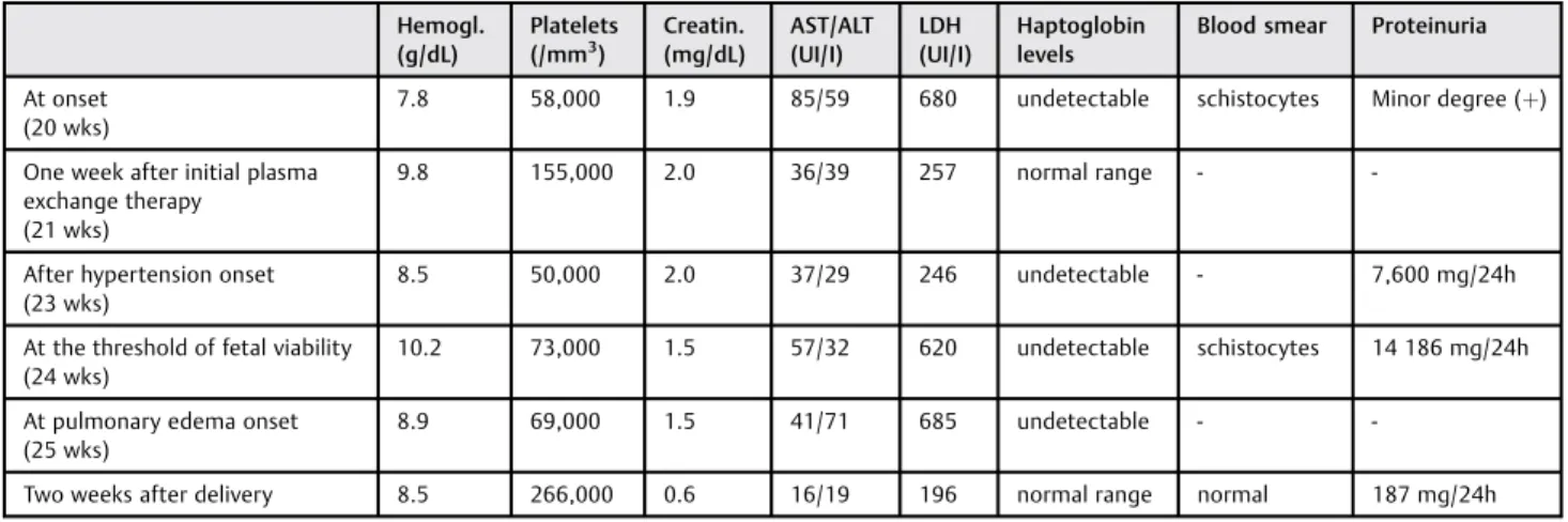
Table 1 Summary of patient’s blood and urine test results throughout the progression of the disease
Hemogl. (g/dL)
Platelets (/mm3)
Creatin. (mg/dL)
AST/ALT (UI/I)
LDH (UI/I)
Haptoglobin levels
Blood smear Proteinuria
At onset (20 wks)
7.8 58,000 1.9 85/59 680 undetectable schistocytes Minor degree (þ)
One week after initial plasma exchange therapy
(21 wks)
9.8 155,000 2.0 36/39 257 normal range -
-After hypertension onset (23 wks)
8.5 50,000 2.0 37/29 246 undetectable - 7,600 mg/24h
At the threshold of fetal viability (24 wks)
10.2 73,000 1.5 57/32 620 undetectable schistocytes 14 186 mg/24h
At pulmonary edema onset (25 wks)
8.9 69,000 1.5 41/71 685 undetectable -
-Two weeks after delivery 8.5 266,000 0.6 16/19 196 normal range normal 187 mg/24h
Abbreviations: ALT, alanine aminotransferase; AST, aspartate aminotransferase; Creatin., creatinine; Hemogl., hemoglobin; LDH, lactate dehydrogenase; wks, weeks.
Rev Bras Ginecol Obstet Vol. 39 No. 4/2017
initial worsening of the acute kidney injury (creatinine 2.5 mg/dl, max), the tests started to improve (►Table 1, line 2). The patient developed hypertension (>160/110 mm Hg)
12 days after admission, requiring diuretic and antihyper-tensive therapies. The laboratory tests (►Table 1, line 3) were consistent with the recurrence of hemolysis and thrombo-cytopenia, mild elevation of transaminases and nephrotic range proteinuria. Complement activity and complement proteins were within the normal range, and the auto-anti-bodies were negative. Later, ADAMTS13 assessment was normal. Hemolytic uremic syndrome with superimposed preeclampsia and HELLP syndrome were considered on the differential diagnosis.
The patient refused to terminate the pregnancy. After the red blood cell transfusion and hypertension control, the tests were stable, and she was transferred to a tertiary-care hospital at 24 weeks of gestation (►Table 1, line 4). A course of betamethasone was administered for fetal lung maturity. An ultrasound scan revealed fetal growth restriction with normal umbilical artery Doppler velocimetry and amniotic fluid index.
At 25 weeks of gestation, she suffered a sudden relapse of microangiopathic hemolysis (►Table 1, line 5) and developed severe hypertension (200/120 mm Hg) and acute pulmonary edema refractory to therapy, suggesting severe preeclampsia and HELLP syndrome. After discussion with the parents, delivery of the fetus was decided upon. While on parenteral magnesium sulfate therapy, an emergency cesarean section was performed. The neonate (600 g, Apgar scores 4–7-7) was admitted at the neonatal ICU, but died the next day.
After the delivery, and without specific therapy, there was remission of the hemolysis, thrombocytopenia, proteinuria and the renal and hepatic injuries (►Table 1, line 6). The patient was discharged two weeks later on antihypertensive therapy, and no further recurrences have been diagnosed.
Discussion
Both HUS and HELLP syndromes are characterized by hemo-lysis and thrombocytopenia, with or without hypertension or proteinuria.3–5Nonetheless, HELLP syndrome is far more frequent than HUS. Preeclampsia complicates 3–5% of preg-nancies, and HELLP syndrome develops in 4–14% of women with severe preeclampsia.2On the other hand, diagnosis of HUS/TTP during pregnancy or postpartum is very rare, probably 1 case in 100,000 pregnancies.1 Furthermore, STEC-HUS represents only 5–10% of the HUS cases in adults, compared with>90% of HUS in children.4
Although both are thrombotic microangiopathies, the microvascular injury in the HELLP syndrome affects mostly the liver, while the kidney is the organ most affected in the HUS.1,2,4,5Hence, transaminases are significantly elevated in the HELLP syndrome, but only mildly elevated in the HUS.3–5 In contrast, significant acute kidney injury is infrequently encountered in patients with HELLP syndrome,2but it is an important feature in the clinical course of HUS.1,4
Hemolytic uremic syndrome / thrombotic thrombocyto-penic purpura should be suspected when hemolysis and
thrombocytopenia are diagnosed during pregnancy in the presence of a normotensive pregnant woman, or when associated with hypertension and/or proteinuria before 24 weeks of gestation, or when the renal injury is worse than the liver injury.1,5Whenever the patient presents with hemorrhagic diarrhea, STEC-HUS should be considered .4In the case reported, the onset of the disease resembled the pathophysiology of STEC-HUS, with hemorrhagic diarrhea preceding hemolytic anemia, thrombocytopenia and acute kidney injury. In the absence of hypertension and severe liver injury, and presenting before 24 weeks of gestation, the diagnosis, although rare, appeared clear. During the diarrheic period, it is important to keep in mind that increased leucocyte count in the blood and increased serum C-reactive protein are risk factors for developing HUS after STEC infec-tion,4as occurred in this case.
Plasma exchange is the first line treatment for aHUS. Conversely, it has no beneficial effect to reduce nephropathy in STEC-HUS.4 Being difficult to distinguish typical HUS/ aHUS/TTP at the onset, plasmapheresis without delay is suggested for most adult patients.4While waiting for culture results, plasmapheresis was, therefore, instituted in our patient. The isolation of STEC serotype O157:H7 further corroborated the initial diagnosis, although it was isolated in the urine but not in the stool culture.
Later development of severe hypertension and nephrotic range proteinuria, associated with the recurrence of hemo-lytic anemia and thrombocytopenia, led to an even greater diagnosis challenge. Within the hypertensive disorders of pregnancy.3 HELLP syndrome has been categorized as a complicated form of severe preeclampsia.2Although hyper-tension can also occur in the HUS, this usually happens during the acute phase of the disease, and does not occur with nephrotic proteinuria.4 Complete remission of the hemolysis, thrombocytopenia, proteinuria and the renal and hepatic injuries without a specific therapy after delivery corroborates the diagnosis of HELLP syndrome.1,2,5To our knowledge, there are no other reports of HELLP superim-posed on HUS.
There are no systematic reviews or randomized trials on HUS during pregnancy, probably due to its rarity. Stella et al5 reported a series of 14 cases of TTP/HUS in pregnancy or postpartum encountered in 4 tertiary-care centers over 8 years, which highlights the difficult differential diagnosis with HELLP. All the patients diagnosed with TTP/HUS before 24 weeks of gestation (n¼6) survived: 3 had term live births, 1 had a missed abortion, and 2 had intrauterine fetal death. On the case here reported, the pregnancy was con-tinued for 5 weeks after the diagnosis of typical HUS, beyond the threshold of fetal viability. Despite this accomplishment, unfortunately, the neonate died.
The differential diagnosis also considered unusual HELLP syndrome postponed by plasma exchange therapy. A few studies investigated the role of plasma exchange in HELLP syndrome, and found that it can be an effective treatment for patients with persistent HELLP syndrome after delivery.6,7 After therapeutic plasma exchange, LDH and AST levels decrease, and platelets count increases. The mechanism of
Rev Bras Ginecol Obstet Vol. 39 No. 4/2017
action is still controversial. In theory, plasmapheresis re-moves from circulation aggregating, procoagulant and va-soactive factors released from activated platelets and endothelial cells, leading to renal and hepatic function improvement.
This case reinforces the difficulty in diagnosing HUS during pregnancy, considering that several clinical and la-boratoryfindings overlap with the far more frequent HELLP syndrome. Hemolysis, elevated liver enzymes, low platelets syndrome superimposed upon STEC-HUS is a unique feature of this case that can impact future research.
References
1 Sibai BM. Imitators of severe pre-eclampsia. Semin Perinatol 2009;33(03):196–205
2 Fang CJ, Richards A, Liszewski MK, Kavanagh D, Atkinson JP. Advances in understanding of pathogenesis of aHUS and HELLP. Br J Haematol 2008;143(03):336–348
3 American College of Obstetricians and Gynecologists; Task Force on Hypertension in Pregnancy. Hypertension in pregnancy. Re-port of the American College of Obstetricians and Gynecologists’ Task Force on Hypertension in Pregnancy. Obstet Gynecol 2013; 122(05):1122–1131
4 Igarashi T, Ito S, Sako M, et al; Study group for establishing guidelines for the diagnosis and therapy of hemolytic uremic syndromeGuidelines for the management and investigation of hemolytic uremic syndrome. Clin Exp Nephrol 2014;18(04): 525–557
5 Stella CL, Dacus J, Guzman E, et al. The diagnostic dilemma of thrombotic thrombocytopenic purpura/hemolytic uremic syn-drome in the obstetric triage and emergency department: lessons from 4 tertiary hospitals. Am J Obstet Gynecol 2009;200(04):381. e1–381.e6
6 Eser B, Guven M, Unal A, et al. The role of plasma exchange in HELLP syndrome. Clin Appl Thromb Hemost 2005;11(02): 211–217
7 Erkurt MA, Berber I, Berktas HB, et al. A life-saving therapy in Class I HELLP syndrome: Therapeutic plasma exchange. Transfus Apheresis Sci 2015;52(02):194–198
Rev Bras Ginecol Obstet Vol. 39 No. 4/2017