C
a s eR
e p o Rt1 9 2 Arq Bras Oftalmol. 2017;80(3):192-5 http://dx.doi.org/10.5935/0004-2749.20170047
INTRODUCTION
Central serous chorioretinopathy (CSC) is a disease characterized by detachment of the serous retinal pigment epithelium (RPE), typically in the macular region, and that is associated with fluid extravasation
into the subretinal space(1). The disease mainly affects middle-aged
men, although women are also commonly affected(2). Classified into
acute and chronic types, the more common acute type usually has a benign course with spontaneous resolution and good final visual
acuity(1). However, persistent complaints of metamorphopsia or of
changes in color perception have been observed after CSC has
re-solved(3). These changes may be explained by injury to the ellipsoid
band, demonstrated by optical coherence tomography (OCT), but
the damage may also be imperceptible(4). Given that OCT alone
pro-vides limited resolution for detailed photoreceptor assessment, it is necessary to use more sensitive methods, like adaptive optics, which can compensate for optical aberrations and individually assess the
photoreceptors(4,5). Herein, we report our experience using adaptive
optics to assess a patient with persistent mild visual alterations in the left eye four years after resolution of CSC.
CASE REPORT
A 47-year-old white woman was seen at the Retina and Vitreous Sector of our hospital in 2012 with symptoms of metamorphopsia
and central scotoma in the left eye (oculus sinister; OS). She had no
comorbidities, but reported that she was going through a period of intense emotional stress at the time. Her best-corrected visual acuity
(BCVA) was 20/20 for the right eye (oculus dexter, OD; +0.5 D, sphere); by
contrast, the BCVA for OS was 20/80 (+0.75 D, sphere). OCT revealed serous retinal detachment in OS (Figure 1), so a diagnosis of CSC was made and the patient’s condition was observed. After three months of persistent signs and symptoms, we then started therapy with 250 mg acetazolamide tablets to be taken orally every 8 h for 20 days,
and applied focal laser to the OS macular region using a PUREPOINT®
Laser (Alcon, USA) set to 100 mW, 50 ms duration, and 33 firings. One month after treatment, clinical improvement was observed, and resolution of serous detachment was confirmed by OCT.
Although the patient suffered no new episodes during follow up, she continued to complain of diminished visual acuity in OS. Objec-tively, her BCVA was 20/20 in OD (+1.5 D sphere) and 20/25 in OS (+1.0 D sphere). Biomicroscopy revealed transparent corneas, trophic irises, transparent crystalline lenses, and intraocular pressures of 12 mmHg in both eyes. Posterior pole fundoscopy was also within normal limits in OD, but macular topography showed some areas with white and yellowish spots in OS (Figure 1). Therefore, OCT was done. This showed a normal OD with preserved foveal depression, an intact inner layer, and an intact outer layer. Although foveal depression was preserved, and the inner and outer layers were intact in OS, there
Photoreceptor assessment using adaptive optics in resolved
central serous chorioretinopathy
Avaliação dos fotorreceptores com adaptive optics na coriorretinopatia serosa central cicatrizada
André Luiz BrAgA MeireLLes1, MuriLo WendeBorn rodrigues1, ALine FernAndA guirAdo¹, rodrigo Jorge1
Submitted for publication: November 18, 2016 Accepted for publication: January 11, 2017
1 Department of Ophthalmology, Otorhinolaringology and Head and Neck Surgery, Universidade de
São Paulo, Ribeirão Preto, SP, Brazil.
Funding: No specific financial support was available for this study.
Disclosure of potential conflicts of interest: None of the authors have any potential conflict of interest to disclose.
Corresponding author: André Luiz Braga Meirelles. Av. Bandeirantes, 3.900 - Ribeirão Preto - SP - 14049-900 - E-mail: [email protected]
ABSTRACT
We present a case of a patient with resolved central serous chorioretinopathy (CSC) in the left eye with persistent mild visual alterations 4 years after the resolution of the disease. Left eye exam revealed a best corrected visual acuity of 20/25 and a slight change of macular pigmentation. Optical coherence tomography revealed only minor irregularities in the topography of retinal pigment epithelium and Bruch’s membrane. Adaptive optics (AO) optics demonstrated lower density, spacing, and changes in the photoreceptor mosaic pattern in the left eye than in the right eye, suggesting that CSC may cause damage to cones after clinical recovery. We conclude that AO can provide additional information to assist in the treatment and follow-up of patients with CSC or other macular pathologies.
Keywords: Central serous chorioretinopathy; Optical imaging/methods tomo-graphy; Optical coherence; Retina
RESUMO
Apresentamos o relato de caso de paciente com coriorretinopatia serosa central (CSC) cicatrizada em olho esquerdo e queixa de discreta alteração visual, mesmo após quatro anos da resolução do quadro. O exame do olho esquerdo apresenta melhor acuidade visual corrigida de 20/25 e discreta alteração de pigmentação macular. Tomografia de coerência óptica (OCT) apresentou apenas pequenas irregularidades em topografia de EPR e Bruch. Foi realizado exame com Adaptive Optics (AO), evidenciando valores inferiores de densidade, espaçamento e alterações no padrão de mosaico dos fotorreceptores em olho esquerdo quando comparado com olho direito, sugerindo que a CSC pode causar danos em cones, mesmo após uma recuperação considerada satisfatória. Concluímos que o AO é uma tecnologia que traz novas informações para auxiliar o tratamento e seguimento dos pacientes com CSC ou outras patologias maculares.
Me i r e l l e s Al, e tA l.
1 9 3 Arq Bras Oftalmol. 2017;80(3):192-5
were some irregular areas on the outer limiting membrane, as well as discrete elevations in both the RPE and Bruch’s membrane (Figure 1). In OS, the laser marks in the macular and inferior macular regions were also evident in infrared.
We then performed assessment by adaptive optics using an RTX-1™ Adaptive Optics Retinal Camera (Imagine Eyes, Orsay, France), based on non-coherence of current illuminated by 850 nm central illumi nation, a wavelength with a 4° × 4° image field (1.2 × 1.2 mm on the retina), and a focus interval of 600 mm. Thus, high-resolution images were obtained of the cones. The images were processed using the software provided by CK V.0.1 and AOdetect V.0.1 (Imagine Eyes, France).
The adaptive optics analysis revealed mean cone densities of 16,121 and of 8,694 in the central regions of OD and OS, respectively (reference value: 20,000 to 30,000). The mean intercellular spacing was 8.65 µm in OD and 11.07 µm in OS (reference value: 15 to 20 µm). The greatest proportion of neighboring cells (Voronoi polygons) was six in both eyes (reference value, six), of which 40.2% were in OD and 35.8% were in OS (Figures 2 and 3).
DISCUSSION
Even though the morphological findings and visual acuity of the patient were within normal limits, her photoreceptor density and the spacing between photoreceptors were below the expected reference values. However, this does not mean that there was a cone deficiency in her eyes, because adaptive optics only detects part of the internal segment of the photoreceptor forming the mosaic. In addition, va-riations in the cone mosaic may occur due to physiological changes in reflectance caused by differences in the phases of photoreceptor
phototransduction, axial length, or even circadian rhythm(6).
Comparison of the two eyes showed that not only were there clearly lower density and lower spacing in OS but that there were also changes in the mosaic pattern of the photoreceptors. This suggested that the resolved CSC caused changes in the cones, even after satisfactory clinical recovery. Ooto et al.(7) reported similar results
in a case series of patients with CSC, and observed that there was a direct relationship between the intensity of photoreceptor changes measured by adaptive optics and the final BCVA.
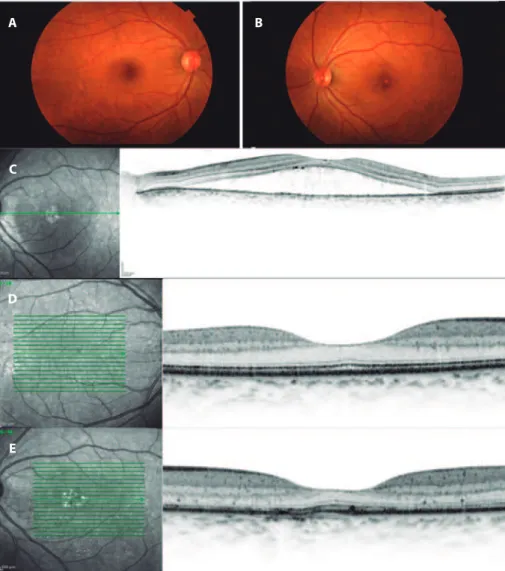
Figure 1. Retinography in OD (A) and OS (B). Yellowish lesions can be seen in the foveal topography of OS. The OCT of OS for the new case (C) shows separation of the photoreceptor layer of the RPE by foveal topography. A recent OCT of OD (D) is normal and in OS (E) there are marks of focal laser in the inferior macular area (infrared) and irregularities of the outer limiting membrane and Bruch/RPE complex in the selected section. OCT= optical coherence tomography; OD= oculus dexter; OS= oculus sinister; RPE= retinal pigment epithelium.
A
C
D
E
Ph o t o r e c e P t o ra s s e s s m e n tu s i n ga d a P t i v eo P t i c si nr e s o lv e dc e n t r a ls e r o u sc h o r i o r e t i n o Pat h y
1 9 4 Arq Bras Oftalmol. 2017;80(3):192-5
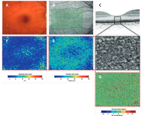
Figure 2. Imaging in OD. Retinography (A), infrared photograph (B), enlarged section obtained by OCT (C), enlarged central area obtained by adaptive optics (D). Also, density mosaics (E), spacing (F), and closeness (G). A near normal pattern of distribution, density, and closeness can be seen. Abbreviations: OCT, Optical coherence tomography; OD, oculus dexter; OS, oculus sinister.
A B C
D
G E
F
Figure 3. Imaging in OS. Retinography (A), infrared photograph (B), enlarged section obtained by OCT (C), and enlarged central area obtained by adaptive optics (D). Also, density mosaics (E), spacing (F), and closeness (G). Compared with OD imaging (Figure 2), there is a more irregular cone mosaic, and there are areas of reduced density and increased spacing, with a worse pattern of photoreceptor closeness. This suggests a loss of cones after CSC. CSC= central serous chorioretinopathy; OCT= optical coherence tomography; OD= oculus dexter; OS= oculus sinister.
A B C
D
G E
Me i r e l l e s Al, e tA l.
1 9 5 Arq Bras Oftalmol. 2017;80(3):192-5
In the present case, we showed that adaptive optics can provide additional data and reveal changes that might otherwise be missed if using OCT alone. Adaptive optics may represent a new assessment tool for use in the treatment and follow-up of patients with CSC or other macular diseases.
REFERENCES
1. Nicholson B, Noble J, Forooghian F, Meyerle C. Central serous chorioretinopathy: update on pathophysiology and treatment. Surv Ophthalmol. 2013;58(2):103-26. 2. Liew G, Quin G, Gillies M, Fraser-Bell S. Central serous chorioretinopathy: a review of
epidemiology and pathophysiology. Clin Exp Ophthalmol. 2013;41(2):201-14.
3. Loo RH, Scott IU, Flynn HW Jr, Gass JD, Murray TG, Lewis ML, et al. Factors associated with reduced visual acuity during long-term follow-up of patients with idiopathic central serous chorioretinopathy. Retina. 2002;22(1):19-24.
4. Ojima Y, Tsujikawa A, Yamashiro K, Ooto S, Tamura H, Yoshimura N. Restoration of outer segments of foveal photoreceptors after resolution of central serous choriore-tinopathy. Jpn J Ophthalmol. 2010;54(1):55-60.
5. Williams DR. Imaging single cells in the living retina. Vision Res. 2011;51(13):1379-96. 6. Pallikaris A, Williams DR, Hofer H. The reflectance of single cones in the living human
eye. Invest Ophthalmol Vis Sci. 2003;44(10):4580-92.
7. Ooto S, Hangai M, Sakamoto A, et al. High-resolution imaging of resolved central serous chorioretinopathy using adaptive optics scanning laser ophthalmoscopy. Ophthalmology. 2010;117(9):1800-9.