DOI: 10.1590/0004-282X20140200
ARTICLE
Aura-like features and photophobia in
sightless migraine patients
Características de fenômenos aura-relacionados e da fotofobia em enxaquecosos
deficientes visuais
Greice Cardoso de Carvalho Silva1, Cristiana Pessoa de Queiroz Faria Góes2, Maurice Borges Vincent2
Migraine is a neurological disorder that frequently in-volves cortical paroxysmal dysfunction expressed as aura1.
he most frequent type of aura is visual, sometimes expe
-rienced as fortiication spectra, known as teichopsia. his typical zigzag, horseshoe-shaped expanding bright percep
-tion, usually perceived as complex interlacing lines named “chevaux de frises” by Gowers, followed by a bean-shaped loss of visual acuity, strongly suggests the involvement of visual processing areas. Clinical, experimental, and neu -roimaging evidence demonstrates that cortical spread-ing depression (CSD) is the phenomenon underlyspread-ing the
pathophysiology of bizarre visual perceptions among
migraineurs. Computerized models simulating a CSD wave
passing through cortical area V1 convincingly reproduce the teichopsia pattern2.
he cortical function is deeply dependent on neuronal in
-put. Depriving the cortex of its natural input may inluence the cerebral function and change the cortical maps both lo -cally and distantly3. In blind individuals, the occipital cortex is
critical for tactile Braille reading4. he visual cortex is pivotal
in the pathophysiology of the migraine aura, the objective of the present study was to investigate whether the phenotype of migraine-related visual phenomena would change in am -aurotic patients.
1Hospital Naval Marcílio Dias, Rio de Janeiro RJ, Brasil;
2Faculdade de Medicina, Hospital Universitário Clementino Fraga Filho, Universidade Federal do Rio de Janeiro, Rio de Janeiro RJ, Brasil.
Correspondence: Maurice Vincent; Av. das Américas, 1155 / 504; 22631-000 Rio de Janeiro RJ, Brasil; E-mail: [email protected] Conflict of interest: There is no conlict of interest to declare.
Received 16 December 2013; Received in inal form 30 August 2014; Accepted 18 September 2014. ABSTRACT
Migraine is a central nervous system disorder frequently expressed with paroxysmal visual dysfunctions. Objective: To test the hypoth-esis that a normal visual input is vital for migrainous aura and photophobia. Methods: We studied the migraine-related visual distur-bances in 8 sightless migraineurs identiied among 200 visually impaired subjects. Results: The main indings were visual aura and photophobia disappearance along with the development of blindness, abnormal aura [too short, colorful (e.g., blue or ire-like), auditory in nature, or different in shape (round forms)], and the lack of photophobia. Conclusions: We propose that the aura duration should be accepted as shorter in visually impaired subjects. The changes in the aura phenotype observed in our patients may be a result of cerebral plasticity induced by visual impairment and/or the lack of a visual input per se. Integrity of visual pathways plays a key role in migraine visual aura and photophobia.
Keywords: migraine with aura, subnormal vision, blindness, visual aura, photophobia
RESUMO
A enxaqueca é doença neurológica frequentemente associada a anormalidades visuais transitórias. Objetivo: Testar a hipótese de que a visão normal é importante para o fenótipo da aura e da fotofobia. Métodos: Estudamos 8 enxaquecosos deicientes visuais identiicados em uma população de 200 indivíduos com visão subnormal. Resultados: Os principais achados foram: o desaparecimento da aura visual e da fotofobia com o início da cegueira; a ocorrência de aura atípica – muito curta, colorida (p. ex. azul, ou cor de fogo) auditiva ou diferente na forma (arredondadas); e a ausência de fotofobia. Conclusões: Propomos que a duração da aura possa ser admitida como mais curta em pessoas com deiciência visual. As mudanças no fenótipo da aura observadas nos nossos pacientes pode ser o resultado da plasticidade cerebral induzida pela deiciência visual e/ou a deiciência visual em si. A integridade da via visual desempenha um papel crucial na aura enxaquecosa e na fotofobia.
950 Arq Neuropsiquiatr 2014;72(12):949-953
METHODS
Two hundred visually impaired adult subjects (VIS) were randomly recruited at the Instituto Benjamin Constant (IBC),
a Brazilian reference treatment and education center for the unsighted located in Rio de Janeiro. One of the authors
(GCCS), a fully trained neurologist, personally interviewed and examined all VIS from October 2010 to May 2011 and from March to July 2012. Study procedures included head
-ache and visual impairment history recording, neurological examination, and review of IBC medical records for access to complete ophthalmological and clinical data. his descrip
-tive, observational study was approved by the Hospital Naval Marcílio Dias (HNMD) ethics committee (56/2010) and au
-thorized by the IBC board on August 10, 2010. All VIS who volunteered to participate signed or ingerprinted an in -formed consent.
A standardized interview was used to espy migraine patients among VIS. he International Classiication of Headache Disorders version II (ICHD-II)5 was applied to distinguish migraineurs fulilling the criteria for the follow
-ing disorders: migraine without aura, typical aura with mi
-grainous headache, typical aura with nonmi-grainous head
-ache, typical aura without head-ache, probable migraine without aura, and probable migraine with aura, present for at least 1 year prior to the interview. Visual phenomena not necessarily meeting the ICHD-II criteria for a typical aura, either atypical in form, color, and/or duration, as well as occurring without, before, or during the headache (being migrainous or not) were also considered. VIS had to suf -fer from acquired or congenital amaurosis according to the
10th revision of the International Classiication of Diseases
(ICD-10) code H54, corresponding to vision impairment categories 3, 4, or 5 (WHO Study Group on the Prevention of Blindness, Geneva, 6-10 November l972, WHO Technical Report Series No. 518, 1973).
Exclusion criteria comprise ICHD-II familial hemiple
-gic migraine, sporadic hemiple-gic migraine, basilar-type mi
-graine, retinal mi-graine, migraine complications, chronic mi
-graine, migrainous status, persistent aura without infarction, migrainous infarction, seizure triggered by migraine, proba
-ble chronic migraine, migraine onset after the age of 50 years, and comorbidities (systemic or not) potentially causing or manifesting as a migraine-like disorder.
RESULTS
From the 200 individuals initially interviewed, 9 were ex
-cluded: three refused to participate, four because of cognitive impairment, one because of a brain tumor, and one because of marijuana addiction. Among the 191 remaining VIS, 63 ful
-illed the criteria for amaurosis. In this subgroup, 35 (55.5%)
reported recurrent headaches, mostly of the migrainous type (23%-37.0%), as opposed to 12 (19%) with other head
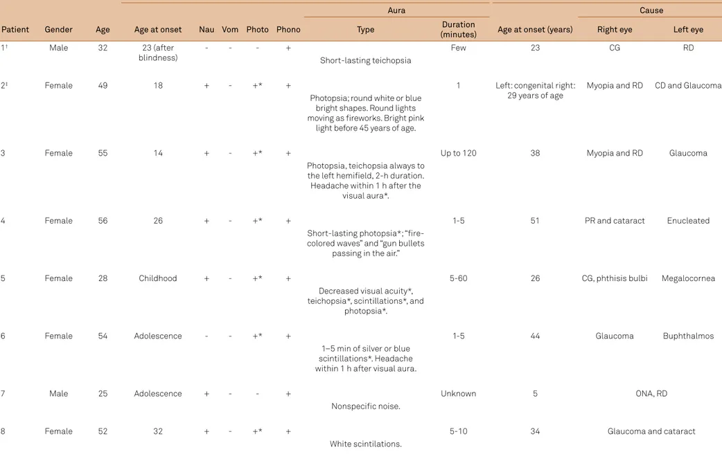
-ache disorders and 28 (44%) head-ache-free subjects. Eight (12.7%, 6 females, 40.00 ± 13.06 years, range 25-56) migraine with aura patients were selected and studied in further detail (Table 1), of which seven presented with visual aura not nec
-essarily fulilling Internacional Headache Society (IHS) diag -nostic criteria5 (in 4 patients, the aura lasted for less than 5 min), and 1 patient reported an auditory aura (uncharacter
-istic noise). In the majority of our population, the headache started before the visual impairment.
Among the seven amaurotic patients with visual aura, ive failed to present an aura following the onset of sightless
-ness. In patient 1, diagnosed as typical aura with headache not fulilling migraine criteria, and patient 7, diagnosed with an atypical auditory aura, the migraine with aura started af
-ter the visual impairment. Patient 4 used to perceive scin
-tillations during some headache attacks prior to blindness,
changing to perceptions in color and forms after the vision
loss. Regardless of the headache type, visual aura symptoms were atypical because of length (too short), colour (blue, sil
-ver, or ire-like), and/or shape (round shapes). Photophobia, reported by all patients in whom the headache preceded the blindness in time (n = 6), disappeared after the visual impair
-ment. he remaining 2 subjects denied this symptom.
DISCUSSION
In this study, we looked for the lifetime prevalence of mi
-graine in a population of visually impaired subjects to address the disease phenotype in this particular population. After blind
-ness, only 1 patient continued to express the aura as before. his suggests that a normal visual input and processing are crucial for aura expression in migraine. Besides, in one patient who became blind years before the migraine attacks, auditory rather than visual phenomena emerged, possibly relecting an aura-like symptom. Photophobia is clearly dependent on vision because it is not present concomitantly with total blindness.
Kowacs et al. looked for migraine among VIS and found a 6-month prevalence ratio of 14.28% (compared with 37% in our material). In their series, 4 subjects had aura: 1 totally blind patient presented an auditory aura and 2 subnormal vi -sion individuals reported an atypical visual aura lasting for 2–4 min. In the last patient aura symptoms disappeared as the vision impairment progressed6. Noseda et al. reported about 6 (2 with an aura) totally blind and 14 (5 with an aura) visually impaired (light perception) migraineurs; however, aura details were not provided7.
Gr
eice Car
doso de Carvalho Silva e
t al
. A
ur
a-lik
e f
ea
tur
es in migr
aine
Table 1. Blind patients with migraine and aura-like symptoms
Headache Blindness
Aura Cause
Patient Gender Age Age at onset Nau Vom Photo Phono Type Duration
(minutes) Age at onset (years) Right eye Left eye
1† Male 32 23 (after
blindness)
- - - +
Short-lasting teichopsia
Few 23 CG RD
2‡ Female 49 18 + - +* +
Photopsia; round white or blue bright shapes. Round lights moving as ireworks. Bright pink
light before 45 years of age.
1 Left: congenital right: 29 years of age
Myopia and RD CD and Glaucoma
3 Female 55 14 + - +* +
Photopsia, teichopsia always to the left hemiield, 2-h duration. Headache within 1 h after the
visual aura*.
Up to 120 38 Myopia and RD Glaucoma
4 Female 56 26 + - +* +
Short-lasting photopsia*; “ire-colored waves” and “gun bullets
passing in the air.”
1-5 51 PR and cataract Enucleated
5 Female 28 Childhood + - +* +
Decreased visual acuity*, teichopsia*, scintillations*, and
photopsia*.
5-60 26 CG, phthisis bulbi Megalocornea
6 Female 54 Adolescence - - +* +
1–5 min of silver or blue scintillations*. Headache within 1 h after visual aura.
1-5 44 Glaucoma Buphthalmos
7 Male 25 Adolescence + - - +
Nonspeciic noise.
Unknown 5 ONA, RD
8 Female 52 32 + - +* +
White scintilations.
952 Arq Neuropsiquiatr 2014;72(12):949-953
particularly responsive to interictal visual stimulation10. here are limited data on CSD provoked by stimuli either than sei
-zure induction or direct chemical/mechanical stimulation; but it is well known that migraine attacks may be triggered by visual stimuli such as sunlight11, red-green lickering12, or
particular striped patterns13. Exercise was used to induce a
migraine aura during which a CSD-like wave was detected in the brain14. In rats, darkness seems to reduce the CSD propa
-gation15. Sound may trigger CSD in rats16, and light has also been reported to precipitate CSD in rabbits hyperexcited by subconvulsive doses of pentylenetetrazol17. hus, there is clin
-ical and experimental evidence that increased neuronal
excit-ability renders the migrainous cortex more vulnerable to CSD. Plasticity is an intrinsic endowment of the brain18, and blindness induces substantial cerebral reorganization19. Blind individuals may compensate for the lack of sight by develop
-ing hyperefective nonvisual senses. Although tactile input ac -tivates the visual cortex in sighted individuals20, blinds activate
their primary visual areas V1 and V2 during Braille reading as a compensatory cross-modal strategy.4 Blind subjects develop
auditory abilities beyond controls21, and the absolute pitch is
more prevalent in blind musicians22. Sightless people detect
much more eiciently moving sounds23.
herefore, in theory, noncortical blindness should not pre
-clude CSD from occurring in a migraineous brain because the cortex remains functional and is activated by various inputs.
Since the occipital cortex may change from processing visual information to processing other sensory modalities in
sight-lessness, we hypothesized that the lack of visual input would render the visual cortex less susceptible to CSD, leading to the cessation of the visual auras. Likewise, as sight deteriorates be
-fore blindness, subnormal vision would partially reduce CSD susceptibility, possibly explaining the atypical visual auras,
mostly too short or expressing color changes not commonly
observed in ordinary migraine. Alternatively, the cortical reor
-ganization induced by the lack of vision could still allow CSD but change just its clinical expression, leading to the discon
-tinuation of visual phenomena. If this hypothesis is correct, the auditory aura referred by patient 7 could be the result of an ab
-errant activation as a result of blindness.
It is noteworthy that visual phenomena were signii
-cantly shorter in many VIS. According to ICHD-II, an aura should develop gradually over 5–20 min and last for less than 60 min. Each aura symptom lasts, by deinition, ≥5 and ≤60
min5. hese time restrictions are maintained in the present ICHD-3 beta version24. In half of our cases, the visual symp
-toms possibly relecting aura were shorter than 5 min. Based on these results, we suggest that the aura limit of 5–60 min should not encompass VIS. More subnormal vision subjects must be studied to conirm this inding. he reason as why aura is shorter among VIS remains unknown; however, it may be the result of cortical plasticity and/or a lack of visual
input to the visual cortex.
Auditory aura is rarely observed in migraine, but audi
-tory hallucination has been reported as a type of acoustic
aura25. Our case and the one described by Kowacs et al.6 in
a small sightless population contrast the rarity of this
phe-nomenon in ordinary migraine, indicating that acoustic aura may be signiicantly more common among blind subjects. Speculatively, if the lack of vision result in the overactivation of cortical areas related to hearing, this could favor an audi -tory aura in this population.
Photophobia is present in >80% of migraine patients26 and seems to be closely inter-related with trigeminal pain. Painful27 and optokinetic simuli28 increase light discomfort in migraineu-rs. In controls, luminous stimulation at certain intensities did not lead to the activation of the visual cortex without concomi
-tant pain as detected by positron emission tomography; how
-ever, in migraineurs, cortical activation occurred at the same lu
-minous stimulation levels without simultaneous pain, which in
turn potentiated the light activation even further29.
he pathophysiology of photophobia is largely unknown and may involve non-image formation visual pathways. he fact that mice lacking rods and cones showed normal sup -pression of pineal melatonin in response to monochromatic
light was the irst evidence of an additional ocular photore
-ception in mammals. Melanopsin, an opsin/vitamin A-based photopigment sensitive to blue stimuli present in some reti -nal ganglion cells30, may play a key role in migraine photo -phobia. In contrast with the present indings, sensitivity to light was reported by blind migraineurs who had preserva -tion of pupillary light response and circadian
photoentrain-ment; however, in patients with bilateral enucleation or dam
-age to the optic nerves, photophobia was absent7, indicating that the image formation input is not obligatory for photo
-phobia. As shown in rats, it is possible that retina-originat
-ed ibers projecting to the thalamus induce retinal photoac
-tivation in a discrete area at the posterior thalamus where dura-sensitive ibers converge7. In our cases, no blind patient admitted pain exacerbation or discomfort by light. his is probably because of the fact that rods, cones, and melanop
-sin perceptions were all damaged in our cases.
he small number of subjects and the lack of congenitally blind subjects are the limitations of the present study. We are completely aware that memory biases could have somehow interfered with our results. Reviewing the medical records was a possible way to minimize this drawback. However, we are conident that the cases reported here in suggest that vi -sual function is crucial for the expression of a migraine aura
and photophobia, regardless of the visual cortex integrity.
Acknowledgments
References
1. Pietrobon D, Moskowitz MA. Pathophysiology of migraine. Annu Rev Physiol. 2013;75(1):365-91. http://dx.doi.org/10.1146/annurev-physiol-030212-183717
2. Dahlem MA, Engelmann R, Lowel S, Muller SC. Does the migraine aura relect cortical organization? Eur J Neurosci. 2000;12(2):767-70. http://dx.doi.org/10.1046/j.1460-9568.2000.00995.x
3. Kujala T, Alho K, Naatanen R. Cross-modal reorganization of human cortical functions. Trends Neurosci. 2000;23(3):115-20.
4. Hamilton RH, Pascual-Leone A. Cortical plasticity associated with Braille learning. Trends Cogn Sci. 1998;2:168-74. http://dx.doi. org/10.1016/s0166-2236(99)01504-0
5. Headache Classiication Subcommittee of the International Headache Society. The International Classiication of Headache Disorders: 2nd ed. Cephalalgia. 2004;24(Suppl 1):S9-160.
6. Kowacs PA, Piovesan EJ, Lange MC, Weneck LC, Tatsio CE, Ribas LC et al. Prevalence and clinical features of migraine in a population of visually impaired subjects in Curitiba, Brazil. Cephalalgia. 2001;21(9):900-5. http://dx.doi.org/10.1046/j.1468-2982.2001.00286.x
7. Noseda R, Kainz V, Jakubowski M, Gooley JJ, Saper CB, Digre K et al. A neural mechanism for exacerbation of headache by light. Nat Neurosci. 2010;13(2):239-45. http://dx.doi.org/10.1038/nn.2475
8. Leão AAP. Spreading depression of activity in cerebral cortex. J Neurophysiol. 1944;7:359-90.
9. Welch KM, D’Andrea G, Tepley N, Barkley G, Ramadan NM. The concept of migraine as a state of central neuronal hyperexcitability. Neurol Clin. 1990;8(4):817-28.
10. Vincent M, Pedra E, Mourao-Miranda J, Bramati IE, Henrique AR, Moll J. Enhanced interictal responsiveness of the migraineous visual cortex to incongruent bar stimulation: a functional MRI visual activation study. Cephalalgia. 2003;23(9):860-8. http://dx.doi. org/10.1046/j.1468-2982.2003.00609.x
11. Bekkelund SI, Hindberg K, Bashari H, Godtliebsen F, Alstadhaug KB. Sun-induced migraine attacks in an Arctic population. Cephalalgia. 2011;31(9):992-8. http://dx.doi.org/10.1177/0333102411409071
12. Cao Y, Welch KM, Aurora S, Vikingstad EM. Functional MRI-BOLD of visually triggered headache in patients with migraine. Arch Neurol. 1999;56(5):548-54. http://dx.doi.org/10.1001/archneur.56.5.548
13. Harle DE, Shepherd AJ, Evans BJ. Visual stimuli are common triggers of migraine and are associated with pattern glare. Headache. 2006;46(9):1431-40. http://dx.doi.org/10.1111/j.1526-4610.2006.00585.x
14. Hadjikhani N, Sanchez del Rio M, Wu O, Schwartz D, Bakker D, Fischi B et al. Mechanisms of migraine aura revealed by functional MRI in human visual cortex. Proc Natl Acad Sci U S A. 2001;98(8):4687-92. http://dx.doi.org/10.1073/pnas.071582498
15. Batinga H, Barbosa PP, Ximenes-da-Silva A. Daytime modulation of cortical spreading depression according to blood glucose levels. Neurosci Lett. 2011;491(1):58-62. http://dx.doi.org/10.1016/j. neulet.2011.01.008
16. Vinogradova LV, Kuznetsova GD, Coenen AM. Unilateral cortical spreading depression induced by sound in rats. Brain Res. 2009;1286:201-7. http://dx.doi.org/10.1016/j.brainres.2009.06.047
17. Van Harreveld A, Stamm JS. Cortical responses to metrazol and sensory stimulation in the rabbit. Electroencephalogr Clin Neurophysiol. 1955;7(3):363-70. http://dx.doi.org/10.1016/0013-4694(55)90005-5
18. Pascual-Leone A, Amedi A, Fregni F, Merabet LB. The plastic human brain cortex. Annu Rev Neurosci. 2005;28:377-401. http://dx.doi. org/10.1146/annurev.neuro.27.070203.144216
19. Collignon O, Dormal G, Albouy G, Vandewall G, Voss P, Phillips C et al. Impact of blindness onset on the functional organization and the connectivity of the occipital cortex. Brain. 2013;136(9):2769-83. http://dx.doi.org/10.1093/brain/awt176
20. Sathian K. Visual cortical activity during tactile perception in the sighted and the visually deprived. Dev Psychobiol. 2005;46(3):279-86. http://dx.doi.org/10.1002/dev.20056
21. Morgan M. Sensory perception: supernormal hearing in the blind? Curr Biol. 1999;9(2):R53-4. http://dx.doi.org/10.1016/S0960-9822(99)80009-8
22. Hamilton RH, Pascual-Leone A, Schlaug G. Absolute pitch in blind musicians. Neuroreport. 2004;15(5):803-6. http://dx.doi. org/10.1097/00001756-200404090-00012
23. Lewald J. Exceptional ability of blind humans to hear sound motion: implications for the emergence of auditory space. Neuropsychologia. 2013;51(1):181-6. http://dx.doi.org/10.1016/j. neuropsychologia.2012.11.017
24. Headache Classiication Committee of the International Headache S. The International Classiication of Headache Disorders, 3rd edition (beta version). Cephalalgia. 2013;33(9):629-808. http://dx.doi. org/10.1177/0333102413485658
25. Feltz-Cornelis CM, Biemans H, Timmer J. Hearing voices: does it give your patient a headache? A case of auditory hallucinations as acoustic aura in migraine. Neuropsychiatr Dis Treat. 2012;8:105-11. http://dx.doi.org/10.2147/NDT.S29300
26. Rasmussen BK, Jensen R, Olesen J. A population-based analysis of the diagnostic criteria of the International Headache Society. Cephalalgia. 1991;11(3):129-34. http://dx.doi.org/10.1046/j.1468-2982.1991.1103129.x
27. Drummond PD, Woodhouse A. Painful stimulation of the forehead increases photophobia in migraine sufferers. Cephalalgia. 1993;13(5):321-4. http://dx.doi.org/10.1046/j.1468-2982.1993.1305321.x
28. Drummond PD. Motion sickness and migraine: optokinetic stimulation increases scalp tenderness, pain sensitivity in the ingers and photophobia. Cephalalgia. 2002;22(2):117-24. http://dx.doi. org/10.1046/j.1468-2982.2002.00332.x
29. Boulloche N, Denuelle M, Payoux P, Fabre N, Trotter Y, Geraud G. Photophobia in migraine: an interictal PET study of cortical hyperexcitability and its modulation by pain. J Neurol Neurosurg Psychiatr. 2010;81(9):978-84. http://dx.doi.org/10.1136/ jnnp.2009.190223