Carotid artery dissection
plus
subdural
hematoma after a roller-coaster ride
Dissecção arterial carotídea e hematoma subdural após passeio em montanha-russa
Ana Luíza Vieira de Araújo
1, Leandro Tavares Lucato
2, Gisela Tinone
1, Hélio Alberto Pereira Leitão-Filho
2,
Fábio Iuji Yamamoto
1, Adriana Bastos Conforto
1,3A 31-year-old woman with history of antiphospholipid
syndrome treated with warfarin reported two days after
a roller-coaster ride, sudden onset of paresthesias in
the right side of the face and in the right hand,
followed by headache. Neuroimaging results are shown in
Figures 1 and 2.
The
“
blood/fluid level
”
represented by the interface between
plasma and sedimented blood (Figure 1) can be seen in
hemor-rhages related to bleeding disorders, among other causes
1. We
suspect that the sudden head and neck movements during the
roller-coaster ride contributed to the remarkably unusual
com-bination of subdural hematoma and carotid artery dissection
(Figure 2) in this patient.
Reference
1. Mittal MK, Burkholder DB, Wijdicks EF. Blood-fluid level on computed tomography head: a sign of warfarin-associated intraparenchymal
hemorrhage. Am J Emerg Med. 2012;30(9):2079.e1-2. http://dx.doi. org/10.1016/j.ajem.2011.10.015
1Divisão de Neurologia Clínica, Hospital das Clínicas, Faculdade de Medicina, Universidade de São Paulo, Sao Paulo SP, Brazil; 2Instituto de Radiologia, Hospital das Clínicas, Faculdade de Medicina, Universidade de São Paulo, Sao Paulo SP, Brazil; 3Hospital Israelita Albert Einstein, Sao Paulo SP, Brazil.
Correspondence:Ana Luiza Vieira de Araujo; Hospital das Clínicas, Faculdade de Medicina, Universidade de São Paulo; Av. Dr. Enéas C. Aguiar, 255 / 5084; 05403-000 São Paulo SP, Brasil; E-mail: [email protected].
Authors’contributions:The authors Ana Luíza Vieira de Araújo, Adriana Bastos Conforto and Leandro Tavares Lucato wrote the manuscript. Gisela Tinone, Fábio Iuji Yamamoto and Hélio Alberto Pereira Leitão Filho critically reviewed the manuscript. All authors had full access to the data.
Conflict of interest:There is no conflict of interest to declare.
Received 02 August 2014; Received in final form 21 August 2014; Accepted 09 September 2014.
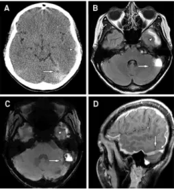
Figure 1.
(A) Initial head CT shows subdural hematomas in the
left temporal region (arrowhead) and in the left tentorium
(arrow), where a blood-fluid level can be observed; (B)
Follow-up brain MR: axial FLAIR; and (C) SWI and sagittal postcontrast
T1-weighted images demonstrate more clearly the same
findings, the blood-fluid level (arrows in B to D) and the
temporal subdural hematoma (arrowheads, B and C).
Figure 2.
Digital subtraction angiography, oblique view, shows
dissection of the proximal left internal carotid artery (arrow).
There is an intimal flap and a patent false lumen.
DOI:10.1590/0004-282X20140169