Exercise Chemosensitivity in Heart Failure: Ventilatory, Chronotropic
and Neurohormonal Responses
Lídia Zytynski Moura, Guilherme Veiga Guimarães, Philippe Vieira Pires, Fátima Cruz, Gabriela Stopa, Edimar
Alcides Bocchi
Instituto do Coração do Hospital de Clínicas/FMUSP, São Paulo, SP - Brazil
Correspondência: Lídia Zytynski Moura •
Pres. Taunay, 1091/72 - Bigorrilho - 80430-000 - Curitiba, PR - Brazil E-mail: [email protected], [email protected]
Manuscript received April 29, 2009; revised manuscript received October 03, 2009; accepted October 23, 2009.
Resumen
Fundamento: Heart failure (HF) is associated with resting increased peripheral and central chemosensitivity which may correlate with an increased ventilatory response to exercise. However, its sensitivity in HF during exercise was never really reported.
Objetivo: We tested if stimulation of central and peripheral chemoreceptors in HF patients could modulate ventilatory, chronotropic, and neurohormonal response during submaximal exercise.
Métodos: We investigated central and peripheral chemosensitivity in 15 HF and 7 control (C) comparing response through three 6 minute walking tests conducted in a treadmill with : room air, hypoxia, and hypercapnia (in a randomic order).
Resultados: RR at room air C and HF was 17±2 and 22±2 (p<.0001); at hypoxia 17±1 and 23±2 (p<.02); at CO25% was 20±2 and 22±5 (p<.02). Tidal volume (TV) at room air was 1.25±0.17 and 1.08±0.19 (p<.01); at hypoxia 1.65±0.34 and 1.2±0.2 (p<.0001); at CO25% 1.55±0.46 and 1.29±0.39 (p<.0001). At rest the increment in HF was higher for VE (C 33±40%, HF 62±94%, p<.01), HR(C 7±10%, HF 10±10%, p<0.05) at rest. During hypoxia exercise increment in HF was higher for RR (C 1±4, HF 11±6,p<.05), HR (C 12±2, HF 14±3, p<.05), VE/VO2 (C -4±18%, HF 24±21%, p<.01), HR/VO2 (C -26±11%, HF 11±5%, p<.01), VE/WD (C 36±10%, 46±14, p<.05%) and HR/WD (C 18±8%, HF
29±11, p<.01). During HF hypoxia exercise NO reduced, and IL-6, aldosterone levels increased. Neurohormonal levels unchanged in C.
Conclusión: Exercise peripheral and central chemosensitivity are increased in HF and may modulate respiratory pattern, cardiac chronotropic, and neurohormonal activity during exercise. (Arq Bras Cardiol 2010; 95(3): 381-391)
Palabras clave: Heart failure; exercise; cardiomyopathies; nitric oxide; Norepinephrine.
Introduction
Peripheral chemoreceptors are accepted as having important modulatory role in the regulation of ventilation during exercise1. Also, central chemoreceptor exerts influence
in pulmonary ventilation, heart rate, blood pressure, and sympathetic activity2.
Many studies have suggested high chemosensitivity in heart failure (HF) patients and consequently ventilation worsening with respiratory patterns alterations3-5.
However, studies about dyspnea mechanisms and the chemoreflex in their great majority are accomplished using chemoreceptor gas sensitization at rest3-6. In fact, few studies
were done with sensitization of peripheral chemoreceptor by hypoxia during exercise, and only in healthy individuals.
In this study we tested the original hypothesis that stimulation of central and peripheral chemoreceptors in HF patients could modulate ventilatory, chronotropic, nitric oxide, systemic blood pressure, and neurohormonal response during submaximal exercise.
Nonstandard abbreviations
O2%, percentage of oxygen; hypoxia, hypoxic isocapnic
test using inspired O2% at 14%; hypercapnia, hypercapnic hyperoxic test using inspired CO2 at 5% and O2 95%; PetCO2, final expiratory CO2 pressure; RR, respiratory rate (rpm); TV, tidal volume(l); VO2, O2 uptake(ml/Kg); slope VE/VCO2,
regression coefficient of the linear regression between the ventilation and VCO2; HR, heart rate(bpm); BNP, B-type natriuretic peptide; WD, walked distance(miles); 6minWT, 6 min treadmill walked test; AV, absolute values; HFNC: HF
group of patients that non conclude de hypoxia; ∆, difference
Methods
Subjects
We studied 22 subjects divided into two groups: Heart failure group with 15 patients and Control group with 7 patients. The inclusion criteria were functional class I or II (NYHA), age at least 21 years old, symptoms and/or ventricular dysfunction for at least six months, optimized medical treatment and stable clinical status for at least three months. The exclusion criteria were ischemic cardiomyopathy, uncontrolled systemic arterial hypertension, obstructive pulmonary disease, diabetes and/or other endocrinopathies, cardiac pacemaker, liver diseases, creatinine serem values
≥ 2.5 mg/dl, stroke in the last six months, osteomuscular
limitations, cardiac cachexia, primary valvular disease, chagasic, restrictive or hypertrophic cardiomyopathy, constrictive pericarditis and primary pulmonary arterial hypertension. The Control group included subjects 21 years old or more, clinically asymptomatic, with no history of heart disease, with no alterations in physical examination or complementary laboratorial exams. All subjects gave written informed consent to participate in this research study, which was approved by institutional ethical board.
Study design
The study was prospective, randomized, case-controlled, and blind for Heart Failure group and Control patients. Before inclusion all subjects underwent an initial clinical and laboratorial evaluation including also transthoracic echocardiogram, myocardial scintigraphy, EKG and cardiopulmonary test.
The six minutes walking test (6minWT)
We measured systolic blood pressure (SBP) and diastolic bloodpressure (DBP) before each exercise test, atthe last minute and at 1-minute recovery.ECG was continuously monitored. Pulmonary ventilation and gasexchange data were determined on a breath-by-breath basis witha computerized system (model Vmax 229 Sensormedics).
The 6minWT was performed using a programmable treadmill without inclination and with patient-controlled
velocity (Series 2000,Marquette Electronics) at least 2 hours after a light meal andwith controlled room temperature (21°C to 23°C). Thepatients were oriented to walk according to Borg’s scale,with exertion level ranging from light to somewhat hard, from11 to 137. The 6-minute walking test associated
with Borg scale, seems to be an accurate measurement of the limitations of a Heart Failure group and has been correlated to individual diary efforts8,9.
Chemosensitization protocol
In the protocol the three 6minWT were performed at the same day in respective three conditions: compressed air (O2 20%) inhalation for room air test, hypoxic test with hypoxia (O2 14%) and balanced nitrogen at 86%, and hypercapnic test with CO2 5% and O2 95% (to blockade peripheral chemoreceptors).
The tree tests were randomly arranged with 20-minute interval between them.
The system used to gas inhalation set to mechanical ventilation Smart model, Takaoka mark, spontaneous mode, which was adapted through rubber elbow in ergoespirometer valve and received the mixture of gazes. The hypercapnia test analysis were performed using data from the mechanical respiratory ventilator and without exhaled gas evaluation due to impossibility of adequate ergoespirometric system evaluation in presence of high CO2 concentrations.
Blood samples were taken at basal and last exercise minute. It was dosed: catecholamine (chromatographic assay)10, BNP
(Biosite)11, aldosterone (radioimmune assay)12, interleukin-6
(Kit Immulite IL-6 (DPC))13, and nitric oxide (NOx, Nitric
Oxide Colorimetric Assay Kit, BioVision, Mountain View, CA).
Statistical analysis
Variables are expressed as mean ±1SD. Data were analyzed using pared Newman-Keuls. The criterion for
significance was p≤0.05.
To compare between room air test and hypoxia or
hypercapnia, we calculated the difference or delta in %(∆) using the equation ∆:(((hypoxia or hypercapnia data) - room
air test)/room air test))x100.
Results
Eleven Heart Failure patients completed all the protocol and 4 patients did not complete the hypoxia protocol (HFNC group). (Table 1) All subjects inspired oxygen fraction approximately 14%, with concomitant reduction in O2% peripheric saturation with constant PetCO2 without the
necessity of CO2 supplementation.
Pre exercise resting isocapnic hypoxic response in comparison with room air test (Figure 1)
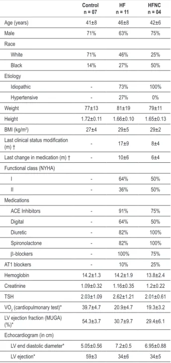
At rest both groups increased ventilation secondary to increment of both respiratory rate and tidal volume. Heart Failure group presented an acute ventilatory response, characterized by superior elevation in ventilation.
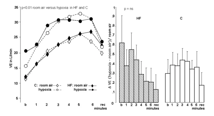
However, the respiratory rate increased was higher in the C whereas tidal volume increase was higher in HF. Control group increased ventilation with greater respiratory rate participation than tidal volume, however HF group increased ventilation with equal tidal volume and respiratory rate contribution.
Both groups demonstrated a pronounced (p<0.02) and equivalent decline in estimated death space.
Control group had higher elevation in oxygen uptake (VO2) than Heart Failure group. Heart Failure group had larger rise in Dheart rate than Control group.The relation between Ventilation/VO2 in Heart Failure group was higher during hypoxia (5.03±1) than room air test (3.13±0.36; p<0.05). Control group didn’t have significant variation between room air test/hypoxia. The relation between heart rate/VO2 was equal during room air test and hypoxia in both groups.
Exercise isocapnic hypoxic test response in comparison with room air test (Table 2) (Figures 1, 2, 3)
Table 1 -Clinical and laboratorial characteristics
Control
n = 07 n = 11HF HFNCn = 04
Age (years) 41±8 46±8 42±6
Male 71% 63% 75%
Race
White 71% 46% 25%
Black 14% 27% 50%
Etiology
Idiopathic - 73% 100%
Hypertensive - 27% 0%
Weight 77±13 81±19 79±11
Height 1.72±0.11 1.66±0.10 1.65±0.13
BMI (kg/m2) 27±4 29±5 29±2
Last clinical status modiication
(m) † - 17±9 8±4
Last change in medication (m) † - 10±6 6±4
Functional class (NYHA)
I - 64% 50%
II - 36% 50%
Medications
ACE Inhibitors - 91% 75%
Digital - 64% 50%
Diuretic - 82% 100%
Spironolactone - 82% 100%
b-blockers - 100% 75%
AT1 blockers - 10% 25%
Hemoglobin 14.2±1.3 14.2±1.9 13.8±2.4
Creatinine 1.09±0.32 1.16±0.35 1.2±0.22
TSH 2.03±1.09 2.62±1.21 2.01±0.61
VO2 (cardiopulmonary test)* 39.7±4.7 20.9±4.7 19.3±3.2
LV ejection fraction (MUGA)
(%)* 54.3±3.7 30.7±9.7 29.4±6.1
Echocardiogram (in cm)
LV end diastolic diameter* 5.05±0.56 7.2±0.5 6.95±0.88
LV ejection* 59±3 34±6 34±5
Values are means ± SD; m - months; VO2 - maximal exercise O2 uptake in ml/kg/ min; TSH - thyroid-stimulating hormone; LV - left ventricular, *p<0.05 between C and HF; †p<0.05 between HF and HFNC.
exercise. However, tidal volume increased more prominently in C than HF, in contrast with respiratory rate which enlarged more in HF. Both groups demonstrated an equivalent decline in estimated dead space with concomitant increase in VO2. HF and C presented more important elevation in heart rate (p<0.006), however HF had larger rise in Dheart rate than C. Both groups presented lowest blood pressure (BP) values at end of exercise and recovery and enhanced slope Ventilation/ VCO2. Walked distance was reduced especially in HF group.
HF group had higher values in Ventilation/VO2 and heart rate/VO2 relations than O2 20%. C didn’t have significant changes in Ventilation/VO2 between them, while heart rate/
VO2 was higher during room air test. Both groups enhanced the relations Ventilation/walked distance and heart rate/ walked distance. Comparing D(room air test/hypoxia) since both groups; all relations were significantly higher in HF group.
Resting hyperoxic hypercapnic test (hypercapnia) response in comparison with room air test
At hypercapnia both groups presented Ventilation lower values, that in C was mainly by tidal volume decrease despite high respiratory rate, but in HF group, it was associated to tidal volume and respiratory rate reduction.
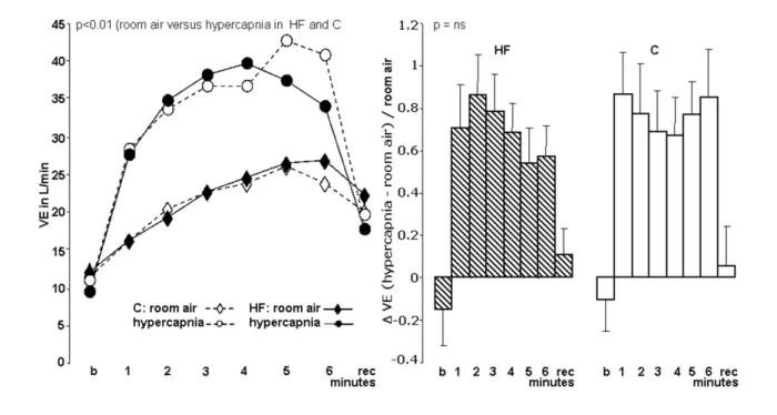
Exercise hyperoxic hypercapnic test (hypercapnia) response in comparison with room air test (Figures 4, 5)
HF and Control group had greater elevation in Ventilation, without significant difference between them. During exercise
both groups increased ∆respiratory rate and ∆tidal volume
(p<0.02), with higher and significant (p<0.01) elevation in Control group than HF group (Figures 5, 6). Both groups hadn’t significant changes in BP and walked distance. Both groups presented a non statistical significant increase in exercise heart rate. HF and Control group course with an acute rise in heart rate in the 1º exercise minute, which was superior to response in hypoxia.
Both groups enhanced the relation Ventilation/walked distance without changes of heart rate/walked distance. Comparing D(room air test/hypercapnia) of Ventilation/walked distance and heart rate/walked distance, they were higher in HF than Control group.
Neurohormonal markers during hypoxia and hypercapnia conditions in comparison with room air test
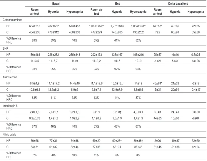
Control group group had not changes in BNP, aldosterone, IL-6 and nitric oxide levels at hypoxia and hypercapnia conditions, but had a tendency to increase catecholamine levels during hypoxia (Table 3).
HF group presented a higher increase in catecholamine and aldosterone levels in room air test in comparison with Control group. During hypoxiaHF group had a significant decrease in NO with concomitant elevation in IL-6 and aldosterone levels. For the hypercapnia test, HF group augmented catecholamine and NO values. HF group increase BNP levels during room air test, however in hypoxia and hypercapnia the BNP levels had a reduction, but all these changes without statistical meaning.
Isocapnic hypoxia -not concluded protocol
The most common symptoms associated to interruption were: dyspnea (3), dizziness (4), pre syncope (4) and “visual darkening” (4), but any of them needs most specific intervention. One patient stops the protocol in the first, one in the second and two of them in the third exercise minute.
Figure 1 -Ventilation of control (C) and heart failure (HF) patients with room air and hypoxia. Values in mean ± SD (percentage %); b - basal minute; rec - recovery
irst minute; VE - pulmonary ventilation; room air - O2 20% test ; hypoxia, hypoxic isocapnic test using inspired O2% at 14%; ∆VE Hypoxia/Room air test: difference(∆) between ventilation in Hypoxia/Room air test data.
Table 2 -Summary of results (Ddelta in percentage)
Parameters
Dhypoxia/room air test Dhypercapnia/Room air test
Rest Exercise Rest Exercise
C HF C HF C HF C HF
VE 33±40† 62±94 37±3 35±13 -10 ±40 -15±50 77±8 69±12
Vt 16±44 24±60 93±47† 26±13 -28±40 -14±40 117±47† 53±6
RR 21±27 6±26 1±4† 9±4 4±40 -8±30 32±10† 14±7
EDS -0,5±52 -18±20 -29±6 -28±7 - - -
-VO2/kg 5±40* 15±68 32±14* 11±6 - - -
-HR 7±10* 10±10 12±2* 14±3 3±2 5±1 9±1* 4±3
BP -0,2±8 -6±11 -1±4 -8±14 4±20 2±10 6±20 1±10
WD - - -5±13† -14±14 - - 20±41 -7±24
Slope VE/VCO2 - 24±31 43±54 - - -
-Relations
VE/VO2 40±8 60±7 -4±18 † 24±21 - - -
-HR/VO2 6±1 5±1 -26±11 † 11±15 - - -
-VE/WD - - 36±10* 46±14 - - 32±21* 53±25
HR/WD - - 18±9† 29±11 - - -5±4† 11±6
Figure 3 -Tidal volume (Vt) of control (C) and heart failure (HF) patients with room air and hypoxia. Values in mean ± SD (percentage %); b - basal minute; rec - recovery
irst minute; room air - O2 20% test; hypoxia - hypoxic isocapnic test using inspired O2% at 14%; ∆VE Hypoxia/Room air test - difference (∆) between ventilation in Hypoxia/Room air test data.
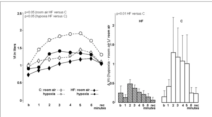
Figure 2 -Respiratory rate (RR) of control (C) and heart failure (HF) patients with room air and hypoxia. Values in mean ± SD (percentage %); b -basal minute; rec -
Figure 4 -Ventilation of control (C) and heart failure (HF) patients with room air and hypercapnia. Values in mean ± SD (percentage %); b -basal minute; rec - recovery
irst minute; VE - pulmonary ventilation; room air - O2 20% test; hypercapnia - hypercapnic hyperoxic test using inspired CO2 at 5% and O2 95%; ∆VE Hypercapnia/Room air test - difference (∆) between ventilation in Hypercapnia/Room air test data.
Figure 5 -Respiratory rate (RR) of control (C) and heart failure (HF) patients with room air and hypercapnia. Values in mean ± SD (percentage %); b - basal minute;
Table 3 -Neurohormonal measurements
Basal End Delta basal/end
Room
air test Hypoxia Hypercapnia Room air test Hypoxia Hypercapnia air testRoom Hypoxia Hypercapnia
Catecholamines
HF 634±215 782±562 573±418 1,061±757† 1,275±813 1,034±931† 67±57* 49±65 72±65
C 454±235 470±312 480±333 477±229 740±205 495±252 7±9 66±91 35±38 %Difference
HF,C 28% 39% 16% 55% 41% 52%
BNP
HF 180±164 226±282 255±348 202±173 138±167 196±216 25±57 -6±46 0.3±35
C 11±3,5 11±6,7 11±9 11±3,2 10±5 12±9 -1±21 5±41 13±28
%Difference
HF,C 93% 95% 95% 94% 92% 93%
Aldosterone
HF 6,5±4,9 14,1±17,2 14,4±19 11,1±12,6 16,3±18‡ 14±19 46±61* 21±28 -2±12
C 10,6±6,1 12,5±8,2 8,9±5 9,6±7,1 13,9±7,9 8,8±5,5 -5±31 20±54 -0.4±17
%Difference
HF,C 63% 11% 38% 13% 14% 37%
Interleukin 6
HF 2,8±1,6 2,6±1,7 3,2±1,6 3±1,9 3±1,8‡ 4,3±3,1 5±43 24±41 33±80
C 0,9±0,78 1,4±1,3 1,9±2,9 1,1±0,9 1,6±1,9 1,4±1,9 44±85 10±60 -6±64
%Difference
HF,C 67% 46% 40% 63% 46% 67%
Nitric oxide
HF 70±28 77±31 74±38 69±20 60±27† 89±38† 2±26 -19±37 32±50
C 64±31 61±32 82±44 77±36 58±31 86±46 31±45 -21±38 12±24 %Difference
HF,C 8% 20% 10% 11% 3% 3%
Values in mean ± SD; Db&e - delta or difference between basal and end; basal - rest; end - recovery; catechol. - catecholamine; *p<0.05 between HF/C(Db&e); †p<0.05 between basal/end; ‡p<0.05 between basal/end.
l/min versus HF group 25.6±5.1 l/min). Concerning ∆ room
air test/hypoxia, HFNC resting Ventilation was reduced, but during exercise it presented an important elevation, similar to an acute ventilatory response, followed by an important drop. HFNC had higher respiratory rate (HFNC: 23±2.4 i/ min versus HF group: 21.4±2.23 i/min, p<0.01) and tidal volume (HFNC: 1.33±0.26 l/min and HF group: 1.14±0.25 l/min p<0.04).
HFNC enhanced VO2 more than HF group in the three first exercise minutes with mean 10.40±5.7 for HFNC and 8.2±4.3 for HF group (p<0.05), with a peak in the 1º minute followed by a decline to a lowest level that room air test. HFNC had globally higher heart rate (106±15.31) than HF group (91.47±12.10; p<0.02), and before the interruption, had an acute heart rate elevation, followed by a decline to a lowest level than room air test.
Discussion
Exercise peripheral and central chemosensitivity are
increased in HF group and may modulate respiratory pattern, cardiac chronotropism, and neurohormonal activity during exercise. Ventilatory response to peripheral chemoreceptors stimulation during exercise in HF group is associated to exacerbation of change in respiratory pattern with more proportional increment in respiratory rate.
Isocapnic hypoxia
Our finding of biphasic hypoxic response in HF group, is in concordance that had been reported in health volunteers during rest and exercise14. The exacerbation of respiratory
rate contribution for ventilation during hypoxic exercise to our knowledge was never reported previously. The peripheral chemoreceptors could be more sensible in HF group, and could determine an abnormal response in ventilatory pattern in HF group5. Alternatively, the hypoxia could have worsened
the pulmonary mechanisms related to respiratory rate response during exercise in HF group such as pulmonary progressive increasing in death space in HF group15,16, respiratory muscles
cycle17. Discordant of this hypothesis is that hypoxia, due
to its selective vasodilator effect, may lead to pulmonary blood flow redistribution, improving alveolar component and ventilation/perfusion disturbance. Also, our estimated dead space reduction during exercise probably by cardiac output improvement in HF group in not favorable16. However, both
mechanisms could be influencing the ventilatory response. Opposite to some authors, in our study the hypoxia VO2 increased progressively, but this discrepancy may be due to different hypoxia and exercise levels (O210-12%)
18,19. A
potential possibility for VO2 increase observed in both groups
is that probably exercise alters dissociation curve of O2 -hemoglobin by discharge of phosphate compounds associated to CO2 releasing increase and acids production by muscles.
The Ventilation/VCO2 slope elevation that we observed is probably related to the primary modification in respiratory pattern in both groups (hyperventilation) and less to PCO2 or estimated dead space increasing as previously described during room air test maximum exercise in HF patients20-26.
We didn’t find studies that used 6minWT concomitant to hypoxia, but despite the lack of comparison, we could suppose that the walked distance reduction observed may be related to great hyperventilation that would lead to thoracic muscle fatigue, and hypoxia of tissues.
The resting heart rate increment similarly observed in both groups confirms that hypoxia is a great chronotropic stimulus, and that peripheral chemoreceptors if stimulated can modulate resting HF group heart rate despite b-blockers3-5,15,20,21.
However, the greater chronotropic response is indicative of higher exercise sensibility of peripheral chemoreceptors in HF group during exercise.
Hyperoxic hypercapnia
The reasons for the discordant reduction of resting Ventilation in both Control group and HF groups in comparison with other reports22-24, is unknown, however, an
possible explanation would be that central chemoreceptors, despite being powerful Ventilation stimulators, are slower than peripheral chemoreceptors3,23, without great influence
in the first minute sensitization. Our exercise results, like others with healthy individuals with hypercapnia at rest, show an acute ventilatory component, possibly due to stimulus of peripheral chemoreceptor by the CO225 plus exercise; and
a slow component, which was related specifically to central chemoreceptor with great Ventilation amplitude increment26.
In our study, like others, the hypercapnic stimulus showed important and global Ventilation increasing, which was superior to the one underwent during hypoxia3.However, the
Ventilation/walked distance ratio higher in HF than Control group, suggests greater central chemosensitization on it.
The explanation for the unexpected limited increment in respiratory rate in HF group during hypercapnic exercise in comparison with Control group that had a bigger response than HF group, suggests that central chemoreceptors could not play a role in ventilatory response during exercise in HF group, mainly when compared to response obtained with 14% O2.
As our study, others have compared the effect of hypercapnic stimulus upon heart rate in healthy and HF
individuals, however only at rest and divergently, demonstrated in HF group an elevation superior to Control group24. Similarly
to us, other study, demonstrated in hypercapnia an heart rate elevation, globally inferior to that one occurred in hypoxia, although it was done with healthy individuals and at rest3.
Without previous descriptions, the acute chronotropic response observed in our results in both groups, immediately after exercise beginning, being superior to the elevation seen in the hypoxia tests. Some factors possibly may influence it: a) peripheral chemoreflex, stimulated by the CO2; b) inadequate blockade of the peripheral chemosensitivity by hyperoxia24,25;
c) important abrupt sympathetic activation and without the attenuation effects of pulmonary stretching vagal reflex, since it’s little stimulated at first exercise minute; d) abrupt withdrawal of parasympathetic activity25.
The attenuated elevation of the heart rate in HF group during exercise when compared with Control group, may be related to less important chronotropic stimulation of hypercapnia, associated with greater significance of parasympathetic vagal stimulus withdraw added to betablockers use24,25. However
when analyzed the relation heart rate/walked distance, it was higher in hypercapnia than room air test in HF group, inversely to Control group where the relation was higher in room air test. These results could suggest for the same effort, great chronotropic exercise answer to central chemostimulation in HF group, despite the attenuated factors cited above.
Catecholamines, BNP, aldosterone, nitric oxide and interleukin-6
The significant elevation in catecholamines levels during room air test and hypercapnia, but just tendency to elevation in hypoxia tests in HF group suggest that the peripheral and central chemoreceptors can modulate neurohormonal activation in HF group, however, with higher sympathetic activation with central stimulus. Studies with animals suggest important catecholamines serum elevation at rest, with chemosensitization using hypoxia and hypercapnia27.
However, in human beings at rest it has been conflicting, some authors describing serum noradrenalin elevation as others describe inexistent or discrete modifications during hypoxia28.
Our data are in accordance to the descriptions in normal individuals where there was elevation in catecholamines serum level during exercise with hypoxia superior to the increment observed with the same exercise load in normoxia29.
The tendency reduction in BNP during exercise under hypoxia and hypercapnia is unexpected in comparison with increment in normoxic exercise30 and increase in ANP after
hypoxia at rest in healthy subjects31. Then, stimulation of
peripheral and central chemoreceptors stimulation during exercise could reduce physiologic effects consequent to BNP secretion.
The reasons for increment in aldosterone during hypoxic exercise is unknown, however, it is compatible with other underlying disorders (septicemia, COPD) or hypoxemic respiratory32.
demonstrated unaffected NO levels during hypoxia and O2 20% during moderate exercise33. We didn’t find studies with
NO and hypoxia in HF patients that help us to justified the reduced levels, but some considerations could be done: a) NO synthesis suggest be reduced in basal conditions in HF group34, b) molecular O
2 is essential substrate for NO synthesis
and possibly limited NO production in hypoxic conditions33,
c) NO is a potent systemic and pulmonary vasodilator and his reduced production has been associated with the development of pulmonary hypertension and potentially with high-altitude pulmonary edema35. We didn’t find studies with
hypercapnia and NO in both groups, but in animal model the hypercapnia course with pulmonary microvessel dilatation and NO elevation like in our results in HF group. This elevation in NO could be related to: a) hyperoxic effect with greater molecular O2 offer
34, b) primary hypercapnic effect with CO
higher seric levels35.
As previously described, we observed in healthy individuals at rest an elevation of IL-6 levels with acute hypoxic stimulus36,37. Like was seen in our results, HF group had
higher baseline IL-6 levels38, but we didn’t observe at rest
hypoxic response. We didn’t find other studies with HF and hypoxic IL-6 answer, but it was described that beta-blockers use repressed IL-6 response even in pathologic situations39.
So, we hypothesized that in our study at rest in HF group IL-6 response could be inhibited by beta-blocker action still that hypoxic stimulus. The physiological significance of IL-6 response to hypoxia remains unknown, once that IL-6 acts not as a mediator of inflammation or acute-phase protein response, since IL-1, TNFα and CRP remain unchanged. Two other possible actions have been suggested as an angiogenic effect plus a modulatory effect on erythropoietin production37.
During submaximal exercise both groups had elevated IL-6 levels, with statistical meaning just in HF group. The exercise has been described as a stressor with IL-6 increase, which was related to effort intensity and potentially justified by lactate levels, endothelial shear stress and muscle damage and less with catecholamine production37,38. In our study we
supposed that despite IL-6 be repressed by beta-blocker action in HF group, when was added exercise stress to hypoxia, this blockade was insufficient and IL-6 course with huge increase37.
Study limitations
The relative small number of subjects could be a limitation; however, it is acceptable the sample based on the relevant results that were obtained in this original study with chemosensibilization during exercise. Pulmonary function tests were not performed before inclusion of patients, but no patient had evidence of pulmonary disease or smoking by history, clinical examination, chest radiograph, and Ventilation/ VCO2 obtained during ergoespirometric tests. Patients did not have resting 3 minutes of hypoxia or hypercapnia before the exercise; however, there was concern about the potential influence of this previous exposure on evaluation of exercise chemoreceptor sensitivity and exercise capacity.
The hypoxia effects could be in part mediated by
hypertensive pulmonary response, however, there is not an experimental investigation that proved it in heart failure, and the hypoxia was moderated. The hypoxic and CO2 inhalation
could in theory stimulated other receptors, however, it is accepted that the main mechanisms is by action in central and peripheral chemoreceptors. CO2 and O2 inspiratory pressure were not continuous determined, but, the frequent monitoring of % of inspired CO2 and O2 gazes counterbalances this lack of information.
It is possible an attenuated stimulation of peripheral chemoreflex by CO2 based on the fast Ventilation response to CO2 despite the hyperoxia to blockade peripheric chemo sensitivity; however, this is the accepted design in most protocols for central chemoreceptors stimulation.
The control group had different age in comparison with HF patients, however, all groups were relatively non older, and there isn’t information about age influence on chemoreflex.
Clinical implications
The knowledge of the enhanced chemosensitivity during exercise may have a potential role in the development of researches about interventions to reduce this abnormality. Also, new strategies of pharmacologic and no-pharmacologic treatment intending chemoreflex modulation could in theory benefit more patients.
Conclusion
This is a demonstration of increased peripheral and central chemoreceptor during exercise in HF group. The abnormal response to peripheral chemoreceptors stimulation in HF group during exercise characterized by exacerbation of increment of respiratory rate and heart rate, increase in IL-6 and aldosterone, reduction in NO and BNP, suggests that peripheral chemoreflex can modulated pattern of ventilatory, cardiac, neurohormonal and vasodilatory response in HF group during exercise. The abnormal response to central chemoreceptors stimulation in HF group during exercise that included higher Ventilation/walked distance, heart rate/walked distance, catecholamine, NO, and no increase in BNP suggests that central chemoreflex could also modulates pattern of ventilatory, cardiac, neurohormonal and vasodilatory response of HF group during exercise.
Potential Conflict of Interest
No potential conflict of interest relevant to this article was reported.
Sources of Funding
This study was funded by FAPESP.
Study Association
References
1. Ciarka A, Cuylits N, Vachiery JL, Lamotte M, Degaute JP, Naeije R, et al. Increased peripheral chemoreceptors sensitivity and exercise ventilation in heart transplant recipients. Circulation. 2006; 113 (2): 252-7.
2. Ainslie PN, Duffin J. - Am J Physiol Regul Integr Comp Physiol. 2009; 296 (5): R1473-95.
3. Somers V, Mark A, Zavala D, Abboud F. Contrasting effects of hypoxia and hypercapnia on ventilation and sympathetic activity in humans. J Appl Physiol. 1989; 67 (5): 2095-101.
4. Agostoni P, Apostolo A, Albert RK. Mechanisms of periodic breathing during exercise in patients with chronic heart failure. Chest. 2008; 133 (1): 197-203.
5. Kara T, Narkiewicz K, Somers VK. Chemoreflexes--physiology and clinical implications. Acta Physiol Scand. 2003; 177 (3): 377-84.
6. Arena R, Myers J, Abella J, Pinkstaff S, Brubaker P, Moore B, et al. The partial pressure of resting end-tidal carbon dioxide predicts major cardiac events in patients with systolic heart failure. Am Heart J. 2008; 156 (5): 982-8.
7. Bocchi EA, Guimarães G, Mocelin A, Bacal F, Belotti G, Ramires JF. Sildenafil effects on exercise, neurohormal activation, and erectile dysfunction in congestive heart. Circulation. 2002; 106 (9): 1097-103.
8. Rubim VS, Drumond Neto C, Romeo JL, Montera MW. Prognostic value of the six-minute walk test in heart failure. Arq Bras Cardiol. 2006; 86 (2): 120-5.
9. Guimarães GV, Bellotti G, Bacal F, Michelin A, Bocchi E. Pode o teste ergoespirometrico de caminhada de seis minutos ser representativo das atividades habituais de pacientes com insuficiência cardíaca? Arq Bras Cardiol. 2002; 78 (6): 557-60.
10. Hansson C, Agrup G, Rorsman H, Rosengren AM, Rosengren E, Edholm LE. Analysis of cysteinyldopas, dopa, dopamine, noradrenaline and adrenaline in serum and urine using high-performance liquid chromatography and electrochemical detection. J Chromatogr. 1979; 162 (1): 7-22.
11. Vogeser M, Jacob K. B-type natriuretic peptide (BNP)--validation of an immediate response assay. Clin Lab. 2001; 47 (1-2): 29-33.
12. Walsh PR, Wang MC, Gitterman ML. A simplified radioimmunoassay for plasma aldosterone. Ann Clin Lab Sci. 1981; 11 (2): 138-45.
13. Timmons BW, Hamadeh MJ, Tarnopolsky MA. Two methods for determining plasma IL-6 in humans at rest and following exercise. Eur J Appl Physiol. 2009; 105 (1): 13-8.
14. Easton PA, Slykerman LJ, Anthonisen NR. Ventilatory response to sustained hypoxia in normal adults. J Appl Physiol. 1986; 61: 906-11.
15. Olson LJ, Arruda-Olson AM, Somers VK, Scott CG, Johnson BD. Exercise oscillatory ventilation: instability of breathing control associated with advanced heart failure. Chest. 2008; 133 (2): 474-81.
16. Wensel R, Georgiadou P, Franas DP, Bayne S, Scott AC, Genth-Zotz S, et al. Differential contribution of dead space ventilation and low arterial pCO2 to exercise hyperpnea in patients with chronic heart failure secondary to ischemic or idiopathic dilated cardiomyopathy. Am J Cardiol. 2004; 93 (3): 318-23.
17. Bocchi EA, Auler Jr JO, Guimarães GV, Carmona MJ, Wajngarten M, Bellotti G, et al. Nitric oxide inhalation reduces pulmonary tidal volume during exercise in severe chronic heart failure. Am Heart J. 1997; 134 (4): 737-44.
18. Fukuoka Y, Endo M, Oishi Y, Ikegami H. Chemoreflex drive and the dynamics of ventilation and gas exchange during exercise at hypoxia. Am J Respir Crit Care Med. 2003; 168 (9): 1115-22.
19. Peltonen JE, Tikkanen HO, Rusko HK. Cardiorespiratory responses to exercise in acute hypoxia, hyperoxia and normoxia. Eur J Appl Physiol. 2001; 85 (1-2): 82-8.
20. Tanabe Y, Hosaka Y, Ito M, Ito E, Suzuki K. Significance of end-tidal PCO2 response to exercise and its relation to functional capacity inpatients with chronic heart failure. Chest. 2001; 119 (3): 811-7.
21. Hopkins SR, Bogaard HJ, Niizeki K, Yamaya Y, Ziegler MG, Wagner PD. Beta-adrenergic or parasympathetic inhibition, heart rate and cardiac output during normoxic and acute hypoxic exercise in humans. J Physiol. 2003; 550 (Pt 2): 605-16.
22. Van de Borne P, Montano N, Narkiewicz K, Degaute J, Malliani A, Somers V. Importance of ventilation in modulation interaction between sympathetic drive and cardiovascular variability. Am J Physiol Heart Circ Physiol. 2001; 280 (2): H722-729.
23. Duffin J. Measuring the ventilatory response to hypoxia. J Physiol. 2007; 584 (Pt 1): 285-93.
24. Narkiewicz K, Pesek C, Van de Borne P, Kato M, Somers V. Enhanced sympathetic and ventilatory responses to central chemoreflex activation in heart failure. Circulation. 1999; 100 (3): 262-7.
25. Ogoh S, Ainslie PN, Miyamoto T. Onset responses of ventilation and cerebral blood flow to hypercapnia in humans: rest and exercise. J Appl Physiol. 2009; 106 (3): 880-6.
26. Janicki JS, Sheriff DD, Robothan JL, Wise RA. Cardiac output during exercise: contribution of the cardiac, circulatory, and respiratory systems. In: Rowell L, Sheperd JT. (eds). Handbook of physiology. Exercise: regulation and integration of multiple systems. Bethesda: Oxford University Press; 1996.
27. Kumar GK, Rai V, Sharma SD, Ramakrishnan DP, Peng YJ, Souvannakitti D, et al. Chronic intermittent hypoxia induces hypoxia-evoked catecholamine efflux in adult rat adrenal medulla via oxidative stress. J Physiol. 2006; 575 (Pt 1): 229-39.
28. Gamboa A, Gamboa JL, Holmes C, Sharabi Y, Leon-Velarde F, Fischman GJ, et al. Plasma catecholamines and blood volume in native Andeans during hypoxia and normoxia. Clin Auton Res. 2006; 16 (1): 40-5.
29. Leuenberger U. Norephinephrine clearance is increased during acute hypoxia in humans. Am J Physiol. 1991; 261 (5 Pt 2): H1659-H1664.
30. Larsen AI, Hall C, Aukrust P, Aarsland T, Faris P, Dickstein K. Prognostic usefulness of an increase of N-terminal proatrial natriuretic peptide during exercise in patients with chronic heart failure. Am J Cardiol. 2003; 92 (1): 91-4.
31. Cargill RI, Mcfarlane LC, Coutie WJ, Lipworth BJ. Acute neurohormonal responses to hypoxemia in man. Eur J Appl Physiol Occup Physiol. 1996; 72 (3): 256-60.
32. Pedersen BK, Steensberg A. Exercise and hypoxia: effects on leukocytes and interleukin-6-shared mechanisms? Med Sci Sports Exerc. 2002; 34 (12): 2004-13.
33. McAllister RM, Newcomer SC, Pope ER, Turk JR, Laughlin MH. Effects of chronic nitric oxide synthase inhibition on responses to acute exercise in swine. J Appl Physiol. 2008; 104 (1): 186-97.
34. Ennezat PV, Van Belle E, Asseman P, Cohen-Solal A, Evans T, Lejemtel TH. Steady endothelial nitric oxide synthase expression in heart failure. Acta Cardiol. 2007; 62 (3): 265-8.
35. Ide K, Worthley M, Anderson T, Poulin MJ. Effects of the nitric oxide synthase inhibitor L-NMMA on cerebrovascular and cardiovascular responses to hypoxia and hypercapnia in humans. J Physiol. 2007; 584 (Pt 1): 321-32.
36. Lundby C, Steensberg A. Interleukin-6 response to exercise during acute and chronic hypoxia. Eur J Appl Physiol. 2004; 91 (1): 88-93.
37. Minetto MA, Rainoldi A, Gazzoni M, Ganzit GP, Saba L, Paccotti P. Interleukin-6 response to isokinetic exercise in elite athletes: relationships to adrenocortical function and to mechanical and myoelectric fatigue. Eur J Appl Physiol. 2006; 98 (4): 373-82.
38. Plenz G, Song ZF, Tjan TD, Koenig C, Baba HA, Erren M, et al. Activation of the cardiac interleukin-6 system in advanced heart failure. Eur J Heart Fail. 2001; 3 (4): 415-21.