Radiology Case. 2017 Jul; 11(7):20-30
Jo
urnal o
f
Rad
io
lo
gy
Case Rep
orts
ww
w.Ra
diol
ogyC
ase
s.c
om
20Mature cystic teratoma with high proportion of solid
thyroid tissue: a controversial case with unusual
imaging findings
Rui Gil
1*, Teresa Margarida Cunha
1, Ines Rolim
21. Serviço de Radiologia, Instituto Portugues de Oncologia de Lisboa Francisco Gentil, Lisboa, Portugal 2. Serviço de Anatomia Patológica, Instituto Portugues de Oncologia de Lisboa Francisco Gentil, Lisboa, Portugal * Correspondence:Rui Gil, Instituto Portugues de Oncologia de Lisboa Francisco Gentil, Rua Professor Lima Basto 1099-023 Lisboa,
Portugal
Radiology Case. 2017 Jul; 11(7):20-30 :: DOI: 10.3941/jrcr.v11i7.2853
ABSTRACT
We describe a case of a mature cystic teratoma of the ovary with high proportion of solid thyroid tissue (< 50% of the entire tumor) in a childbearing woman. The patient presented with non-specific abdominal bloating. Pelvic ultrasound and magnetic resonance imaging revealed a complex cystic-solid tumor confined to the left ovary with an anterior fat-containing locus compatible with mature cystic teratoma and a posterior predominantly solid component with low signal intensity on T2-weighted images that was histopatologically diagnosed as benign thyroid tissue. Thyroglobulin levels were in normal range. Although thyroid tissue is present in up to 20% of mature cystic teratomas, with exception of struma ovarii, it is not usually macroscopically nor radiologically identified. The differential diagnosis should include T2-hypointense adnexal lesions associated with mature cystic teratoma, malignant transformation of mature teratoma, and immature teratoma.
CASE
REPORT
A routine ultrasound (US) examination revealed a complex cystic-solid left adnexal tumor in a 37-year-old woman complaining of diffuse abdominal bloating. The patient was admitted in our institution for further investigation. Her personal and family medical histories were unremarkable. On vaginal bimanual examination, an 80mm hard-elastic, mobile and painless mass was detected in the left adnexal area. The remainder of the physical examination was unremarkable. Routine laboratory data, thyroglobulin, and cancer antigen 125 (CA-125) were in the normal range.
Imaging findings
Pelvic transabdominal and transvaginal ultrasounds were performed and revealed a complex cystic-solid left adnexal tumor measuring approximately 85 mm x 60 x 60mm. The tumor had a larger cystic locus containing multiple thin, echogenic bands and a densely echogenic tubercle suggestive of a Rokitansky nodule (Fig. 1A), associated with a posterior solid echogenic mass with a small peripheral cystic area (Fig. 1B). The uterus and the right ovary were unremarkable. CASE REPORT
Radiology Case. 2017 Jul; 11(7):20-30
Jo
urnal o
f
Rad
io
lo
gy
Case Rep
orts
ww
w.Ra
diol
ogyC
ase
s.c
om
21 Although suggestive of mature cystic teratoma (MCT), thelesion was considered indeterminate on US, so magnetic resonance imaging (MRI) was performed.
MRI confirmed a complex cystic-solid tumor confined to the left ovary. The tumor was well defined and had two different components: an anterior demonstrated high signal intensity on T2-weighted (T2W) images (Figs. 2A and 2B) and high signal intensity on T1-weighted (T1W) images (Fig. 2C), with signal drop on fat-suppressed T1W (FST1W) images (Fig. 2D); and a posterior component that was predominantly solid and demonstrated low signal intensity on T2W images (Figs. 2A and 2B) and low signal intensity on T1W images (Fig. 2C). On diffusion-weighted images (DWI), the tumor (solid and fatty components) showed low signal intensity at a b value of 1000 sec/mm2 (Fig. 3A), with correspondent low signal intensity on the apparent diffusion coefficient (ADC) map (Fig. 3B). After gadolinium administration, the solid component showed moderate enhancement (Fig. 3C), with a peak followed by a plateau (type 2 time-intensity curve (TIC)), on dynamic contrast-enhanced gadolinium (DCE) study (Fig. 3D). This posterior solid component had a peripheral cystic locus that presented intermediate signal intensity on T1W images (Fig. 2C) with signal drop on T2W images – “shading” sign – (Fig. 2A), and no enhancement after gadolinium administration (Fig. 3C). A small amount of physiological fluid was detected in the rectouterine pouch (Fig. 2A). No metastases, nor lymphadenopathy or peritoneal implants were detected.
Management and follow up
Considering the aforementioned lesion demonstrated no features of malignancy and was considered resectable, only left salpingo-oophorectomy was performed. The pelvic and abdominal cavities were explored during surgery, and neither peritoneal implants nor lymphadenopathy were found.
The left ovary measured about 65mm x 65mm x 50mm and weighted 41 g; the capsule was intact and the ovary was almost completely replaced by a complex cystic-solid tumor. Macroscopically, the cystic component represented about 70% of the entire tumor and had irregular walls and viscous content with hair and sebum. The solid component represented the remaining 30% of the tumor and was brown and bright resembling thyroid tissue. Microscopically, the cystic component was lined by squamous epithelium with adnexal structures (Fig. 4A). The solid component was composed of numerous small follicles filled with colloid. The ectodermal derivates from the cystic wall were clearly separated from thyroid tissue (Fig. 4B).
The left fallopian tube was unremarkable.
Peritoneal fluid cytology was negative for neoplastic cells. The diagnosis of MCT with high proportion of thyroid tissue (< 50%) was made.
No further treatment was performed and the thyroglobulin levels were in normal range. To date the patient has undergone 3 years of follow-up without complications.
We report a rare case of MCT with high proportion of thyroid tissue that was macroscopically identified but represented less than 50% of the entire tumor. Furthermore, this mature thyroid tissue showed a predominantly solid appearance as opposed to the multilocular cystic appearance with some solid components and variable signal intensity within cysts (owing to viscous colloid material) that macroscopically recognized thyroid tissue in ovary usually presents 1,2.
Complex mature cystic teratoma – differential diagnoses. Mature teratomas are the most common ovarian neoplasm in women with less than 45 years old and are usually well characterized on US, computed tomography (CT), and MRI
3. However, according to their variable composition and possible complications (from rupture, torsion or malignancy), MCTs may have a variety of unusual clinical and imaging manifestations, thereby leading to misdiagnosis 3,4. Furthermore, in rare cases MCTs show atypical imaging findings because they do not contain fat or it is present as a combination or as a collision tumor. A combination tumor identifies a tumor in which intermixed varying histological components result from a common stem cell (e.g., mixed germ cell tumors), and a collision tumor identifies a tumor with two adjacent but histologically distinct tumors without histological admixture at the interface 3,5.
In our case, we described a complex cystic-solid tumor of the left ovary with an anterior fatty component compatible with a mature cystic teratoma on US and MRI associated with a prominent and indeterminate posterior solid mass. At MRI, there was a well-defined interface between the fatty component and the solid component, the capsule was intact and there were no signs of torsion or extra-ovarian malignancy. The posterior solid component showed low signal intensity on T2W (comparable to skeletal muscle) and on T1W images, which was suspicious for a stromal ovarian tumor. The moderate enhancement after contrast administration was suggestive of a Brenner tumor because fibroma and thecoma are usually hypovascular with weak enhancement 6,7. Therefore, the diagnosis of a collision tumor with mature cystic teratoma and Brenner tumor was considered. Although Brenner tumors can be associated with mucinous cystadenomas or other epithelial tumors in up to 20% of cases, the association with germ cell tumors is very rare 6,8.
Additionally, a prominent soft tissue component in a cystic teratoma, detected by any imaging modality should raise suspicion of a malignancy once the gross appearance of MCTs with malignant transformation is similar to the benign MCTs but with a more solid component 3,4. Approximately 1-2% of mature teratomas may undergo malignant degeneration in one of its elements. Squamous cell carcinoma is the most common subtype, occurring more frequently than basal cell carcinoma, melanoma, adenocarcinoma, sarcoma, neuroectodermal tumor, and thyroid cancer 9-11. However, malignant transformation in MCTs usually occurs in the 6th or 7th decade of life and the solid component usually tends to extend transmurally with direct invasion of neighboring pelvic
Radiology Case. 2017 Jul; 11(7):20-30
Jo
urnal o
f
Rad
io
lo
gy
Case Rep
orts
ww
w.Ra
diol
ogyC
ase
s.c
om
22 organs 4. In this case the tumor was confined to the leftovary, and no other signs of malignancy were present. Moreover, the solid component had low signal intensity on T2W images and low signal intensity on DWI obtained with a b value of 1000 sec/mm2, an important aspect that predicts benignity 12-14. Only a small amount of physiological fluid was detected in the rectouterine pouch and there were no peritoneal implants or lymphadenopathy. Therefore, malignant transformation was considered unlikely.
Another tumor that could be included in the differential diagnosis is immature teratoma (IT) because ITs may have a prominent solid component (usually with scattered calcifications) and cystic areas filled with serous or mucinous fluid, or even with fatty sebaceous material. However, ITs usually demonstrate malignant behavior, they are much less common than MCTs (representing only about 1% of ovarian teratomas), they mainly affect a younger age group (usually during the first 2 decades of life), and they are histologically distinguished by the presence of immature and/or embryonic tissues. ITs are also typically larger (14-25 cm) than mature teratomas at initial presentation, and typically demonstrate rupture of the capsule 3,4. According to these characteristics, IT was also considered unlikely.
Finally, the presence of small cysts with T2 “shading” sign (T2 shortening in an adnexal cyst that is hyperintense on T1W images) within a solid component that showed strong enhancement after contrast administration could raise the suspicion of the presence of thyroid tissue 6,15. However, in our case, the solid component only had one recognizable small cystic space, and the “shading” sign may also be found in several benign and malignant adnexal tumors, more frequently in endometriomas or endometrioid tumors 15.
Etiology & Demographics:
MCTs are the most common germ cell neoplasms in the ovary accounting for 20% to 30% of all ovarian tumors. MCTs are more likely to occur in the second and third decades of life (as in our case), and occur bilaterally in 10-15% of cases
4,16. MCTs are cystic tumors composed of well-differentiated derivations from at least two of the three germ cell layers (ectoderm, mesoderm, and endoderm). While ectodermal tissue is invariably present and mesodermal tissue is present in the majority of cases, endodermal tissue is much less common 3,4.
Thyroid tissue originates from cells of endodermal tissue and can be identified histologically in up to 20% cases of MCTs. However, it is macroscopically recognized in less than 3% of MCTs, mainly corresponding to the variant form of teratoma denominated as struma ovarii (SO) 1,11. SO is the most common form of monodermal teratomas and is defined as an ovarian mature teratoma that is composed exclusively or predominantly of thyroid tissue (representing more than 50% of the entire tumor) 4. SO is characterized by the presence of macroscopically and histologically detectable thyroid tissue containing variable-sized follicles with colloid material separated by a prominent edematous to less often fibromatous
stroma 3,4. Also included in this category, are cases of MCT having less than 50% of thyroid tissue but contain functional or histologically malignant thyroid tissue 17. SO has a peak incidence in the 5th decade of life and accounts for 0.30%-0.65% of ovarian tumors and 2% of ovarian teratomas 1,6.
Reviewing the literature, there is no clear definition when benign thyroid tissue is macroscopically recognizable in MCTs but constitutes less than 50% of the ovarian tumor and has no functional nor malignant behavior. While some authors consider it as a MCT with high proportion of thyroid tissue, others consider it as a SO associated with a MCT. Interpreting the 4th Edition of World Health Organization of Tumors of Female Reproductive Organs, we decided to consider this case as a mature cystic teratoma with high proportion of thyroid tissue (representing less than 50% of the entire tumor) 8.
Clinical & Imaging findings:
Most MCTs are asymptomatic and they are often discovered incidentally at routine pelvic examination. Abdominal pain or other non-specific symptoms may be present in a minority of patients. The diagnosis of simple MCTs is generally easily made through imaging. On US, MCTs usually vary from a classic cystic lesion with a densely echogenic tubercle (that represents the Rokitansky nodule) projecting into the cystic lumen, to a diffusely or partially echogenic mass with the echogenic area usually producing sound attenuation owing to sebaceous material and hair within the cystic cavity, up to multiple thin, echogenic bands caused by hair in the cystic cavity. On CT, fat attenuation within a cyst, with or without calcification in the wall, is diagnostic for MCT, whereas at MRI, the sebaceous component is specifically identified with fat-suppressed techniques 3,4. The components of benign teratomas display variable enhancement patterns after intravenous contrast administration and may rarely show malignant type 3 TIC on DCE-MRI 18. Because it is rarely macroscopically or radiological identified, the presence of thyroid tissue in the ovary is essentially described when it is the predominant component as in cases of SO 1-3. As well as patients with MCTs, most patients with SO are asymptomatic or present with non-specific symptoms 1. Clinical evidence of hyperthyroidism occurs in about 5% of cases of SO 6,19. Imaging findings are usually non-specific. On US, SO usually presents as a multilocular cystic ovarian mass with solid components of various amounts 1. CT demonstrates the complex appearance of the tumor with multiple cystic and solid areas, with foci of high attenuation on non-contrast images 2. At MRI, the cystic spaces usually demonstrate variable appearance, with some cysts presenting low signal intensity on both T1W and T2W images, and other cysts presenting low signal intensity on T2W images and intermediate signal intensity on T1W images due to thick, highly viscous, and gelatinous colloid material. There are also cysts with high signal intensity on T1W images owing to hemorrhage 1,2,20,21. The solid components, corresponding microscopically to thyroid tissue with microfollicular appearance, are of low signal intensity on T2W images and intermediate signal intensity on T1W images
Radiology Case. 2017 Jul; 11(7):20-30
Jo
urnal o
f
Rad
io
lo
gy
Case Rep
orts
ww
w.Ra
diol
ogyC
ase
s.c
om
23 and should be considered on the differential diagnosis ofT2-hypointense lesions 1,6. Following administration of intravenous contrast material, the solid components usually exhibit strong enhancement on enhanced FST1W images and enhance at the early phase of DCE-MRI 1,18. SO are benign in 95% of cases and usually occur in premenopausal women. The rare cases of malignant SO are usually papillary thyroid cancer and the diagnosis is based on histological features of the resected ovary, as no specific imaging features are available to detect malignant struma, despite the same criteria applied to detect malignancy in teratomas could be considered
1,11.
Treatment & Prognosis:
Once diagnosed, MCTs have routinely been removed by elective surgery. However, expectant management is feasible in asymptomatic women because the risk of cysts becoming symptomatic is low. Intervention should be considered in younger multiparous women, particularly if the cysts are large and bilateral 22. MCTs of the ovaries may be removed by simple ovarian cystectomy rather than salpingo-oophorectomy. Although malignant degeneration is quite rare, the cyst should be removed in its entirety and if immature elements are found, the patient should undergo a standard staging procedure
23,24. Laparoscopic management of MCTs is a safe and efficient procedure, offering a shorter hospital stay, a quicker recovery, and allows a conservative treatment, very important in premenopausal women who want to preserve their fertility
25. In the last decade, single port laparoscopic surgery has been introduced as a further development of laparoscopy, and recently single port robotic surgery has been successfully performed in ovary cystectomy, with potential advantages on the superiority of magnified 3-dimensional view, more precise dissection, and improved homeostasis 26.
According to the few reports available, the rate of post-surgical recurrence of MCT is usually 3-4%. Younger age (< 30 years), large cyst size (diameter > 80 mm), and bilaterality, are predictive risk factors for recurrence, with the risk of recurrence being especially high in the presence of more than one of these factors 27,28.
Although the diagnosis of mature cystic teratomas is in general easily made through imaging, according to their different composition and possible complications they may have a variety of unusual clinical and imaging manifestations, thereby leading to misdiagnosis. At MRI, mature thyroid tissue in the ovary may have a predominantly solid appearance with low signal intensity on T2-weighted images resembling stromal tumors and should be included in the differential diagnoses of T2-hypointense adnexal lesions, particularly when associated with fatty components.
1. Dujardin MI, Sekhri P, Turnbull LW. Struma ovarii: role of imaging. Insights Imaging 2014 Feb;5(1):41-51. PMID: 24357453
2. Ikeuchi T, Koyama T, Tamai K, et al. CT and MR features of Struma ovarii. Abdom Imaging 2012 Oct;37(5):904-10. PMID: 22052450
3. Saba L, Guerriero S, Sulcis R, Virgilio B, Melis G, Mallarini G. Mature and immature ovarian teratomas: CT, US and MR imaging characteristics. Eur J Radiol 2009 Dec;72(3):454-63. PMID: 18804932
4. Outwater EK, Siegelman ES, Hunt JL. Ovarian Teratomas: Tumor Types and Imaging Characteristics. RadioGraphics 2001 Mar-Apr;21(2):475-90. PMID: 11259710
5. Pal SJ, Lobo FD, Nayak SR, Rao A, Bansal D. Collision Tumors of Ovary: A Rare Entity. International Journal of Innovative Research & Development 2014;3(7):425-27.
Available at:
www.ijird.com/index.php/ijird/article/viewFile/51818/41853. Accessed March 2, 2016.
6. Khashper A, Addley HC, Abourokbah N, Nougaret S, Sala E, Reinhold C. T2-Hypointense Adnexal Lesions: An Imaging Algorithm. RadioGraphics 2012 Jul-Aug;32(4);1047-64. PMID: 22786993
7. Spencer JA, Ghattamaneni S. MR Imaging of the Sonographically Indeterminate Adnexal Mass. Radiology 2010 Sep;256(3):677-94. PMID: 20720065
8. Prat J, Cao D, Carinelli SG, Nogales FF, Vang R, Zaloudek CJ. Germ cell tumours. In: WHO Classification of Tumours of female reproductive organs, 4th ed. Lyon 2014; IARC; 60-64. 9. Wolff E, Hughes M, Merino M, et al. Expression of Benign and Malignant Thyroid Tissue in Ovarian Teratomas and the Importance of Multimodal Management as Illustrated by a BRAF-Positive Follicular Variant of Papillary Thyroid Cancer. Thyroid 2010 Sep;20(9):981-7. PMID: 20718682 10. Petousus S, Kalogiannidis I, Margioula-Siarkou C, et al. Mature ovarian teratoma with carcinoid tumor in a 28-year-old patient. Case Rep Obstet Gynecol. 2013;2013:108582. PMID: 23984130
11. Leite I, Cunha TM, Figueiredo JP, Félix A. Papillary carcinoma arising in Struma ovarii versus ovarian metastasis from primary thyroid carcinoma: a case report and review of the literature, J Radiol Case Rep. 2013 Oct; 7(10):24-33. PMID: 24421920
12. Thomassin-Naggara I, Toussaint I, Perrot N, et al. Characterization of complex adnexal masses: value of adding
perfusion- and diffusion weighted
MR imaging to conventional MR imaging. Radiology 2011 Mar;258(3):793-803. PMID 21193596
REFERENCES
Radiology Case. 2017 Jul; 11(7):20-30
Jo
urnal o
f
Rad
io
lo
gy
Case Rep
orts
ww
w.Ra
diol
ogyC
ase
s.c
om
24 13. Thomassin-Naggara I, Aubert E, Rockall A, et al. AdnexalMasses: Development and Preliminary Imaging Scoring System. Radiology 2013 May;267(2):432-43. PMID: 21193596
14. Takeuchi M, Matsuzaki K, Nishitani H. Diffusion-weighted magnetic resonance imaging of ovarian tumors: differentiation of benign and malignant solid components of ovarian masses. J Comput Assist Tomogr. 2010 Mar-Apr;34(2):173-6. PMID: 20351498
15. Dias JL, Gomes FV, Lucas R, Cunha TM. The shading sign: is it exclusive of endometriomas?. Abdom Imaging 2015 Oct;40(7):2566-72. PMID: 26063071
16. Desouki M, Fadare O, Chamberlain B, Shakir N, Shaki A. Malignancy associated with ovarian teratomas: frequency, histotypes, and diagnostic accuracy of intraoperative consultation. Ann Diagn Pathol 2015 Jun;19(3):103-6. PMID: 25773307
17. Roth LM, Talerman A. The enigma of Struma ovarii. Pathology 2007 Feb;39(1):139-46. PMID: 17365830
18. Poncelet E, Delpierre C, Kerdraon O, Lucot JP, Colinet P, Bazot M. Value of dynamic contrast-enhanced MRI for tissue characterization of ovarian teratomas: Correlation with histopathology. Clin Radiol 2013 Sep;68(9):909-16. PMID: 23726654
19. Kumar SS, Rema P, R AK, Varghese BT. Thyroid type papillary carcinoma arising in a mature teratoma. Indian J Surg Oncol. 2014 Sep;5(3):168-70. PMID: 25419058
20. Qiao PF, Gao Y, Niu GM. Struma ovarii accompanied by mature cystic teratoma of the other ovary: A case report and literature review. Oncol Lett. 2015 May;9(5):2053-5. PMID: 26137011
21. Peyrot H, Montoriol PF, Canis M. Spontaneous T1-Hyperintensity Within an Ovarian Lesion: Spectrum of Diagnoses. Can Assoc Radiol J. 2015 May;66(2):115-20. PMID: 25578742
22. Hoo WL, Yazbek J, Holland T, Mavrelos D, Tong ENC, Jurkovic D. Expectant management of ultrasonically diagnosed ovarian dermoid cysts: is it possible to predict outcome? Ultrasound Obstet Gynecol. 2010 Aug;36(2):235-40. PMID: 20201114
23. Nor N, Kusumoto T, Inoue S, et al. Three Cases of Struma Ovarii Underwent Laparoscopic Surgery with Definitive Preoperative Diagnosis. Acta Med. Okayama 2013;67(3):191-5. PMID: 23804143
24. Hamilton C, Ellison M. Cystic teratoma Treatment &
Management. Available at:
www.http://emedicine.medscape.com/article/281850-treatment. Accessed Mar 27, 2016.
25. Târcoveanu E, Vasilescu A, Georgescu S, et al. Laparoscopic approach to ovarian dermoid cysts. Chirurgia (Bucur) 2012 Jul-Aug;107(4):461-8. PMID: 23025112 26. Gungor M, Kahraman K, Ozbasli E, Genim C. Ovarian cystectomy for a dermoid cyst with the new single-port robotic system. Minim Invasive Ther Allied Technol. 2015 Apr;24(2):123-6. PMID: 25715356
27. Harada M, Osuga Y, Fujimoto A, Fujimoto T, Yano T, Kozuma S. Predictive factors for recurrence of ovarian mature cystic teratomas after surgical excision. Eur J Obstet Gynec Reprod Biol. 2013 Dec;171(2): 325-8. PMID: 24070501 28. Chang CF, Lin CK. A case of recurrent, bilateral ovarian mature teratoma in a young woman. BMC Womens Health. 2014 Apr 13;14:57. PMID: 24726009
Radiology Case. 2017 Jul; 11(7):20-30
Jo
urnal o
f
Rad
io
lo
gy
Case Rep
orts
ww
w.Ra
diol
ogyC
ase
s.c
om
25Figure 1: A 37-year-old woman with mature cystic teratoma of the left ovary with high proportion of thyroid tissue.
Pelvic transvaginal ultrasound with a 10-MHz endocavitary probe.
FINDINGS: A complex cystic-solid tumor was detected in the left adnexal area. The tumor had a larger cystic locus containing multiple thin and echogenic bands (white arrows; 1A), and a densely echogenic tubercle suggestive of Rokitansky nodule (white-bordered arrow; 1B). The cystic locus was associated with a posterior solid echogenic mass (thin white arrow; 1C) that contained a small peripheral cystic area (white asterisks; 1C).
Radiology Case. 2017 Jul; 11(7):20-30
Jo
urnal o
f
Rad
io
lo
gy
Case Rep
orts
ww
w.Ra
diol
ogyC
ase
s.c
om
26Figure 2: A 37-year-old woman with mature cystic teratoma of the left ovary with high proportion of thyroid tissue.
Pelvic magnetic resonance imaging.
FINDINGS: Axial T2W image (2A), sagittal T2W image (2B), axial T1W image (2C), and axial FST1W image (2D), showed a well-defined complex cystic-solid tumor of the left ovary. The larger anterior cystic component demonstrated high signal intensity on T2W images (white arrow; 2A and 2B) and high signal intensity on T1W image (white arrow; 2C) with signal drop on FST1W images (white arrow; 2D). The posterior solid component demonstrated low signal intensity on T2W images (white-bordered arrow; 2A and 2B) and intermediate signal intensity on T1W image (white-bordered arrow; 2C). The posterior component had one small peripheral cystic area with low signal intensity on T2W images (white asterisks; 2A) and intermediate signal intensity on T1W images, slightly higher than the solid area (white asterisks; 2C). On T2W images was possible to see peripheral normal ovarian follicles (white thin arrow; 2A) and a small amount of physiological fluid in the rectouterine pouch (white square dot arrow; 2A).
(Philips Intera Pulsar 1.5T: axial T2W image TR= 3500, TE= 100 ; sagittal T2W image TR= 3750, TE= 110 ; axial T1W image TR= 643, TE= 10 ; axial FST1W images TR= 576, TE= 10 )
Radiology Case. 2017 Jul; 11(7):20-30
Jo
urnal o
f
Rad
io
lo
gy
Case Rep
orts
ww
w.Ra
diol
ogyC
ase
s.c
om
27Figure 3: A 37-year-old woman with mature cystic teratoma of the left ovary with high proportion of thyroid tissue.
Pelvic magnetic resonance imaging.
FINDINGS: Axial DWI obtained with a b value of 1000 s/mm2 showed low signal intensity of the entire tumor (white arrows; 3A), while the ADC map showed low signal intensity of the fatty component (white arrow; 3B) and intermediate signal intensity of the posterior solid component (white-bordered arrow; 3B). Axial contrast-enhanced FST1W image showed moderate enhancement of the posterior solid component (white-bordered arrow; 3C), without enhancement of the cystic (white asterisks; 3C) and fatty components (white arrow; 3C). TIC obtained with perfusion-weighted images acquired with DCE FST1W, performed through the posterior solid component (pink line; 3D) and the peripheral myometrium (white line; 3D) revealed a type 2 TIC with a moderate initial increase in the signal intensity of solid tissue relative to that of myometrium followed by a plateau. (Philips Intera Pulsar 1.5T: axial DWI TR= 2882, TE= 67; flip angle 90º performed with b values of 1000 s/mm2 ; DCE images obtained with a 3-dimensional gradient-recalled echo FST1W sequence after the administration of 0.1 mmol/kg of gadopentate dimeglumine at a rate of 2 mL/s)
Radiology Case. 2017 Jul; 11(7):20-30
Jo
urnal o
f
Rad
io
lo
gy
Case Rep
orts
ww
w.Ra
diol
ogyC
ase
s.c
om
28Etiology Germ cell tumor of the ovary
Incidence Mature cystic teratoma accounts for 10-20% of all ovarian neoplasms
Thyroid tissue is present in up to 20% of cases of MCTs but is macroscopically recognized in less than 3%.
Gender ratio Women
Age predilection < 45 years old
Risk factors Unknown
Treatment Ovarian cystectomy / Salpingo-oophorectomy
Prognosis Two major complications: torsion (mean diameter 11 cm) and malignant transformation (risk 1-2%)
Findings on imaging
Ultrasound:
MCT: cystic lesion with a densely echogenic tubercle (that represents the Rokitansky nodule); thin band-like echoes; fat-fluid level.
Thyroid tissue in ovary: hyperechoic mass often vascularized on Doppler flow, with areas of cystic degeneration.
Computed Tomography:
MCT: fat within the tumor (typically measuring -20 HU or lesser); fat-fluid levels; sometimes a raised protuberance (Rokitansky nodule) is visible and may have bone or teeth.
Thyroid tissue in ovary: complex appearance with cystic and solid areas, and foci of high attenuation on non-contrast images.
Magnetic Resonance Imaging:
MCT: hyperintense on T1W images, with signal drop on FST1W images; intermediately intense to
hyperintense on T2W images; variable signal on DWI (epidermoid components may have high signal on high b values and a low ADC value).
Thyroid tissue in ovary: multiloculated cystic mass with solid components of various amounts; cystic areas might have variable signal on T1W and T2W images due to colloid and/or hemorrhage; solid components with low signal intensity on T2W images and intermediate signal intensity on T1W images.
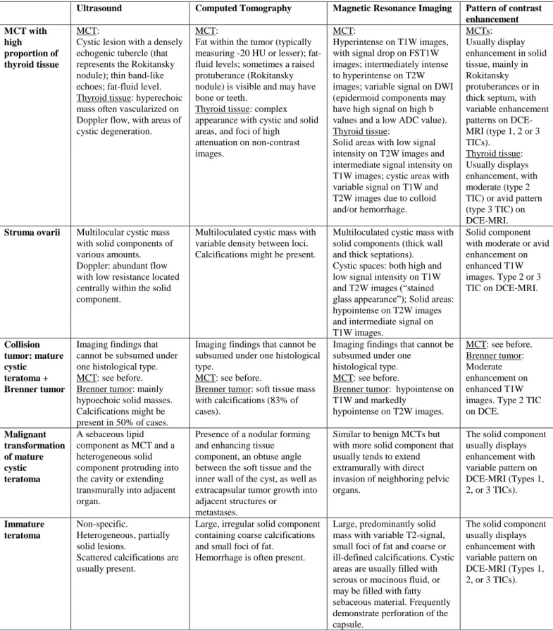
Table 1: Summary table of mature cystic teratoma with high proportion of thyroid tissue characteristics.
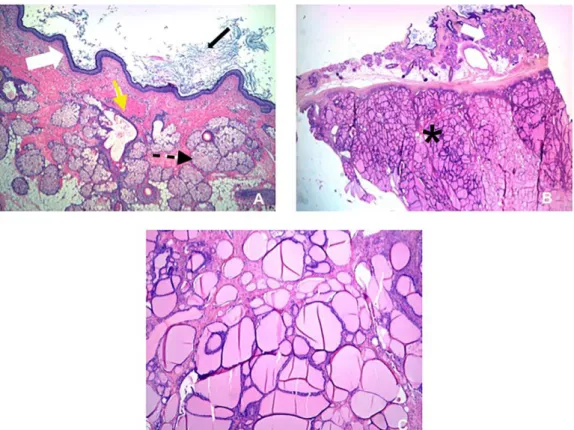
Figure 4: A 37-year-old woman with mature cystic teratoma of the left ovary with high proportion of thyroid tissue.
Microscopic examination of hematoxylin-eosin stained sections.
4A - Photomicrograph of the cyst wall showed squamous cell lining (white arrow), sebaceous glands (black dotted arrow), and intervening muscle (yellow arrow). Sebaceous material and cell debris were filling the lumen (black arrow). 4B - The ectodermal derivates from the cystic wall (white arrow) were clearly separated from thyroid tissue (black asterisks). 4C - Thyroid tissue was composed of numerous small follicles filled with colloid.
Radiology Case. 2017 Jul; 11(7):20-30
Jo
urnal o
f
Rad
io
lo
gy
Case Rep
orts
ww
w.Ra
diol
ogyC
ase
s.c
om
29Ultrasound Computed Tomography Magnetic Resonance Imaging Pattern of contrast enhancement MCT with high proportion of thyroid tissue MCT:
Cystic lesion with a densely echogenic tubercle (that represents the Rokitansky nodule); thin band-like echoes; fat-fluid level. Thyroid tissue: hyperechoic mass often vascularized on Doppler flow, with areas of cystic degeneration.
MCT:
Fat within the tumor (typically measuring -20 HU or lesser); fat-fluid levels; sometimes a raised protuberance (Rokitansky nodule) is visible and may have bone or teeth.
Thyroid tissue: complex appearance with cystic and solid areas, and foci of high
attenuation on non-contrast images.
MCT:
Hyperintense on T1W images, with signal drop on FST1W images; intermediately intense to hyperintense on T2W images; variable signal on DWI (epidermoid components may have high signal on high b values and a low ADC value). Thyroid tissue:
Solid areas with low signal intensity on T2W images and intermediate signal intensity on T1W images; cystic areas with variable signal on T1W and T2W images due to colloid and/or hemorrhage. MCTs: Usually display enhancement in solid tissue, mainly in Rokitansky protuberances or in thick septum, with variable enhancement patterns on DCE-MRI (type 1, 2 or 3 TICs). Thyroid tissue: Usually displays enhancement, with moderate (type 2 TIC) or avid pattern (type 3 TIC) on DCE-MRI.
Struma ovarii Multilocular cystic mass with solid components of various amounts. Doppler: abundant flow with low resistance located centrally within the solid component.
Multiloculated cystic mass with variable density between loci. Calcifications might be present.
Multiloculated cystic mass with solid components (thick wall and thick septations). Cystic spaces: both high and low signal intensity on T1W and T2W images (“stained glass appearance”); Solid areas: hypointense on T2W images and intermediate signal on T1W images.
Solid component with moderate or avid enhancement on enhanced T1W images. Type 2 or 3 TIC on DCE-MRI. Collision tumor: mature cystic teratoma + Brenner tumor
Imaging findings that cannot be subsumed under one histological type. MCT: see before. Brenner tumor: mainly hypoechoic solid masses. Calcifications might be present in 50% of cases.
Imaging findings that cannot be subsumed under one histological type.
MCT: see before.
Brenner tumor: soft tissue mass with calcifications (83% of cases).
Imaging findings that cannot be subsumed under one
histological type. MCT: see before.
Brenner tumor: hypointense on T1W and markedly hypointense on T2W images. MCT: see before. Brenner tumor: Moderate enhancement on enhanced T1W images. Type 2 TIC on DCE. Malignant transformation of mature cystic teratoma A sebaceous lipid component as MCT and a heterogeneous solid component protruding into the cavity or extending transmurally into adjacent organ.
Presence of a nodular forming and enhancing tissue
component, an obtuse angle between the soft tissue and the inner wall of the cyst, as well as extracapsular tumor growth into adjacent structures or
metastases.
Similar to benign MCTs but with more solid component that usually tends to extend extramurally with direct invasion of neighboring pelvic organs.
The solid component usually displays enhancement with variable pattern on DCE-MRI (Types 1, 2, or 3 TICs). Immature teratoma Non-specific. Heterogeneous, partially solid lesions.
Scattered calcifications are usually present.
Large, irregular solid component containing coarse calcifications and small foci of fat.
Hemorrhage is often present.
Large, predominantly solid mass with variable T2-signal, small foci of fat and coarse or ill-defined calcifications. Cystic areas are usually filled with serous or mucinous fluid, or may be filled with fatty sebaceous material. Frequently demonstrate perforation of the capsule.
The solid component usually displays enhancement with variable pattern on DCE-MRI (Types 1, 2, or 3 TICs).
Radiology Case. 2017 Jul; 11(7):20-30
Jo
urnal o
f
Rad
io
lo
gy
Case Rep
orts
ww
w.Ra
diol
ogyC
ase
s.c
om
30 ADC – apparent diffusion coefficientCA-125 – Cancer antigen 125 CT – Computed tomography DCE – Dynamic contrast-enhanced DWI – Diffusion-weighted images IT – immature teratoma
MCT – Mature cystic teratoma MRI – Magnetic resonance imaging US – ultrasound
TIC – Time-intensity curve W – Weighted
Urogenital neoplasms; Ovary; Teratoma; Thyroid gland; Magnetic Resonance Imaging
Online access
This publication is online available at:
www.radiologycases.com/index.php/radiologycases/article/view/2853
Peer discussion
Discuss this manuscript in our protected discussion forum at: www.radiolopolis.com/forums/JRCR
Interactivity
This publication is available as an interactive article with scroll, window/level, magnify and more features.
Available online at www.RadiologyCases.com Published by EduRad
www.EduRad.org ABBREVIATIONS