Echocardiographic Assessment of Cardiac Resynchronization
Therapy: Two-Year Follow-up Period
Viviane Cordeiro Veiga
1,2, Salomón Soriano Ordinola Rojas
1,2, Fernando Sérgio Oliva de Souza
2, Reinaldo Wilson Vieira
1,
Amilton Silva Junior
2, Marcelo Luiz Patrício
2, Elias César Hauy Marum
2, Henry Abensur
2Universidade Estadual de Campinas (UNICAMP)1; Real e Benemérita Associação Portuguesa de Beneficência2 - São Paulo, SP - Brazil
Abstract
Background: The cardiac resynchronization therapy (CRT) is an effective option for patients with advanced heart failure (HF). Clinical, electrocardiographic and echocardiographic criteria have been studied in an attempt to find the patients that will benefit from the CRT, considering that the echocardiogram is the method that is used both in the selection and in the assessment of such therapy.
Objective: The objective of this work is to analyze the use of echocardiogram to assess the CRT, in a ten-day follow-up period and after two years of evolution.
Method: The assessment considered 0 patients subjected to CRT, for a period of two years, 80% of which were male.
The Minnesota Living with Heart Failure Questionnaire (MLWHF) was filled out. Patients underwent a six-minute walking test. Then, the two-dimensional echo-Doppler-cardiogram was performed. The initial assessment was repeated ten days after and two year after the implantation of the biventricular pacemaker.
Results: In two years, 5 patients (5%) died; 4 had cardiomyopathy caused by the Chagas’s disease. There was no statistically significant change in the ejection fraction between the pre-operation period and the following ten days, but there was a significant change between the pre-operation period and two years after that, and the ten-day period and two years after that. In the ten-day follow-up period, there was the worsening of the intraventricular dyssynchrony, as evaluated by the tissue Doppler method, and the “living with heart failure” score was higher in the group of deaths. Conclusion: Out of the echocardiographic parameters assessed, only the intraventricular dyssynchrony assessment through the tissue Doppler method, after the procedure, was capable of predict the CRT efficiency with respect to the death rate. (Arq Bras Cardiol 00; 94() : -8)
Key Words: echocardiography; heart failure / therapy; stroke volume.
Mailing address: Viviane Cordeiro Veiga •
Alameda Itália, 430 - Alphaville Resid 1 - 06474-140 – Barueri, SP – Brazil E-mail: [email protected], [email protected]
Manuscript received Januart 20, 2009; revised manuscript received June 05, 2009; accepted June 22, 2009.
Introduction
Heart failure (HF) is a medical condition with a high morbidity-mortality rate that affects approximately 23 million people in the world1. According to DATASUS2, in Brazil, in
the period between January and July 2008, 147,348 hospital admissions caused by HF.
Intraventricular conduction disturbances with extension of the QRS complex are present in 25-50% of HF patients 3-6, in which the most frequent case is the left bundle branch
block (LBBB)6,7.
The conduction of the electrical stimulus is associated with the functional cardiac efficiency. In patients in whom there is a change in the regular conduction of the stimulus, there may be some interference in the ventricular and/or atrial contractile coordination – called cardiac dyssynchrony. That may cause a change in the myocardial function8,9.
Cazeau et al10, in 1994, described the stimulation of the
left ventricle through the coronary sinus, with a four-chamber pacemaker. In 2001, the cardiac resynchronization therapy (CRT) was approved for clinical use by the Food and Drug Administration (FDA). After that, more than 270,000 patients have already undergone the procedure11. In Brazil, according
to Pachón et al12, in the period from 1994 to 2006, 2,180
resynchronizers were implanted. Nowadays, the CRT, through heart stimulation with a biventricular pacemaker, has been used as an auxiliary therapy in patients that are refractory to optimized medication therapy13,14.
Bakker et al15, in an analysis of 5 patients with dilated
cardiomyopathy, with large QRS and who were refractory to medication, were the first authors to demonstrate the correlation between biventricular stimulation and the improvement in heart performance. After that, many studies were published to assess the effects of biventricular stimulation16-18.
Despite the proven benefits of CRT16-18, approximately
20-30% of patients do not respond to this therapy (they are called “non-responders”19,20). That makes it necessary to adopt
from cardiac resynchronization21,22.
According to the Brazilian Guidelines for Implantable Electronic Cardiac Devices (2007)23, recommendation I and
the evidence level A for CRT is: Patients in functional class III or IV (NYHA), with optimized medication therapy, ejection fraction ≤ 35%, sinus rhythm and duration of the QRS complex > 150 ms or QRS between 120 and 150 ms, with proof of dyssynchrony by the imaging method.
The use of echocardiography as a supplemental method for indication of the CRT was described for the first time in 2005, in the CARE-HF (Cardiac Resynchronization - Heart Failure)24 study, which compared the effect of CRT on the
risk of complications and death. Countless echocardiographic techniques have been proposed to quantify the ventricular dyssynchrony, with the purpose of optimizing the selection of patients for the CRT25.
Pitzalis et al26 used the monodimensional mode (M mode)
to assess the intraventricular dyssynchrony, considering values above 130 ms as abnormal. The pulsed Doppler is an echocardiographic method used to assess the intraventricular dyssynchrony24, by the electromechanical delay difference
between the right and left ventricles, indicating dyssynchrony when the values above 40 ms19,25.
The echocardiogram is also used in the evaluation of the mitral regurgitation. Breithardt et al27 studied its acute effects
on CRT, assessing 24 patients subjected to resynchronization, who did not show significant reduction in the level of mitral insufficiency, directly associated with the dP/dt increase.
The tissue Doppler allows identifying and measuring the myocardial motion speed, by positioning the cursor in the segment that you wish to evaluate28. Bax et al19,in a study
with 85 patients for a 6-month follow-up period after the CRT, demonstrated, among four basal segments, sensitiveness and specificity of 80% as predictor of clinical improvement and 92% as predictor of reverse modeling, in the presence of interval greater than 65 ms and evaluated by the tissue Doppler.
Objective
The objective of this work is to analyze the use of the echocardiogram in the evaluation of the cardiac resynchronization therapy, in patients with refractory HF, in a short follow-up period (ten days) and after two years of evolution.
Case selection and method
The study was based on 20 patients, to whom a biventricular pacemaker implant had been prescribed, through the coronary sinus, for cardiac resynchronization therapy, aged 59.70 ± 12.59 years on average, 16 of which (80%) were male, monitored for a two-year period. The cardiomyopathy had been caused by ischemia in 10 patients (50%), by Chagas’s disease in 6 patients (30%) and by other unknown factors in 4 patients (20%). Fifteen patients (75%) were in functional class III and 5 (25%) in functional class IV, when the resynchronization therapy was prescribed to them.
Study dynamics
When the CRT was prescribed, the patients’ medical history was prepared. Then, they underwent clinical tests and were classified according to the New York Heart Association (NYHA), for a functional evaluation. After the initial clinical evaluation, the Minnesota Living with Heart Failure Questionnaire (MLWHFQ) was filled out. The distance traveled was determined (in meters, through a six-minute walking test) and the two-dimensional echo-Doppler-cardiogram was performed, with the use of a 3.5 Hz transducer in the Nemio equipment (Toshiba). Ten days after the biventricular pacemaker was implanted, the entire initial evaluation was repeated, and the same process was repeated after two years.
Echocardiographic evaluation
Evaluation of the left ventricular function
The ejection fraction of the left ventricle was evaluated through the two-dimensional method (Simpson’s method). In addition, the myocardial performance index (or Tei index) was calculated, for evaluating the systole and diastole functions of the left ventricle, calculated by the sum of isovolumetric contraction and relaxation times divided by the ejection time, considering values below 0.40 as normal.
Evaluation of the reverse remodeling
The reverse remodeling is characterized by a reduction of more than 15% of the final systolic volume, evaluated by the echocardiogram, between the pre-operation and post-operation periods.
Evaluation of the mitral regurgitation
The mitral regurgitation evaluation was conducted with the use of the color flow mapping in the four-chamber apical and parasternal cross-section slices. The mitral regurgitation was quantified by the ratio of the regurgitating jet area to the left atrium area, considering that the regurgitation is minor when the percentage area is smaller than 20% and important when it is larger than 40%.
Evaluation of the interventricular dyssynchrony
The pulsed Doppler method was used to analyze the interventricular dyssynchrony, through the electromechanical delay difference between left and right ventricles, by measuring the time interval between the R wave of the electrocardiogram and the beginning of the speed curve of the aortic flow and pulmonary flow. Interval above 40 ms indicates interventricular dyssynchrony.
Evaluation of the intraventricular dyssynchrony
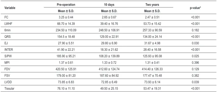
Table 1 - Mean and standard deviation in the comparison of clinical, electrocardiographic and echocardiographic variables throughout time
Variable Pre-operation 10 days Two years p-value*
Mean ± S.D. Mean ± S.D. Mean ± S.D.
FC 3.25 ± 0.44 2.65 ± 0.67 2.47 ± 0.51 <0.001
LWHF 66.70 ± 14.39 39.40 ± 16.76 53.73 ± 15.42 <0.001
6min 234.50 ± 110.09 248.50 ± 106.91 257.33 ± 90.59 0.182
QRS 154.5 ± 18.48 129.00 ± 22.91 134.00 ± 24.14 <0.001
EJ 27.90 ± 5.51 29.80 ± 6.90 31.67 ± 4.98 0.030
INTER 41.90 ± 22.21 18.30 ± 21.62 28.40 ± 16.58 <0.001
S/PW 185.90 ± 95.21 108.20 ± 139.89 125.93 ± 95.08 0.025
MPI 1.37 ± 0.61 1.33 ± 0.72 1.31 ± 0.41 0.396
FDV 420.50 ± 125.91 412.60 ± 124.74 414.40 ± 126.33 0.129
FSV 179.00 ± 81.20 187.60 ± 84.92 177.47 ± 70.48 0.382
LVDD 73.85 ± 6.83 72.85 ± 6.49 73.93 ± 6.14 0.039
Tissular 76.10 ± 11.10 49.50 ± 25.15 53.47 ± 19.31 <0.001
FC – functional class; LWHF – living with heart failure score; 6 min – distance traveled in the six-minute walking test; QRS – QRS complex time length; EF – ejection fraction; INTER – interventricular dyssynchrony; S/PW – time between the maximum contraction of the septum and the posterior wall; MPI – myocardial performance index; FDV – inal diastolic volume; FSV – inal systolic volume; LVDD – left ventricle diastolic diameter; Tissular – use of tissue-Doppler to assess the intraventricular
dyssynchrony.
interval between the maximum contraction of the septum and the posterior wall of the left ventricle (S/PW), considering as dyssynchrony values above 130 ms. In the tissue-Doppler evaluation, the myocardial speeds are obtained in the apical plane of the basal segments of septal, lateral, anterior and inferior walls, and the time interval between the beginning of the QRS complex and the peak of the myocardial systolic wave of the tissue-Doppler is measured in the different segments. There is significant dyssynchrony when the time interval difference is higher than 65 ms between any segments assessed. The values considered were obtained by the average of four consecutive heart beats.
Statistical analysis
The Statistical Package for Social Sciences (SPSS) for Windows, version 11.5, was used for analyzing the data. To run the tests, bilateral hypotheses and a significance level α = 5% were taken into account. Descriptive statistics were used to assess the frequency, average and standard deviation of the variables of interest. The t-test for independent samples was used for the comparison of the age-variable averages. The Mann-Whitney test was used to analyze clinical, electrocardiographic and echocardiographic variables, when they were quantitative variables.
The Chi-square test was used to check if the proportions of the categories of clinical and echocardiographic variables, when they were qualitative variables, were homogenous in groups of interest. For comparing the functional class, “living with heart failure” score and traveled distance, QRS time length variables and echocardiographic variables (ejection fraction, mitral insufficiency, interventricular dyssynchrony, posterior wall/septum distance, myocardial performance index, final diastolic and systolic volume, diastolic diameter of the left ventricle and tissue-Doppler throughout time [before
operation, 10 days and two years]), the NON-PARAMETRIC ANOVA (Analysis of Variance) was used.
Results
In this work, 20 patients in whom a biventricular pacemaker was implanted were monitored for two years. There were neither complications with respect to the surgical procedure nor deaths in the 10-day follow-up period after operation.
An analysis was conducted to assess the behavior of clinical, electrocardiographic and echocardiographic variables, comparing them in the period before and after the operation (10 days after and two days after). When the pacemaker was implanted, 15 patients were in functional class III; 5 in functional class IV; 13 patients (65%) showed improvement in the functional class in the first post-operation evaluation; 6 (30%) kept the same functional class; and there was worsening of the functional class in one patient (5%). In the two-year follow-up period, 10 patients showed no change in the functional class, compared to the previous evaluation, and there was improvement in three patients and worsening in two of them.
In the assessment of variables throughout time, it was possible to find statistically significant differences for the following variables: functional class, living with heart failure score, QRX complex time length, ejection fraction, inter and intraventricular dyssynchrony (time between contraction of septum and the posterior wall – M mode and tissue-Doppler) and diastolic diameter of left ventricle. Table 1 shows the mean and the standard deviation for each one of the variables in the pre-operation period, in the 10 days after the surgery and two years after the CRT.
Table 2 - Mean, standard deviation and p-value referring to the comparison of the means of the functional class variable in the groups of interest in the pre-operation periods, 10 days after and two years after surgery
Variables QRS ≤ 160 QRS > 160 p-value
Mean ± S.D Mean ± S.D
FC pre-operation 3.07 ± 0.26 3.67 ± 0.51 0.006
FC 10 days 2.67 ± 0.69 2.50 ± 0.71 0.853
FC two years 2.43 ± 0.41 3.00 ± 0.0
-FC – functional class; S.D. - standard deviation.
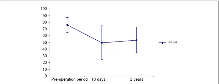
Chart 1 - Use of tissue-Doppler to evaluate the intraventricular dyssynchrony in pre-operation period, after ten days and in the two-year follow-up period (p < 0,001). in the 10-day evaluation; and 134.0 ± 24.14 ms in the
two-year evaluation, with p < 0,001.
When the QRS complex time length and functional class were compared, it was possible to notice that patients with QRS time length, showed a higher functional class, in the pre-operation evaluation (p = 0.006), but there was not statistically significant difference in the post-operation periods (Table 2).
For the functional class, QRX complex time length and tissue-Doppler variables, it was possible to notice statistically significant differences between the pre-operation period and the evaluation 10 days after the procedure, as well as between the pre-operation period and two years after the post-implantation. However, there was no difference between the 10-day and two-year periods. For these variables, it was possible to see a reduction in the measures after the surgical intervention, which remained stable after two years. In Chart 1, it is possible to analyze the variation noticed throughout time by the evaluation of the tissue-Doppler.
For the ejection fraction, we noticed no statistically significant difference between the pre-operation period and the ten days after the operation, but there were differences between the pre-operation period and the next two years and between the ten days and the two subsequent years.
It was possible to find statistically significant differences between all periods in the assessment of the following variables: Interventricular dyssynchrony, posterior wall/septum distance and living with heart failure score. Therefore, there was a significant reduction when we compare the operation period and the 10 days after that and the pre-operation period and the two subsequent years in the three variables analyzed. However, there was a statistically significant increase in the values found between the 10-day period and two-year period of these variables.
In the assessment of the LVDD, there was significant reduction between the pre-operation period and the 10-day period and significant increase between the 10-day period and the two-year period, so there is no statistically significant difference between initial and final measurements.
In our case selection, 5 patients (25%) died. In Table 3, it is possible to see that the proportion of men in the death group to men in the non-death group is homogeneous (86.7% and 60%, respectively), with predominance of men in both groups, and with no significant difference between the proportion of men to women in the two groups (p = 0.249).
For the cause (etiology) variable, in the non-death group, the number of patients with dilated and ischemic etiology is larger than in the death-group. In Chagas-disease etiology, the situation is the opposite: 80% of patients are in the death group, and there is a statistically significant difference in the proportions of the etiology types according to death (p = 0.018).
There was no statistically significant difference between the death and non-death groups for the mitral insufficiency variable and QRS complex time length variable, both in the pre-operation period and after 10 days.
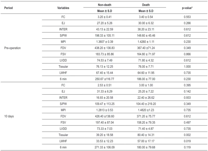
Table 4 - Mean, standard deviation and p-value for the comparison of the averages of variables of interest, in death and non-death groups, in the pre-operation period and 10 days after surgery
Period Variables Non-death Death p-value*
Mean ± S.D Mean ± S.D
Pre-operation
FC 3.20 ± 0.41 3.40 ± 0.54 0.553
EJ 27.20 ± 5.26 30.00 ± 6.32 0.266
INTER 43.13 ± 22.59 38.20 ± 23.11 0.612
S/PW 198.33 ± 105.11 148.60 ± 45.46 0.612
MPI 1.3607 ± 0.39 1.4260 ± 1.11 0.230
FDV 438.20 ± 136.83 367.40 ±71.24 0.349
FSV 183.73 ± 85.86 164.80 ± 71.97 0.866
LVDD 74.53 ± 7.49 71.80 ± 4.32 0.612
Tissular 76.13 ± 12.25 76.00 ± 7.71 1.000
LWHF 67.40 ± 15.44 64.60 ± 11.95 0.735
6 min 250.67 ±116.77 186.00 ± 77.00 0.230
10 days
FC 2.53 ± 0.51 3.00 ± 1.00 0.395
EJ 31.33 ± 6.29 25.20 ± 7.22 0.142
INTER 16.93 ± 20.58 22.40 ± 26.62 0.933
S/PW 109.47 ± 113.25 104.40 ± 219.20 0.349
MPI 1.2813 ± 0.53 1.4820 ±1.23 0.735
FDV 426.40 ±136.60 371.20 ± 75.77 0.612
FSV 197.40 ± 87.04 158.20 ± 79.35 0.497
LVDD 73.33 ± 7.03 71.40 ± 4.87 0.735
Tissular 39.20 ± 18.58 80.40 ± 14.31 0.002
LWHF 33.53 ± 12.23 57.00 ± 17.17 0.019
6 min 271.33 ± 106.09 180.00 ± 79.68 0.119
*Mann-Whitney. (FC – functional class; EF – ejection fraction; INTER – assessment of the interventricular dyssynchrony; S/PW – distance between the maximum contraction of the septum and the posterior wall; IPM – myocardial performance index; FDV – inal diastolic volume; FSV – inal systolic volume; LVDD – left ventricle
diastolic diameter; Tissural – tissue-Doppler, LWHF – living with heart failure score; 6 min – distance traveled in the six-minute walking test).
Table 3 - Frequency and percentage of patients in the death group and non-death group, according to gender and cause (etiology)
Non-death Death
p-value*
N (%) N (%)
Gênero
Female 2
(13.3)
2 (40.0)
0.249
Male 13
(86.7)
3 (60.0)
Etiology (cause)
Dilated 4
(26.7)
0 (0.0)
0.018
Ischemia 9
(60.0)
1 (20.0)
Chagas’s disease
2 (13.3)
4 (80.0)
*Chi-square of Pearson.
There was no statistically significant difference between the death and non-death group, in the assessment of pre-operation variables. However, in the 10-day assessment, there was a statistically significant difference in the living with heart failure score, and the living with heart failure score of death group patients is higher than that of non-death group patients. The same thing happened for the tissue-Doppler variable, in which the values of the death group suggested greater intraventricular dyssynchrony.
Discussion
HF is a serious public health problem, with high morbidity-mortality rate and costs above 33 billion dollars a year1.
et al7, assessed, in 2002, the link between the LBBB and the
mortality rate and demonstrated that LBBB is an unfavorable prognostic marker and regardless of age, HF level and use of therapy.
Nowadays, the CRT has been used as an auxiliary therapy in patients that are refractory to optimized medication therapy16,18.
In our case selection, we assessed 20 patients, 15 of which were in functional class III and 5 in functional class IV, who had undergone the CRT through the coronary sinus, and without complications inherent in the procedure. All patients show LBBB in the electrocardiogram, with QRS complex time length above 120 ms.
Despite the proven benefits of CRT16-18, approximately
20-30% of patients do not respond to this therapy19,20. That
makes it necessary to implement additional criteria to identify patients that will be entitled to this benefit21,22.
In this case selection, in the follow-up period of 10 days after implantation of the biventricular pacemaker, there was an improvement in the functional class in 13 patients (65%), worsening in one of them (5%) and in 6 patients (30%) the functional class remained unchanged. After two years of evolution, 8 patients were in functional class II and 7 in functional class III. The mortality in this period was of 25% (5 patients), with no correlation between the death and the worsening of the functional class.
In the assessment of the causes of the cardiomyopathy, Reuter et al29 associated the dilated cardiomyopathy with
the better clinical response of CRT. Martinelli Filho et al30,
in a study including patients whose heart failure resulted from Chagas’s disease, there was a significant reduction in the functional class in patients with dilated cardiomyopathy, besides the fact that this etiology had been a predictor, regardless of clinical improvement.
Among the patients of this study, the ones whose cardiomyopathy had been caused by Chagas’s disease were linked to the higher death rate, with statistically significant when compared to patients with ischemic and dilated etiology.
Shamim et al8 assessed, in a 36-month follow-up period,
the association between the QRS duration and the mortality, where patients with QRS below 120 ms showed a mortality of 20%; QRS between 120 and 160 ms, 36%; and above 160 ms, the mortality was of 58%31.
In our case selection, comparing the functional class and the QRS time length, a statistically significant difference was found between the groups in the pre-CRT period, where the group with QRS ≥ 160 ms showed a worse functional class31.
In the 10-day follow-up period, there was no statistically significant difference between the two groups. In the two-year assessment, there was only one patient alive with QRS time length above 160 ms.
Many echocardiographic techniques have been used to quantify the interventricular and intraventricular dyssynchronies, with the purpose of optimizing the selection of patients for CRT.
The assessment of the interventricular dyssynchrony
by using the M-mode of the echocardiogram (S/PW) was proposed by Pitzalis et al26 – in a group of 20 patients. All
responders had an S/PW distance of more than 130 ms. In this test, we had a statistically significant reduction in the values of the S/PW value, when the three analysis periods were compared. However, there was no statistical difference in the death group compared to the non-death group in the pre-operation evaluation and in the 10-day follow-up period. In patients with ischemic cardiomyopathy, the S/PW variable analysis may be impaired by segmental contraction changes.
With the pulsed Doppler, Chung et al32 showed the link
between this variable and the clinical improvement and reverse remodeling.
In our group, there was a statistically significant difference in the pre-operation period and in the two post-operation evaluation periods. However, when we compared the values of the death group and non-death group, there was no statistically significant difference.
In the mitral regurgitation evaluation, in 10 days, 11 patients showed no changes in the mitral regurgitation level. In the two-year follow-up period, 10 patients had the same level of regurgitation that they had in the pre-operation period in the first follow-up period (10 days) and subsequent worsening in the two-year assessment. In the comparison of the death-group and non-death group, the mitral insufficiency value was not statistically significant.
In a study with 85 patients, in a 6-month follow-up period after the CRT, Bax et al19 demonstrated the sensitiveness and
specificity of 80% as a predictor of clinical improvement and 92% of sensitiveness and specificity as a predictor of reverse remodeling, when the dyssynchrony value assessed by the tissue-Doppler was greater than 65 ms.
In our group, we had a statistically significant difference in the variation of the tissue-Doppler of the pre-operation and post-operation periods. And, it was possible to consider that the tissue-Doppler evaluation in the first days after the procedure is a useful tool to assess the CRT’s efficiency. When compared to the death and non-death groups, the tissue-Doppler revealed an increase associated with the death group.
In this study, more advanced echocardiography techniques, such as the three-dimensional echocardiogram and the bidimensional strain, were not used. The use of such techniques, in the future, may expand the application of echocardiography in the evaluation of patients for CRT.
Study limitations
- Small case selection, with absence in the control group; - Echocardiographic evaluation without intra and interobserver reproducibility study.
Conclusion
References
1. McAlister FA, Teo KK, Taher M. Insights into the contemporary epidemiology and outpatient management of congestive heart failure. Am Heart J. 1999; 138: 87-94.
2. Ministério da Saúde. DATASUS. Sistema de Informações hospitalares do SUS. [Acesso em 2009 jan 8]. Disponível em: http://www.datasus.gov.br.
3. Furman S, Robinson G. Use of intracardiac pacemaker in correction of total heart block. Surg Forum. 1958; 9: 245.
4. Horwich T, Foster E, De Marco T, Tseng Z, Saxon L. Effects of resynchronization therapy on cardiac function in pacemaker patients “upgraded” to biventricular devices. J Cardiovasc Electrophysiol. 2004; 15 (11): 1284-9.
5. Aaronson KD, Schwartz JS, Chen TM, Wong KL, Goin JE, Mancini DM. Development and prospective validation of a clinical index to predict survival in ambulatory patients referred for cardiac transplant evaluation. Circulation. 1997; 95 (12): 2660-7.
6. Leclercq C, Hare JM. Ventricular resynchronization: current state of the art. Circulation. 2004; 109 (3): 296-9.
7. Baldasseroni S, Opasich C, Gorini M, Lucci D, Marchionni M, Marini M, et al. Left bundle-branch block is associated with increased 1-year sudden and total mortality rate in 5517 outpatients with congestive heart failure: a report from the Italian Network on congestive heart failure. Am Heart J. 2002; 143 (3): 398-405.
8. Shamim W, Francis DP, Yousufuddin M, Varney S, Pieopli MF, Anker SD, et al. Intraventricular conduction delay: a prognostic marker in chronic heart failure. Int J Cardiol. 1999; 70 (2): 171-8.
9. Rosenqvist M, Isaaz K, Botvinick EH, Dae MW, Cockrell J, Aboot JA, et al. Relative importance of activation sequence compared to atrioventricular synchrony in left ventricular function. Am J Cardiol. 1991; 67: 148-56.
10. Cazeau, S, Ritter P, Bakdach S. Four chamber pacing in dilated cardiomyopathy. Pacing Clin Electrophysiol. 1994; 17 (11 Pt 2): 1974-9.
11. Aranda JM, Woo GW, Schofield RS, Handberg EM, Hill JA, Curtis AB, et al. Management of heart failure after cardiac resynchronization therapy: integrating advanced heart failure treatment with optimal device function. J Am Coll Cardiol, 2005; 46 (12): 2193-8.
12. Pachón MJC, Mosquéra JAP, Pachón MJC, Vargas RNA, Campos Neto CM, Costa ARB. Aspectos epidemiológicos da estimulação cardíaca no Brasil - 12º ano do RBM – Registro brasileiro de marcapassos, desfibriladores e ressincronizadores cardíacos. Relampa. 2008; 21 (1): 5-12.
13. Díaz-Infante E, Hernández-Madrid A, Brugada-Terradellas J, Férnandez-Lozano I, García-Bolao I, Del Ojo JL, et al. Consenso sobre la terapia de resincronización cardíaca. Rev Esp Cardiol. 2005; 5 (Supl): 3B-11B.
14. Brito Jr HL, Bianchi FN, Nascimento LEP, Toledo RM, Barral MM. Estimulação cardíaca artificial como tratamento dos pacientes com miocardiopatia dilatada e insuficiência cardíaca. Novos conceitos, novas técnicas e necessidades de atualização. Reblampa. 2000; 13 (4): 185-93.
15. Bakker PF, Meijburg H, Dejonge N, Mechelen RV, Wittkampf F, Mower M, et al. Beneficial effects of biventricular pacing in congestive heart failure. [abstract] Pacing Clin Electrophysiol. 1994; 17: 820.
16. Abraham WT, Fisher WG, Smith AL, Delurgio DB, Leon AR, Loh E, et al. for the Multicenter InSync randomized clinical evaluation (MIRACLE) study group.
Cardiac resynchronization in chronic heart failure. N Engl J Med. 2002; 346 (24): 1845-53.
17. Bristow MR, Saxon LA, Boehmer J, Krueger S, Kass DA, De Marco T, et al. for the Comparison of Medical Therapy, Pacing, and Defibrillation in Heart Failure (COMPANION) Investigators. Cardiac-resynchronization therapy with or without an implantable defibrillator in advanced chronic heart failure. N Engl J Med. 2004; 350 (21): 2140-50.
18. Cazeau S, Leclercq C, Lavergne T, Walker S, Varma C, Linde C, et al. for the Multisite Stimulation in Cardiomyopathies (MUSTIC) Study Investigators. Effects of multisite biventricular pacing in patients with heart failure and intraventricular conduction delay. N Engl J Med. 2001; 344 (12): 873-80.
19. Bax JJ, Bleeker GB, Marwick TH, Molhoek SG, Boersma E, Steendijk P, et al. Left ventricular dyssynchrony predicts response and prognosis after cardiac resynchronization therapy. J Am Coll Cardiol. 2004; 44: 1834-40.
20. Saxon LA, Ellenbogen KA. Resynchronization therapy for the treatment of heart failure. Circulation. 2003; 108: 1044-8.
21. Galvão Filho SS, Vasconcelos JTM, Barcelos CB, Rabelo AC. Seleção de pacientes e modos de estimulação cardíaca no tratamento da disfunção ventricular. Rev Soc Cardiol Estado de São Paulo. 2004; 1: 43-54.
22. Díaz-Infante E, Mont L, Leal J, García-Bolao I, Fernández-Lozano I, Hernández-Madrid A, et al. of the Spanish Cardiac Resynchronization Study (SCARS). Predictors of lack of response to resynchronization therapy. Am J Cardiol. 2005; 95: 1436-40.
23. Sociedade Brasileira de Cardiologia. Diretrizes brasileiras de dispositivos cardíacos eletrônicos implantáveis. Arq Bras Cardiol. 2007; 89 (6): e210-e238.
24. Cleland JGF, Daubert JC, Erdmann E, Freemantle N, Gras D, Kappenberger L, et al. for the Cardiac Resynchronization – Heart failure (CARE-HF) Study Investigators. The effect of cardiac resynchronization on morbidity and mortality in heart failure. N Engl J Med. 2005; 352: 1539-49.
25. Gorcsan IIII J, Abraham T, Agler DA, Bax JJ, Derumeaux G, Grimm RA, et al. Echocardiography for cardiac resynchronization therapy: recommendations for performance and reporting – a report from the American Society of Echocardiography Dyssynchrony writing group endorsed by the Heart Rhythm Society. J Am Soc Echocardiogr. 2008; 21 (3): 191-213.
26. Pitzalis MV, Iacoviello M, Romito R, Massari F, Rizzon B, Luzzi G, et al. Cardiac resynchronization therapy tailored by echocardiographic evaluation of ventricular asynchrony. J Am Coll Cardiol. 2002; 40: 1615-22.
27. Breithdardt OA, Sinha AM, Schwammenthal E, Bidaoui N, Markus KU, Franke A, et al. Acute effects of cardiac resynchronization therapy on functional mitral regurgitation in advanced systolic heart failure. J Am Coll Cardiol. 2003; 41: 765-70.
28. Silva CES, Barreto ACP. Avaliação ecocardiográfica da terapia de ressincronização cardíaca. Arq Bras Cardiol. 2005; 84 (6): 503-7.
29. Reuter S, Garrigue S, Barold SS, Jais P, Hocini M, Haissaguerre M, et al. Comparison of characteristics in responders versus nonresponders with biventricular pacing for drug-resistant congestive heart failure. Am J Cardiol. 2002; 89: 346-50.
30. Martinelli Filho M, Baggio Jr JM, Nishioka SAD, Pedrosa A, Torres GG, Escarião A, et al. Ressincronização cardíaca em seguimento tardio: análise capable of predicting the cardiac resynchronization efficiency
with respect to mortality. The echocardiographic parameters are not associated with the clinical improvement.
Potential Conflict of Interest
No potential conflict of interest relevant to this article was reported.
Sources of Funding
There were no external funding sources for this study.
Study Association
de preditores de resposta clínica. Reblampa. 2006; 19 (1): 45-52.
31. Achilli A, Sassara M, Ficili S, Pontillo D, Achilli P, Alessi C, et al. Long-term effectiveness of cardiac resynchronization therapy in patients with refractory heart failure and “narrow” QRS. J Am Coll Cardiol. 2003; 42 (12):
2117-24.